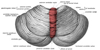
3C syndrome

Last updated
3C syndrome
Other namesCCC dysplasia, Craniocerebellocardiac dysplasia [1] or Ritscher–Schinzel syndrome, [2]
PBB GE KIAA0196 201985 at tn.png
Gene expression pattern of the KIAA0196 gene.
Specialty Medical genetics   OOjs UI icon edit-ltr-progressive.svg

3C syndrome is a rare condition whose symptoms include heart defects, cerebellar hypoplasia, and cranial dysmorphism. It was first described in the medical literature in 1987 by Ritscher and Schinzel, for whom the disorder is sometimes named.

Contents

Signs and symptoms

The classical triad of symptoms that defines 3C syndrome includes certain heart defects, hypoplasia (underdevelopment) of the cerebellum, and cranial dysmorphisms, which can take various forms. The heart defects and cranial dysmorphisms are heterogeneous in individuals who are all classed as having Ritscher-Schinzel syndrome. [2]

Heart defects commonly seen with Ritscher-Schinzel syndrome are associated with the endocardial cushion and are the most important factor in determining a diagnosis. The mitral valve and tricuspid valve of the heart can be malformed, the atrioventricular canal can be complete instead of developing into the interatrial septum and interventricular septum, and conotruncal heart defects, which include tetralogy of Fallot, double outlet right ventricle, transposition of the great vessels, [2] and hypoplastic left heart syndrome. Aortic stenosis and pulmonary stenosis have also been associated with 3C syndrome. [3]

The cranial dysmorphisms associated with 3C syndrome are heterogeneous and include a degree of macrocephaly, a large anterior fontanel, a particularly prominent occiput and forehead, ocular hypertelorism (wide-set eyes), slanted palpebral fissures, cleft palate, a depressed nasal bridge, cleft palate with associated bifid uvula, [2] low-set ears, micrognathia (an abnormally small jaw), [4] brachycephaly (flattened head), and ocular coloboma. [3] Low-set ears are the most common cranial dysmorphism seen in 3C syndrome, and ocular coloboma is the least common of the non-concurrent symptoms (cleft lip co-occurring with cleft palate is the least common). [5]

Cranial dysplasias associated with 3C syndrome are also reflected in the brain. Besides the cerebellar hypoplasia, cysts are commonly found in the posterior cranial fossa, the ventricles and the cisterna magna are dilated/enlarged, and Dandy–Walker malformation is present. These are reflected in the developmental delays typical of the disease. [4] [5] 75% of children with 3C syndrome have Dandy-Walker malformation and hydrocephalus. [6]

Signs and symptoms in other body systems are also associated with 3C syndrome. In the skeletal system, ribs may be absent, and hemivertebrae, syndactyly (fusion of fingers together), and clinodactyly (curvature of the fifth finger) may be present. [3] [6] In the GI and genitourinary systems, anal atresia, hypospadia (misplaced urethra), and hydronephrosis may exist. Adrenal hypoplasia and growth hormone deficiency are associated endocrine consequences of Ritscher-Schinzel syndrome. [3] Some immunodeficiency has also been reported in connection with 3C syndrome. [6] Many children with the disorder die as infants due to severe congenital heart disease. [4] The proband of Ritscher and Schinzel's original study was still alive at the age of 21. [7] A fetus with 3C syndrome may have an umbilical cord with one umbilical artery instead of two. [3] [7]

Genetics

3C syndrome has an autosomal recessive pattern of inheritance. This means that two parents with one copy of the gene each will not have the disease themselves, but can pass on the gene to their children. Statistically, one in four of these children will inherit both copies of the recessive gene and develop the disease. Autorecessive.svg
3C syndrome has an autosomal recessive pattern of inheritance. This means that two parents with one copy of the gene each will not have the disease themselves, but can pass on the gene to their children. Statistically, one in four of these children will inherit both copies of the recessive gene and develop the disease.

3C syndrome is an autosomal recessive disease, caused by a mutation on the long arm of chromosome 8 at 8q24.13, the locus for KIAA0196, [4] the gene for the protein strumpellin. Strumpellin is highly expressed in skeletal muscle cells and mutations in it are also associated with spastic paraplegia. Strumpellin is involved in endosomal transport and cell death processes. [8] The mutation occurs at a splice site and causes a substantial decrease in the amount of strumpellin produced by the cell. The phenotype is similar to 6pter-p24 deletion syndrome and 6p25 deletion syndrome but has a different etiology. [4] [7]

Screening for the condition prenatally may be done with ultrasound.
  • First-trimester ultrasounds can detected nuchal abnormalities
  • Second-trimester ultrasounds can pick up characteristic major structural abnormalities. [9]
Prenatal diagnosis is possible through genetic testing.
  • Chorionic villus sampling or chorionic villus biopsy (CVS) in the first-trimester.
  • Amniocentesis in the second-trimester.

Because 3C syndrome is an autosomal recessive disorder, parents with one child with the disorder have a 25% chance of having another child with the disorder. [4]

Diagnosis

Differential diagnosis

There is an overlap in symptoms between 3C syndrome and Joubert syndrome. Joubert syndrome often manifests with similar cerebellar hypoplasia and its sequelae, including hyperpnea, ataxia, changes in eye movement, and cleft lip and palate. Occasionally, Joubert syndrome will include heart malformations. Brachmann–de Lange syndrome must also be differentiated from 3C syndrome. It presents with similar craniofacial and heart abnormalities and can include Dandy–Walker phenotype, making it difficult to distinguish. Dandy-Walker malformation is also occasionally seen in Ellis–Van Creveld syndrome, which is characterized by heart defects and malformed alveolar ridge. [5] Many disorders include the Dandy–Walker phenotype and thus it is not pathognomonic for 3C syndrome. [10]

CHARGE syndrome can also be misdiagnosed. This is because both CHARGE syndrome and 3C syndrome share symptoms of ocular colobomas, cardiac defects, growth retardation, and minor facial abnormalities. [2]

Coffin–Siris syndrome presents with fifth-finger deformities and congenital heart defects. It is distinguished from 3C syndrome by differences in facial dysmorphisms. [6]

Management

The outcome of this disease is dependent on the severity of the cardiac defects. Approximately 1 in 3 children with this diagnosis require shunting for the hydrocephaly that is often a consequence. Some children require extra assistance or therapy for delayed psychomotor and speech development, including hypotonia. [2]

Prognosis

Prognoses for 3C syndrome vary widely based on the specific constellation of symptoms seen in an individual. Typically, the gravity of the prognosis correlates with the severity of the cardiac abnormalities. For children with less severe cardiac abnormalities, the developmental prognosis depends on the cerebellar abnormalities that are present. Severe cerebellar hypoplasia is associated with growth and speech delays, as well as hypotonia and general growth deficiencies. [5]

Epidemiology

3C syndrome is very rare, occurring in less than 1 birth per million. [2] Because of consanguinity due to a founder effect, it is much more common in a remote First Nations village in Manitoba, where 1 in 9 people carries the recessive gene. [4]

History

The syndrome was first reported in 1987 in two sisters who had similar craniofacial abnormalities, Dandy–Walker phenotype, and congenital heart abnormalities. Neither of the parents was affected, indicating that the disorder was transmitted in an autosomal recessive pattern. [4] [11] The syndrome's symptoms were further refined in 1989 when the third case of the syndrome was reported, with similar craniofacial abnormalities to the first two cases, ventricular septal defect, and enlargement of the cisterna magna and fourth ventricle of the brain. [5]

Other animals

Animal models of 3C syndrome have not been created; however, strumpellin is a highly conserved protein, with 12 known homologs and 83 known orthologs. [8]

Related Research Articles

<span class="mw-page-title-main">Cerebellar hypoplasia</span> Medical condition

Cerebellar hypoplasia is characterized by reduced cerebellar volume, even though cerebellar shape is (near) normal. It consists of a heterogeneous group of disorders of cerebellar maldevelopment presenting as early-onset non–progressive congenital ataxia, hypotonia and motor learning disability.

<span class="mw-page-title-main">Dandy–Walker malformation</span> Congenital malformation of the cerebellar vermis

Dandy–Walker malformation (DWM), also known as Dandy–Walker syndrome (DWS), is a rare congenital brain malformation in which the part joining the two hemispheres of the cerebellum does not fully form, and the fourth ventricle and space behind the cerebellum are enlarged with cerebrospinal fluid. Most of those affected develop hydrocephalus within the first year of life, which can present as increasing head size, vomiting, excessive sleepiness, irritability, downward deviation of the eyes and seizures. Other, less common symptoms are generally associated with comorbid genetic conditions and can include congenital heart defects, eye abnormalities, intellectual disability, congenital tumours, other brain defects such as agenesis of the corpus callosum, skeletal abnormalities, an occipital encephalocele or underdeveloped genitalia or kidneys. It is sometimes discovered in adolescents or adults due to mental health problems.

<span class="mw-page-title-main">Larsen syndrome</span> Medical condition

Larsen syndrome (LS) is a congenital disorder discovered in 1950 by Larsen and associates when they observed dislocation of the large joints and face anomalies in six of their patients. Patients with Larsen syndrome normally present with a variety of symptoms, including congenital anterior dislocation of the knees, dislocation of the hips and elbows, flattened facial appearance, prominent foreheads, and depressed nasal bridges. Larsen syndrome can also cause a variety of cardiovascular and orthopedic abnormalities. This rare disorder is caused by a genetic defect in the gene encoding filamin B, a cytoplasmic protein that is important in regulating the structure and activity of the cytoskeleton. The gene that influences the emergence of Larsen syndrome is found in chromosome region, 3p21.1-14.1, a region containing human type VII collagen gene. Larsen syndrome has recently been described as a mesenchyme disorder that affects the connective tissue of an individual. Autosomal dominant and recessive forms of the disorder have been reported, although most cases are autosomal dominant. Reports have found that in Western societies, Larsen syndrome can be found in one in every 100,000 births, but this is most likely an underestimate because the disorder is frequently unrecognized or misdiagnosed.

Abruzzo–Erickson syndrome is an extremely rare disorder characterized by deafness, protruding ears, coloboma, a cleft palate or palatal rugosity, radial synostosis, and short stature. It was first characterized by Abruzzo and Erickson in 1977 as a CHARGE like syndrome as variably expressed among a family of two brothers, their mother, and their maternal uncle. Members of this family exhibited many of the CHARGE symptoms, but notably did not have choanal atresia and the brothers experienced typical genital development. Due to the recent discovery of this disorder, its etiology is not fully known but it is understood that it arises from mutations on the TBX22 gene on the X-chromosome. The disorder is inherited in an X-linked recessive manner. There is currently no known cure but its symptoms can be treated.

<span class="mw-page-title-main">Young–Simpson syndrome</span> Medical condition

Young–Simpson syndrome (YSS) is a rare congenital disorder with symptoms including hypothyroidism, heart defects, facial dysmorphism, cryptorchidism in males, hypotonia, intellectual disability, and postnatal growth retardation.

<span class="mw-page-title-main">Frontonasal dysplasia</span> Medical condition

Frontonasal dysplasia (FND) is a congenital malformation of the midface. For the diagnosis of FND, a patient should present at least two of the following characteristics: hypertelorism, a wide nasal root, vertical midline cleft of the nose and/or upper lip, cleft of the wings of the nose, malformed nasal tip, encephalocele or V-shaped hair pattern on the forehead. The cause of FND remains unknown. FND seems to be sporadic (random) and multiple environmental factors are suggested as possible causes for the syndrome. However, in some families multiple cases of FND were reported, which suggests a genetic cause of FND.

<span class="mw-page-title-main">Fryns syndrome</span> Medical condition

Fryns syndrome is an autosomal recessive multiple congenital anomaly syndrome that is usually lethal in the neonatal period. Fryns (1987) reviewed the syndrome.

<span class="mw-page-title-main">Roberts syndrome</span> Medical condition

Roberts syndrome, or sometimes called pseudothalidomide syndrome, is an extremely rare autosomal recessive genetic disorder that is characterized by mild to severe prenatal retardation or disruption of cell division, leading to malformation of the bones in the skull, face, arms, and legs.

<span class="mw-page-title-main">Goldberg–Shprintzen syndrome</span> Medical condition

Goldberg–Shprintzen is a very rare connective tissue condition associated with mutations in KIAA1279 gene which encodes KIF-binding protein (KBP), a protein that may interact with microtubules and actin filaments. KBP may play a key role in cytoskeleton formation and neurite growth.

<span class="mw-page-title-main">13q deletion syndrome</span> Medical condition

13q deletion syndrome is a rare genetic disease caused by the deletion of some or all of the large arm of human chromosome 13. Depending upon the size and location of the deletion on chromosome 13, the physical and mental manifestations will vary. It has the potential to cause intellectual disability and congenital malformations that affect a variety of organ systems. Because of the rarity of the disease in addition to the variations in the disease, the specific genes that cause this disease are unknown. This disease is also known as:

<span class="mw-page-title-main">Pascual-Castroviejo syndrome type 1</span> Medical condition

Pascual-Castroviejo syndrome type 1 is a rare autosomal recessive condition characterized by facial dysmorphism, cognitive impairment and skeletal anomalies.

Fine-Lubinsky syndrome is a rare genetic disorder which is characterized by ocular and hearing problems, speech and developmental delay, short stature, intellectual disabilities and facial dysmorphisms.

<span class="mw-page-title-main">Porencephaly-cerebellar hypoplasia-internal malformations syndrome</span> Medical condition

Porencephaly-cerebellar hypoplasia-internal malformations syndrome is a rare autosomal recessive syndrome that mainly affects the central nervous system. It causes cardiac defects, brain anomalies, and craniofacial dysmorphisms. It has been reported in a pair of German siblings of the opposite sex born to consanguineous Turkish parents.

<span class="mw-page-title-main">Sloping forehead</span> Medical condition

Sloping forehead is a condition in which the forehead is objectively vertically inclined more than two standard deviations, or the forehead shows subjectively excessive posterior sloping when viewed in profile.

References

  1. Disease ID 5666 at NIH's Office of Rare Diseases
  2. 1 2 3 4 5 6 7 "3C syndrome". Orphanet. Retrieved 11 April 2014.
  3. 1 2 3 4 5 Kniffin, Cassandra L.; Jackson, John F. (6 January 2014). "Ritscher-Schinzel Syndrome - Clinical Synopsis". Online Mendelian Inheritance in Man. Johns Hopkins University. Retrieved 12 April 2014.
  4. 1 2 3 4 5 6 7 8 Kniffin, Cassandra L.; McCusick, Victor A. (6 January 2014). "Ritscher-Schinzel Syndrome". Online Mendelian Inheritance in Man. Johns Hopkins University. Retrieved 11 April 2014.
  5. 1 2 3 4 5 Leonardi, Michael L.; Pai, G. Shashidhar; Wilkes, Beth; Lebel, Robert Roger (15 August 2001). "Ritscher-Schinzel cranio-cerebello-cardiac (3C) syndrome: Report of four new cases and review". American Journal of Medical Genetics. 102 (3): 237–242. doi:10.1002/ajmg.1449. PMID   11484200.
  6. 1 2 3 4 Gorlin, Robert J.; Cohen Jr., Michael; Hennekam, Raoul C.M. (2001). Syndromes of the Head and Neck (4 ed.). Oxford University Press. ISBN   9780199747726.
  7. 1 2 3 Jones, Kenneth Lyons; Jones, Marilyn Crandall; del Campo, Miguel (2013). Smith's Recognizable Patterns of Human Malformation (7th ed.). Elsevier Health Sciences. ISBN   9780323186681.
  8. 1 2 "KIAA0196". NIH. Retrieved 12 April 2014.
  9. Rusnak, A. J., Hadfield, M. I., Chudley, A. E., Marles, S. L., Reid, G. J., & Chodirker, B. N. (2008). Increased Nuchal Translucency Thickness: A Potential Indicator for Ritscher-Schinzel Syndrome. Fetal Diagnosis & Therapy, 24(4), 395-399. doi : 10.1159/000165697
  10. Albright, A. Leland; Pollack, Ian F. (2011). Principles and Practice of Pediatric Neurosurgery. Thieme. ISBN   9781604064605.
  11. Ritscher, D.; Schinzel, A.; Boltshauser, E.; Briner, J.; Arbenz, U.; Sigg, P. (February 1987). "Dandy-Walker(like) malformation, atrio-ventricular septal defect and a similar pattern of minor anomalies in 2 sisters: a new syndrome?". American Journal of Medical Genetics. 26 (2): 481–491. doi:10.1002/ajmg.1320260227. PMID   3812597.