3D cell culturing by magnetic levitation

Last updated
3D Cell Culturing by Magnetic Levitation Introduction Picture.jpg

3D cell culturing by Magnetic LevitationMethod (MLM) is the application of growing 3D tissue by inducing cells treated with magnetic nanoparticle assemblies in spatially varying magnetic fields, using neodymium magnetic drivers and promoting cell-to-cell interactions by levitating the cells up to the air/liquid interface of a standard petri dish. The magnetic nanoparticle assemblies consist of magnetic iron oxide nanoparticles, gold nanoparticles, and the polymer polylysine. 3D cell culturing is scalable, with the capability of culturing as few as 500 cells up to millions of cells, or from a single dish to high-throughput low volume systems. [1] [2] [3] Once magnetized cultures are generated, they can also be used as the building block material, or the "ink" for the magnetic 3D bioprinting process.

Contents

Overview

Standard monolayer cell culturing on tissue culture plastic does not replicate the complex 3D architecture of in vivo tissue, and it can significantly modify cell properties. This often compromises experiments in basic life science, leads to misleading drug-screening results on efficacy and toxicity, and produces cells that may lack the characteristics needed for developing tissue regeneration therapies. [1] [2] [4] [5] [6] [7] [8] [9]

The future of cell culturing for fundamental studies and biomedical applications lies in the creation of multicellular structure and organization in three dimensions. [4] [5] [7] [8] [10] [11] [12] Many schemes for 3D culturing are being developed or marketed, such as bio-reactors [13] or protein-based gel environments. [7] [14]

A 3D cell culturing system known as the Bio-Assembler uses biocompatible polymer-based [1] reagents to deliver magnetic nanoparticles to individual cells so that an applied magnetic driver can levitate cells off the bottom of the cell culture dish and rapidly bring cells together near the air-liquid interface. This initiates cell-cell interactions in the absence of any artificial surface or matrix. Magnetic fields are designed to rapidly form 3D multicellular structures in as little as a few hours, including expression of extracellular matrix proteins. The matrix, protein expression, and response to exogenous agents of resulting tissue show great similarity to in vivo results. [1]

History

3D cell culturing by magnetic levitation method (MLM) was developed from collaboration between scientists at Rice University and University of Texas MD Anderson Cancer Center in 2008. [1] Since then, this technology has been licensed and commercialized by Nano3D Biosciences. [15]

The magnetic levitation process

3D Cell Culturing by Magnetic Levitation, The Magnetic Levitation Process.png

Above is a picture showing 3D cell culturing through magnetic levitation with the Bio-Assembler cell culturing system. The figure's letters refer to the following:

(A) A magnetic iron oxide nanoparticle assembly known as the "Nanoshuttle" is added and dispersed over cells, and the mixture is incubated.

(B) After incubation with Nanoshuttle, cells are detached and transferred to a petri dish.

(C) A magnetic drive is then placed on top of a petri dish top.

(D) The magnetic field causes cells to rise to the air–medium interface.

(E) Human umbilical vein endothelial cells (HUVEC) levitated for 60 minutes (left images) and 4 hours (right images) (Scale bar, 50 μm).

The onset of cell-cell interaction takes place as soon as cells levitate, and 3D structures start to form. At 1 hour, the cells are still relatively dispersed, but they are already showing some signs of stretching. Formation of 3D structures is visible after 4 hours of levitation (arrows). [1] [2]

Protein expression

Protein expression in levitated cultures shows striking similarity to in-vivo patterns. N-cadherin expression in levitated human glioblastoma cells was identical to the expression seen in human tumor xenografts grown in immunodeficient mice, while standard 2D culture showed much weaker expression that did not match xenograft distribution as shown in the picture below. [1] The transmembrane protein N-cadherin is often used as an indicator of in-vivo-like tissue assembly in 3D culturing. [1]

3D Cell Culturing by Magnetic Levitation Protein Expression.jpg

The above picture shows the distribution of N-cadherin (red) and nuclei (blue) in human brain cancer mouse xenograft (left, human brain cancer cells grown in a mouse brain), brain cancer cells cultured by 3D magnetic levitation for 48 h. (middle), and cells cultured on a glass slide cover slip (2D, right). The 2D system shows N-cadherin in the cytoplasm and nucleus and notably absent from the membrane, while in the levitated culture and mouse, N-cadherin is clearly concentrated in the membrane, and also present in cytoplasm and cell junctions. [1]

Applications

Co-culturing, magnetic manipulation, and invasion assays

One of the challenges in generating in vivo like cultures or tissue in vitro is the difficulty in co-culturing different cell types. Because of the ability of 3D cell culturing by magnetic levitation to bring cells together, co-culturing different cell types is possible. Co-culturing of different cell types can be achieved at the onset of levitation, by mixing different cell types in before levitation or by magnetically guiding 3D cultures in an invasion assay format. [1]

The unique ability to manipulate cells and shape tissue magnetically offers new possibilities for controlled co-culturing and invasion assays. Co-culturing in a realistic tissue architecture is critical for accurately modeling in vivo conditions, such as for increasing the accuracy of cellular assays as shown in the figure below. [1]

3D Cell Culturing by Magnetic Levitation Invasion Assay.jpg

Shown in the picture above is an invasion assay of magnetically levitated multicellular spheroids; [1] fluorescence images of human glioblastoma (GBM) cells (green; GFP-expressing cells) and normal human astrocytes (NHA) (red; mCherry-labelled) cultured separately and then magnetically guided together (left, time 0). Invasion of GBM into NHA in 3D culture provides a powerful new assay for basic cancer biology and drug screening (right, 12h to 252h). [1] [2]

Vascular simulation with stem cells

By facilitating assembly of different populations of cells using the MLM, consistent generation of organoids termed adipospheres capable of simulating the complex intercellular interactions of endogenous white adipose tissue (WAT) can be achieved. [16]

Co-culturing 3T3-L1 pre-adipocytes in 3D with murine endothelial bEND.3 cells creates a vascular-like network assembly with concomitant lipogenesis in perivascular cells. See figure below. [16]

In addition to cell lines, WAT organogenesis can be simulated from primary cells. [16]

Adipocyte-depleted stromal vascular fraction (SVF) containing adipose stromal cells (ASC), endothelial cells, and infiltrating leukocyte derived from mouse white adipose tissue (WAT) were cultured in 3D. This revealed organoids striking in hierarchical organization with distinct capsule and internal large vessel-like structures lined with endothelial cells, as well as perivascular localization of ASC. [16]

Upon adipogenesis induction of either 3T3-L1 adipospheres or adipospheres derived from SVF, the cells efficiently formed large lipid droplets typical of white adipocytes in vivo, whereas only smaller lipid droplet formation is achievable in 2D. This indicates intercellular signaling that better recapitulates WAT organogenesis. [16]

This MLM for 3D co-culturing creates adipospheres appropriate for WAT modeling ex vivo and provides a new platform for functional screens to identify molecules bioactive toward individual adipose cell populations. It can also be adopted for WAT transplantation applications and aid other approaches to WAT-based cell therapy. [16]

Organized co-culturing to create in vivo-like tissue

Using the MagPen (a Nano3D Biosciences, Inc. product), organized 3D co-cultures similar to native tissue architecture can be rapidly created. Endothelial cells (PEC), smooth muscle cells (SMC), fibroblasts (PF), and epithelial cells (EpiC) cultured with the Bio-Assembler can be sequentially layered in a drag-and-drop manner to create bronchioles that maintain phenotype and induce extracellular matrix formation. [17]

Organized bronchiole created with MagPen and Bio-Assembler. Endothelial cells (PEC), smooth muscle cells (SMC), fibroblasts (PF), and epithelial cells (EpiC) can be sequentially layered. Scale bar: 100um. Organized bronchiole created with MagPen(tm) and Bio-Assembler(tm).tiff
Organized bronchiole created with MagPen and Bio-Assembler. Endothelial cells (PEC), smooth muscle cells (SMC), fibroblasts (PF), and epithelial cells (EpiC) can be sequentially layered. Scale bar: 100um.
IHC reveals epithelial markers on A549 after culturing in 3D with the MLM IHC reveals epithelial markers on A549 after culturing in 3D with the MLM.jpg
IHC reveals epithelial markers on A549 after culturing in 3D with the MLM

Cell types cultured

Listed below are the cell types (primary and cell lines) that have been successfully cultured by the magnetic levitation method. The second table is the same but with images included.

CellsCell lineOrganismOrgan tissue
Murine endothelial [16] Cell lineMouseVessel
Murine adipocyte [16] Cell lineMouseAdipose
Rattus norvegicus hepatomaCell lineRatLiver
Pulmonary fibroblasts (HPF) [17] PrimaryHumanLung
Pulmonary endothelial (HPMEC) [17] PrimaryHumanLung
Small airway epithelial (HSAEpiC) [17] PrimaryHumanLung
Bronchial epithelial [17] PrimaryHumanLung
Human alveolar adenocarcinoma [17] A549HumanLung
Type II alveolar [17] PrimaryHumanLung
Tracheal smooth muscle (HTSMC) [17] PrimaryHumanLung
Mesenchymal stem cells (HMSC)PrimaryHumanBone marrow
Bone marrow endothelial cells (HBMEC)PrimaryHumanBone marrow
Dental pulp stem cellsPrimaryHuman
Human umbilical vein endothelial cells (HUVEC)PrimaryHuman
Murine chondrocytesPrimaryMouseBone
Murine adipose tissue [16] PrimaryMouse
Heart valve endothelialPrimaryPorcine
Pre-adipocytes fibroblasts [16] 3T3Mouse
Neural stem cellsC17.2MouseBrain
Human embryonic kidney cellsHEK293HumanKidney
MelanomaB16MouseSkin
Astrocytes [1] NHAHumanBrain
Glioblastomas [1] LN229HumanBrain
T-cells and antigen presenting cellsHuman
Mammary epithelialMCF10AHumanBreast
Breast cancerMDA231HumanBreast
OsteosarcomaMG63HumanBone

Same table as above, but with images.

CellsCell lineOrganismOrgan tissueImage
Murine endothelial [16] Cell lineMouseVessel [16]
Murine adipocyte [16] Cell lineMouseAdipose
Adipocytes cultured by MLM acquire in vivo morphology in adipospheres Adipocytes cultured by MLM acquire in vivo morphology in adipospheres.tiff
Adipocytes cultured by MLM acquire in vivo morphology in adipospheres
[16]
Rattus norvegicus hepatomaCell lineRatLiver
Hepatoma cultured by MLM for different cell numbers at 24 hours Hepatoma cultured by MLM for different cell numbers at 24 hours.tiff
Hepatoma cultured by MLM for different cell numbers at 24 hours
Pulmonary fibroblasts (HPF) [17] PrimaryHumanLung
Primary pulmonary fibroblasts cultured by MLM compared to in vivo Primary pulmonary fibroblasts cultured by MLM compared to in vivo.tiff
Primary pulmonary fibroblasts cultured by MLM compared to in vivo
[17]
Pulmonary endothelial (HPMEC) [17] PrimaryHumanLung
Small airway epithelial (HSAEpiC) [17] PrimaryHumanLung
Primary small airway epithelial cells cultured by MLM compared to in vivo Primary small airway epithelial cells cultured with Bio-Assembler compared to in vivo.tiff
Primary small airway epithelial cells cultured by MLM compared to in vivo
[17]
Bronchial epithelial [17] PrimaryHumanLung
Human alveolar adenocarcinoma [17] A549HumanLung
Human alveolar adenocarcinoma cell line - A549 cultured by MLM at different time points and cell numbers Human alveolar adenocarcinoma cell line - A549 cultured by MLM at different time points and cell numbers.tiff
Human alveolar adenocarcinoma cell line - A549 cultured by MLM at different time points and cell numbers
[17]
Type II alveolar [17] PrimaryHumanLung
Tracheal smooth muscle (HTSMC) [17] PrimaryHumanLung
SMC cultured by MLM with IHC staining SMC cultured by MLM with IHC staining.tiff
SMC cultured by MLM with IHC staining
[17]
Mesenchymal stem cells (HMSC)PrimaryHumanBone marrow
Human mesenchymal stem cells cultured by MLM Human mesenchymal stem cells cultured by MLM.tiff
Human mesenchymal stem cells cultured by MLM
Bone marrow endothelial cells (HBMEC)PrimaryHumanBone marrow
Dental pulp stem cellsPrimaryHuman
Dental pulp cultured by MLM (3D vs. 2D) - 5 days Dental Pulp cultured by MLM (3D vs. 2D) - 5 Days.tiff
Dental pulp cultured by MLM (3D vs. 2D) - 5 days
Human umbilical vein endothelial cells (HUVEC)PrimaryHuman
HUVEC cultured by MLM in 24 Well format HUVEC cultured by MLM in 24 Well format.tiff
HUVEC cultured by MLM in 24 Well format
Murine chondrocytesPrimaryMouseBone
Chondrocytes cultured in 3D by MLM at different time points Chondrocytes cultured in 3D by MLM at different time points.tiff
Chondrocytes cultured in 3D by MLM at different time points
Murine adipose tissue [16] PrimaryMouse
Primary WAT cells cultured by MLM assemble into vascularized adipospheres Primary WAT cells cultured by MLM assemble into vascularized adipospheres.tiff
Primary WAT cells cultured by MLM assemble into vascularized adipospheres
[16]
Heart valve endothelialPrimaryPorcine
Pre-adipocytes fibroblasts [16] 3T3Mouse
Neural stem cellsC17.2MouseBrain
Levitating neural stem cells cultured by MLM Levitating Neural Stem cells cultured by MLM.jpg
Levitating neural stem cells cultured by MLM
Human embryonic kidney cellsHEK293HumanKidney
HEK 293 cultured by MLM and tested with ibuprofen using bioassay HEK 293 cultured by MLM and tested with Ibuprofen using BiOAssay.tiff
HEK 293 cultured by MLM and tested with ibuprofen using bioassay
MelanomaB16MouseSkin
Astrocytes [1] NHAHumanBrain
Astrocyte and glioblastoma invasion assay performed after 3D culturing by MLM Astrocyte and Glioblastoma Invasion Assay performed after 3D culturing by MLM.tiff
Astrocyte and glioblastoma invasion assay performed after 3D culturing by MLM
[1]
Glioblastomas [1] LN229HumanBrain
Glioblastoma cultured with Bio-Assembler vs. Matrigel at 24 hours Glioblastoma cultured with Bio-Assembler vs. Matrigel at 24 hours.tiff
Glioblastoma cultured with Bio-Assembler vs. Matrigel at 24 hours
[1]
T-cells and antigen presenting cellsHuman
Mammary epithelialMCF10AHumanBreast
Bio-Assembler vs. Matrigel with human mammary epithelial cells Bio-Assembler vs. Matrigel with Human Mammary Epithelial Cells.tiff
Bio-Assembler vs. Matrigel with human mammary epithelial cells
Breast cancerMDA231HumanBreast
OsteosarcomaMG63HumanBone

Related Research Articles

<span class="mw-page-title-main">Tissue engineering</span> Biomedical engineering discipline

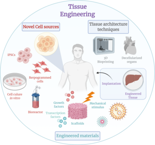
Tissue engineering is a biomedical engineering discipline that uses a combination of cells, engineering, materials methods, and suitable biochemical and physicochemical factors to restore, maintain, improve, or replace different types of biological tissues. Tissue engineering often involves the use of cells placed on tissue scaffolds in the formation of new viable tissue for a medical purpose, but is not limited to applications involving cells and tissue scaffolds. While it was once categorized as a sub-field of biomaterials, having grown in scope and importance, it can is considered as a field of its own.

<span class="mw-page-title-main">Hepatocyte</span> Liver cell type

A hepatocyte is a cell of the main parenchymal tissue of the liver. Hepatocytes make up 80% of the liver's mass. These cells are involved in:

<span class="mw-page-title-main">Cell adhesion</span> Process of cell attachment

Cell adhesion is the process by which cells interact and attach to neighbouring cells through specialised molecules of the cell surface. This process can occur either through direct contact between cell surfaces such as cell junctions or indirect interaction, where cells attach to surrounding extracellular matrix, a gel-like structure containing molecules released by cells into spaces between them. Cells adhesion occurs from the interactions between cell-adhesion molecules (CAMs), transmembrane proteins located on the cell surface. Cell adhesion links cells in different ways and can be involved in signal transduction for cells to detect and respond to changes in the surroundings. Other cellular processes regulated by cell adhesion include cell migration and tissue development in multicellular organisms. Alterations in cell adhesion can disrupt important cellular processes and lead to a variety of diseases, including cancer and arthritis. Cell adhesion is also essential for infectious organisms, such as bacteria or viruses, to cause diseases.

<span class="mw-page-title-main">Cell culture</span> Process by which cells are grown under controlled conditions

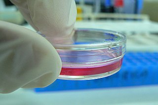
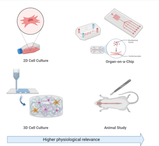
Cell culture or tissue culture is the process by which cells are grown under controlled conditions, generally outside of their natural environment. After cells of interest have been isolated from living tissue, they can subsequently be maintained under carefully controlled conditions. They need to be kept at body temperature (37 °C) in an incubator. These conditions vary for each cell type, but generally consist of a suitable vessel with a substrate or rich medium that supplies the essential nutrients (amino acids, carbohydrates, vitamins, minerals), growth factors, hormones, and gases (CO2, O2), and regulates the physio-chemical environment (pH buffer, osmotic pressure, temperature). Most cells require a surface or an artificial substrate to form an adherent culture as a monolayer (one single-cell thick), whereas others can be grown free floating in a medium as a suspension culture. This is typically facilitated via use of a liquid, semi-solid, or solid growth medium, such as broth or agar. Tissue culture commonly refers to the culture of animal cells and tissues, with the more specific term plant tissue culture being used for plants. The lifespan of most cells is genetically determined, but some cell-culturing cells have been 'transformed' into immortal cells which will reproduce indefinitely if the optimal conditions are provided.

Matrigel is the trade name for the solubilized basement membrane matrix secreted by Engelbreth-Holm-Swarm (EHS) mouse sarcoma cells produced by Corning Life Sciences. Matrigel resembles the laminin/collagen IV-rich basement membrane extracellular environment found in many tissues and is used by cell biologists as a substrate for culturing cells.

Stromal cells, or mesenchymal stromal cells, are differentiating cells found in abundance within bone marrow but can also be seen all around the body. Stromal cells can become connective tissue cells of any organ, for example in the uterine mucosa (endometrium), prostate, bone marrow, lymph node and the ovary. They are cells that support the function of the parenchymal cells of that organ. The most common stromal cells include fibroblasts and pericytes. The term stromal comes from Latin stromat-, "bed covering", and Ancient Greek στρῶμα, strôma, "bed".

<span class="mw-page-title-main">Perlecan</span>

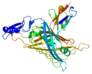
Perlecan (PLC) also known as basement membrane-specific heparan sulfate proteoglycan core protein (HSPG) or heparan sulfate proteoglycan 2 (HSPG2), is a protein that in humans is encoded by the HSPG2 gene. The HSPG2 gene codes for a 4,391 amino acid protein with a molecular weight of 468,829. It is one of the largest known proteins. The name perlecan comes from its appearance as a "string of pearls" in rotary shadowed images.

Actibind is an actin-binding fungal T(2)-RNase protein that is produced by the black mold Aspergillus niger, a microorganism used in biotechnology and food technology. In plants, actibind binds actin, a major component of the cytoskeleton, interfering with the plants' pollen tubes and halting cell growth. Research published in the journal Cancer on 15 May 2006 reports evidence that actibind has antiangiogenic and anticarcinogenic characteristics. In human colon cancer, breast cancer and melanoma, increasing the level of actibind was found to reduce the ability of these cells to form tumorogenic colonies. In animal models, increased actibind inhibited the growth of colon cancer-derived tumors, metastases and blood vessel formation. During the completion of the Human Genome Project, the gene encoding for RNaseT2, the human actibind-like protein, was found on chromosome 6.

<span class="mw-page-title-main">T-cadherin</span> GPI-anchored signaling protein

T-cadherin, also known as cadherin 13, H-cadherin (heart), and CDH13, is a unique member of the cadherin superfamily of proteins because it lacks the transmembrane and cytoplasmic domains common to all other cadherins and is instead anchored to the cell's plasma membrane by the GPI anchor.

<span class="mw-page-title-main">3T3-L1</span> Cell line used in biological research

3T3-L1 is a sub clonal cell line derived from the original 3T3 Swiss albino cell line of 1962. The 3T3 original cell line was isolated from a mouse embryo and propagated for this specific line of 3T3 cells is used to study adipose tissue-related diseases and dysfunctions. The 3T3-L1 Swiss subclone line has been widely utilized, since its development, due to its affinity for lipid droplet deposition in vitro. 3T3-L1 cells have a fibroblast-like morphology, but, under appropriate conditions, the cells differentiate into an adipocyte-like phenotype, providing an exemplar model for white adipocytes. 3T3-L1 cells can be utilized to study a number of cellular and molecular mechanisms related to insulin-resistance, obesity, and diabetes in vitro. Aside from its usages, this cell line is widely developed and can be purchased for continuous propagation for numerous research studies. 3T3-L1 cells of the adipocyte morphology increase the synthesis and accumulation of triglycerides and acquire the signet ring appearance of adipose cells. These cells are also sensitive to lipogenic and lipolytic hormones, as well as drugs, including epinephrine, isoproterenol, and insulin.

<span class="mw-page-title-main">PTPRM</span> Protein-coding gene in the species Homo sapiens

Receptor-type tyrosine-protein phosphatase mu is an enzyme that in humans is encoded by the PTPRM gene.

Angiogenesis is the process of forming new blood vessels from existing blood vessels, formed in vasculogenesis. It is a highly complex process involving extensive interplay between cells, soluble factors, and the extracellular matrix (ECM). Angiogenesis is critical during normal physiological development, but it also occurs in adults during inflammation, wound healing, ischemia, and in pathological conditions such as rheumatoid arthritis, hemangioma, and tumor growth. Proteolysis has been indicated as one of the first and most sustained activities involved in the formation of new blood vessels. Numerous proteases including matrix metalloproteinases (MMPs), a disintegrin and metalloproteinase domain (ADAM), a disintegrin and metalloproteinase domain with throbospondin motifs (ADAMTS), and cysteine and serine proteases are involved in angiogenesis. This article focuses on the important and diverse roles that these proteases play in the regulation of angiogenesis.

An organ-on-a-chip (OOC) is a multi-channel 3-D microfluidic cell culture, integrated circuit (chip) that simulates the activities, mechanics and physiological response of an entire organ or an organ system. It constitutes the subject matter of significant biomedical engineering research, more precisely in bio-MEMS. The convergence of labs-on-chips (LOCs) and cell biology has permitted the study of human physiology in an organ-specific context. By acting as a more sophisticated in vitro approximation of complex tissues than standard cell culture, they provide the potential as an alternative to animal models for drug development and toxin testing.

<span class="mw-page-title-main">Tumor microenvironment</span> Surroundings of tumors including nearby cells and blood vessels

The tumor microenvironment is a complex ecosystem surrounding a tumor, composed of cancer cells, stromal tissue and the extracellular matrix. Mutual interaction between cancer cells and the different components of the tumor microenvironment support its growth and invasion in healthy tissues which correlates with tumor resistance to current treatments and poor prognosis. The tumor microenvironment is in constant change because of the tumor's ability to influence the microenvironment by releasing extracellular signals, promoting tumor angiogenesis and inducing peripheral immune tolerance, while the immune cells in the microenvironment can affect the growth and evolution of cancerous cells.

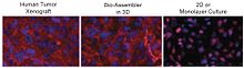
A 3D cell culture is an artificially created environment in which biological cells are permitted to grow or interact with their surroundings in all three dimensions. Unlike 2D environments, a 3D cell culture allows cells in vitro to grow in all directions, similar to how they would in vivo. These three-dimensional cultures are usually grown in bioreactors, small capsules in which the cells can grow into spheroids, or 3D cell colonies. Approximately 300 spheroids are usually cultured per bioreactor.

Magnetic 3D bioprinting is a methodology that employs biocompatible magnetic nanoparticles to print cells into 3D structures or 3D cell cultures. In this process, cells are tagged with magnetic nanoparticles that are used to render them magnetic. Once magnetic, these cells can be rapidly printed into specific 3D patterns using external magnetic forces that mimic tissue structure and function.

<span class="mw-page-title-main">Vasculogenic mimicry</span>

Vasculogenic mimicry (VM) is a strategy used by tumors to ensure sufficient blood supply is brought to its cells through establishing new tumor vascularization. This process is similar to tumor angiogenesis; on the other hand vascular mimicry is unique in that this process occurs independent of endothelial cells. Vasculature is instead developed de novo by cancer cells, which under stress conditions such as hypoxia, express similar properties to stem cells, capable of differentiating to mimic the function of endothelial cells and form vasculature-like structures. The ability of tumors to develop and harness nearby vasculature is considered one of the hallmarks of cancer disease development and is thought to be closely linked to tumor invasion and metastasis. Vascular mimicry has been observed predominantly in aggressive and metastatic cancers and has been associated with negative tumor characteristics such as increased metastasis, increased tissue invasion, and overall poor outcomes for patient survival. Vascular mimicry poses a serious problem for current therapeutic strategies due to its ability to function in the presence of Anti-angiogenic therapeutic agents. In fact, such therapeutics have been found to actually drive VM formation in tumors, causing more aggressive and difficult to treat tumors to develop.

Physical oncology (PO) is defined as the study of the role of mechanical signals in a cancerous tumor. The mechanical signals can be forces, pressures. If we generalize we will speak of "stress field" and "stress tensor".

<span class="mw-page-title-main">Invasion (cancer)</span> Direct extension and penetration by cancer cells into neighboring tissues

Invasion is the process by which cancer cells directly extend and penetrate into neighboring tissues in cancer. It is generally distinguished from metastasis, which is the spread of cancer cells through the circulatory system or the lymphatic system to more distant locations. Yet, lymphovascular invasion is generally the first step of metastasis.

Cultrex Basement Membrane Extract (BME) is the trade name for a extracellular protein mixture secreted by Engelbreth-Holm-Swarm (EHS) mouse sarcoma cells and manufactured into a hydrogel by R&D Systems, a brand of Bio-Techne. Similar to Matrigel, this hydrogel is a natural extracellular matrix that mimics the complex extracellular environment within complex tissues. It is used as a general cell culture substrate across a wide variety of research applications.

References

  1. 1 2 3 4 5 6 7 8 9 10 11 12 13 14 15 16 17 18 19 Souza, G. R. et al. Three-dimensional Tissue Culture Based on Magnetic Cell Levitation. Nature Nanotechnol.5, 291-296, doi:10.1038/nnano.2010.23 (2010).
  2. 1 2 3 4 Molina, J., Hayashi, Y., Stephens, C. & Georgescu, M.-M. Invasive glioblastoma cells acquire stemness and increased Akt activation. Neoplasia12, 453-463 (2010).
  3. "Bio-Assembling in 3-D with Magnetic Levitation - Technology Review." Technology Review. N.p., n.d. Web. 20 Aug. 2012. <http://www.technologyreview.com/view/426363/bio-assembling-in-3-d-with-magnetic-levitation/>.
  4. 1 2 Pampaloni, F., Reynaud, E. G. & Stelzer, E. H. K. The third dimension bridges the gap between cell culture and live tissue. Nat. Rev. Mol. Cell Biol.8, 839-845 (2007).
  5. 1 2 Cukierman, E., Pankov, R., Stevens, D. R. & Yamada, K. M. Taking Cell-Matrix Adhesions to the Third Dimension. Science294, 1708-1712 (2001).
  6. Abbott, A. Biology's new dimension. Nature424, 870-872 (2003).
  7. 1 2 3 Prestwich, G. D. Simplifying the extracellular matrix for 3-D cell culture and tissue engineering: A pragmatic approach. J. Cell. Biochem.101, 1370-1383, doi:10.1002/jcb.21386 (2007).
  8. 1 2 Boudreau, N. & Weaver, V. Forcing the Third Dimension. Cell125, 429-431 (2006).
  9. Griffith, L. G. & Swartz, M. A. Capturing complex 3D tissue physiology in vitro. Nat. Rev. Mol. Cell Biol.7, 211-224 (2006).
  10. Birgersdotter, A., Sandberg, R. & Ernberg, I. Gene expression perturbation in vitro--a growing case for three-dimensional (3D) culture systems. Semin. Cancer Biol.15, 405- 412 (2005).
  11. Yamada, K. M. & Cukierman, E. Modeling tissue morphogenesis and cancer in 3D. Cell130, 601-610 (2007).
  12. Atala, A. Engineering tissues, organs and cells. J. Tissue Eng. Regen. Med.1, 83-96 (2007).
  13. Bilodeau, K. & Mantovani, D. Bioreactors for Tissue Engineering: Focus on Mechanical Constraints. A Comparative Review. Tissue Eng.12, 2367-2383 (2006).
  14. Prestwich, G. D., Liu, Y., Yu, B., Shu, X. Z. & Scott, A. 3-D culture in synthetic extracellular matrices: new tissue models for drug toxicology and cancer drug discovery. Adv. Enzyme Regul. 47, 196-207 (2007).
  15. "N3D Biosciences, Inc. » ABOUT US." N3D Biosciences, Inc. » ABOUT US. N.p., n.d. Web. 20 Aug. 2012. <http://www.n3dbio.com/about/ Archived 2013-12-14 at the Wayback Machine >.
  16. 1 2 3 4 5 6 7 8 9 10 11 12 13 14 15 16 17 Daquinag, A. C., Souza, G. R., Kolonin, M. G. Adipose Tissue Engineering in Three-Dimensional Levitation Tissue Culture System Based on Magnetic Nanoparticles. Tissue Eng. Part C. -Not available-, ahead of print. doi:10.1089/ten.tec.2012.0198 (2012).
  17. 1 2 3 4 5 6 7 8 9 10 11 12 13 14 15 16 17 18 19 Tseng, H., Gage, J. A., Raphael, R., Moore, R. H., Killian, T. C., Grande-Allen, K. J., Souza, G. R. Assembly of a three-dimensional multitype bronchiole co-culture model using magnetic levitation. Tissue Eng. Part C. -Not available-, ahead of print. doi:10.1089/ten.TEC.2012.0157 (2013)