5S ribosomal RNA

Last updated
5S ribosomal RNA
RF00001.jpg
Predicted secondary structure and sequence conservation of 5S ribosomal RNA
Identifiers
Symbol5S_rRNA
Rfam RF00001 CL00113
Other data
RNA type Gene; rRNA
Domain(s) Eukaryota; Bacteria; Archaea
GO GO:0005840 GO:0003735
SO SO:0000652
PDB structures PDBe

The 5S ribosomal RNA (5S rRNA) is an approximately 120 nucleotide-long ribosomal RNA molecule with a mass of 40 kDa. It is a structural and functional component of the large subunit of the ribosome in all domains of life (bacteria, archaea, and eukaryotes), with the exception of mitochondrial ribosomes of fungi and animals. The designation 5S refers to the molecule's sedimentation velocity in an ultracentrifuge, which is measured in Svedberg units (S). [1]

Contents

Figure 1: A 3D representation of a 5S rRNA molecule. This structure is of the 5S rRNA from the Escherichia coli 50S ribosomal subunit and is based on a cryo-electron microscopic reconstruction. PDB 1c2x EBI.png
Figure 1: A 3D representation of a 5S rRNA molecule. This structure is of the 5S rRNA from the Escherichia coli 50S ribosomal subunit and is based on a cryo-electron microscopic reconstruction.

Biosynthesis

In prokaryotes, the 5S rRNA gene is typically located in the rRNA operons downstream of the small and the large subunit rRNA, and co-transcribed into a polycistronic precursor. [3] A particularity of eukaryotic nuclear genomes is the occurrence of multiple 5S rRNA gene copies (5S rDNA) clustered in tandem repeats, with copy number varying from species to species. [4] [5] Eukaryotic 5S rRNA is synthesized by RNA polymerase III, whereas other eukaryotic rRNAs are cleaved from a 45S precursor transcribed by RNA polymerase I. In Xenopus oocytes, it has been shown that fingers 4–7 of the nine-zinc finger transcription factor TFIIIA can bind to the central region of 5S RNA. [6] [7] Binding between 5S rRNA and TFIIIA serves to both repress further transcription of the 5S RNA gene and stabilize the 5S RNA transcript until it is required for ribosome assembly. [8] s

Structure

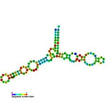
The secondary structure of 5S rRNA consists of five helices (denoted I–V in roman numerals), four loops (B-E), and one hinge (A), which form together a Y-like structure. Loops C and D are terminal hairpins and loops B and E are internal. [4] According to phylogenetic studies, helices I and III are likely ancestral. [9] Helix III includes two highly conserved adenosines. [10] Helix V, with its hairpin structure, is thought to interact with TFIIIA. [4]

Location within the ribosome

Figure 2: Atomic 3D structure of the 50S subunit from Haloarcula marismortui, PDB 1FFK. Proteins are shown in blue, 23S rRNA in orange and 5S rRNA in yellow. 5S rRNA together with the ribosomal proteins L5 and L18 and the domain V of 23S rRNA constitute the bulk of the central protuberance (CP). 010 large subunit-1FFK.gif
Figure 2: Atomic 3D structure of the 50S subunit from Haloarcula marismortui , PDB 1FFK. Proteins are shown in blue, 23S rRNA in orange and 5S rRNA in yellow. 5S rRNA together with the ribosomal proteins L5 and L18 and the domain V of 23S rRNA constitute the bulk of the central protuberance (CP).

Using a variety of molecular techniques, including immuno-electron microscopy, cryo-electron microscopy, intermolecular chemical cross-linking, and X-ray crystallography, the location of the 5S rRNA within the large ribosomal subunit has been determined to great precision. In bacteria and archaea, the large ribosomal subunit (LSU) itself is composed of two RNA moieties, the 5S rRNA and another larger RNA known as 23S rRNA, along with numerous associated proteins. [3] [12]

In eukaryotes, the LSU contains 5S, 5.8S, and 28S rRNAs and even more proteins. [13] [14] The structure of LSU in 3-dimensions shows one relatively smooth surface and the opposite surface having three projections, notably the L1 protuberance, the central protuberance (CP), and the L7/L12 stalk. The L1 protuberance and L7/L12 stalk are arranged laterally surrounding CP. The 5S rRNA is located in the CP and participates in formation and structure of this projection. The other major constituents of the central protuberance include the 23S rRNA (or alternatively 28S in eukaryotes) and several proteins including L5, L18, L25, and L27. [15]

Ribosomal functions

The exact function of 5S rRNA is not yet clear. In Escherichia coli , 5S rRNA gene deletions reduce the protein synthesis rate and have a more profound detrimental effect on cell fitness than deletions of a comparable number of copies of other (16S and 23S) rRNA genes. [16] Crystallographic studies indicate that 5S rRNA-binding proteins and other proteins of the central protuberance of the LSU plays a role in binding tRNAs. [15] Also, the topographical and physical proximity between 5S rRNA and 23S rRNA, which forms the peptidyl transferase and GTPase-associating center, suggests that 5S rRNA acts as a mediator between the two functional centers of the ribosome by forming, together with 5S rRNA-binding proteins and other components of the central protuberance, intersubunit bridges and tRNA-binding sites. [15]

Roles in ribosomal assembly

In eukaryotes, the cytosolic ribosome is assembled from four rRNAs and over 80 proteins. [14] [17] Once transcribed, the 3' ends of 5S rRNA can only be trimmed to mature length by functional homologues of RNase T, for example Rex1p in Saccharomyces cerevisiae . [18] The 60S and 40S ribosomal subunits are exported from the nucleus to the cytoplasm where they join to form the mature and translation-competent 80S ribosome. When exactly 5S rRNA is integrated into the ribosome remains controversial, [4] but it is generally accepted that 5S rRNA is incorporated into the 90S particle, which is a precursor to 60S particle, as part of a small ribosome-independent RNP complex formed by 5S rRNA and ribosomal protein L5. [17]

Interactions with proteins

Several important proteins which interact with 5S rRNA are listed below.

La protein

Interaction of 5S rRNA with the La protein prevents the RNA from degradation by exonucleases in the cell. [19] La protein is found in the nucleus in all eukaryotic organisms and associates with several types of RNAs transcribed by RNA pol III. La protein interacts with these RNAs (including the 5S rRNA) through their 3' oligo-uridine tract, aiding stability and folding of the RNA. [4] [20]

L5 protein

In eukaryotic cells, ribosomal protein L5 associates and stabilizes the 5S rRNA forming a pre-ribosomal ribonucleoprotein particle (RNP) that is found in both cytosol and the nucleus. L5 deficiency prevents transport of 5S rRNA to the nucleus and results in decreased ribosomal assembly. [4]

Other ribosomal proteins

In prokaryotes the 5S rRNA binds to the L5, L18 and L25 ribosomal proteins, whereas in eukaryotes 5S rRNA is only known to bind the L5 ribosomal protein. [21] In T. brucei, the causative agent of sleeping sickness, 5S rRNA interacts with two closely related RNA-binding proteins, P34 and P37, whose loss results in a lower global level of 5S rRNA. [4]

Presence in organelle ribosomes

Permuted mitochondrial genome encoded 5S rRNA
Identifiers
SymbolmtPerm-5S
Rfam RF02547 CL00113
Other data
RNA type Gene; rRNA
Domain(s) Eukaryota;
GO GO:0005840 GO:0003735
SO SO:0000652
PDB structures PDBe
Figure 3: Consensus secondary structure models of 5S rRNA based on the covariance models used to search for 5S rRNA genes. Models for: A) bacteria, archaea, and eukaryotic nuclei, B) plastids, and C) mitochondria. The IUPAC code letters and circles indicate conserved nucleotides and positions with variable nucleotide identity, respectively. Conserved and covariant substitutions in canonical (Watson-Crick) base-pairs are shaded. Consensus secondary structure models of 5S rRNAs.tiff
Figure 3: Consensus secondary structure models of 5S rRNA based on the covariance models used to search for 5S rRNA genes. Models for: A) bacteria, archaea, and eukaryotic nuclei, B) plastids, and C) mitochondria. The IUPAC code letters and circles indicate conserved nucleotides and positions with variable nucleotide identity, respectively. Conserved and covariant substitutions in canonical (Watson-Crick) base-pairs are shaded.

Translation machineries of mitochondria and plastids (organelles of endosymbiotic bacterial origin), and their bacterial relatives share many features but also display marked differences. Organelle genomes encode SSU and LSU rRNAs without exception, yet the distribution of 5S rRNA genes (rrn5) is most uneven. Rrn5 is easily identified and common in genomes of most plastids. In contrast, mitochondrial rrn5 initially appeared to be restricted to plants and a small number of protists. [22] [23] Additional, more divergent organellar 5S rRNAs were only identified with specialized covariance models that incorporate information on the pronounced sequence composition bias and structural variation. [24] This analysis pinpointed additional 5S rRNA genes not only in mitochondrial genomes of most protist lineages, but also in genomes of certain apicoplasts (non-photosynthetic plastids of pathogenic protozoa such as Toxoplasma gondii and Eimeria tenella ).

Figure 4: Comparison of the conventional and permuted secondary structure models of 5S rRNA. 5S-rRNA-topologies.tiff
Figure 4: Comparison of the conventional and permuted secondary structure models of 5S rRNA.

Mitochondrial 5S rRNAs of most stramenopiles comprise the largest diversity of secondary structures. [24] The permuted mitochondrial 5S rRNAs in brown algae represent the most unconventional case, where the closing helix I, which otherwise brings together the molecule's 5′ and 3′ ends, is replaced by a (closed) hairpin resulting in an open three-way junction.

Current evidence indicates that mitochondrial DNA of only a few groups, notably animals, fungi, alveolates and euglenozoans lacks the gene. [24] The central protuberance, otherwise occupied by 5S rRNA and its associated proteins (see Figure 2), was remodeled in various ways. In the fungal mitochondrial ribosomes, 5S rRNA is replaced by LSU rRNA expansion sequences. [25] In kinetoplastids (euglenozoans), the central protuberance is made entirely of evolutionarily novel mitochondrial ribosomal proteins. [26] Lastly, animal mitochondrial ribosomes have coopted a specific mitochondrial tRNA (Val in vertebrates) to substitute the missing 5S rRNA. [27] [28]

See also

Related Research Articles

<span class="mw-page-title-main">Messenger RNA</span> RNA that is read by the ribosome to produce a protein

In molecular biology, messenger ribonucleic acid (mRNA) is a single-stranded molecule of RNA that corresponds to the genetic sequence of a gene, and is read by a ribosome in the process of synthesizing a protein.

<span class="mw-page-title-main">Nucleolus</span> Largest structure in the nucleus of eukaryotic cells

The nucleolus is the largest structure in the nucleus of eukaryotic cells. It is best known as the site of ribosome biogenesis, which is the synthesis of ribosomes. The nucleolus also participates in the formation of signal recognition particles and plays a role in the cell's response to stress. Nucleoli are made of proteins, DNA and RNA, and form around specific chromosomal regions called nucleolar organizing regions. Malfunction of nucleoli can be the cause of several human conditions called "nucleolopathies" and the nucleolus is being investigated as a target for cancer chemotherapy.

<span class="mw-page-title-main">Ribosome</span> Intracellular organelle consisting of RNA and protein functioning to synthesize proteins

Ribosomes are macromolecular machines, found within all cells, that perform biological protein synthesis. Ribosomal RNA is found in the ribosomal nucleus where this synthesis happens. Ribosomes link amino acids together in the order specified by the codons of messenger RNA molecules to form polypeptide chains. Ribosomes consist of two major components: the small and large ribosomal subunits. Each subunit consists of one or more ribosomal RNA molecules and many ribosomal proteins. The ribosomes and associated molecules are also known as the translational apparatus.

<span class="mw-page-title-main">RNA polymerase</span> Enzyme that synthesizes RNA from DNA

In molecular biology, RNA polymerase, or more specifically DNA-directed/dependent RNA polymerase (DdRP), is an enzyme that catalyzes the chemical reactions that synthesize RNA from a DNA template.

<span class="mw-page-title-main">Translation (biology)</span> Cellular process of protein synthesis

In biology, translation is the process in living cells in which proteins are produced using RNA molecules as templates. The generated protein is a sequence of amino acids. This sequence is determined by the sequence of nucleotides in the RNA. The nucleotides are considered three at a time. Each such triple results in addition of one specific amino acid to the protein being generated. The matching from nucleotide triple to amino acid is called the genetic code. The translation is performed by a large complex of functional RNA and proteins called ribosomes. The entire process is called gene expression.

<span class="mw-page-title-main">Ribonuclease H</span> Enzyme family

Ribonuclease H is a family of non-sequence-specific endonuclease enzymes that catalyze the cleavage of RNA in an RNA/DNA substrate via a hydrolytic mechanism. Members of the RNase H family can be found in nearly all organisms, from bacteria to archaea to eukaryotes.

The 5′ untranslated region is the region of a messenger RNA (mRNA) that is directly upstream from the initiation codon. This region is important for the regulation of translation of a transcript by differing mechanisms in viruses, prokaryotes and eukaryotes. While called untranslated, the 5′ UTR or a portion of it is sometimes translated into a protein product. This product can then regulate the translation of the main coding sequence of the mRNA. In many organisms, however, the 5′ UTR is completely untranslated, instead forming a complex secondary structure to regulate translation.

<span class="mw-page-title-main">Ribosomal RNA</span> RNA component of the ribosome, essential for protein synthesis in all living organisms

Ribosomal ribonucleic acid (rRNA) is a type of non-coding RNA which is the primary component of ribosomes, essential to all cells. rRNA is a ribozyme which carries out protein synthesis in ribosomes. Ribosomal RNA is transcribed from ribosomal DNA (rDNA) and then bound to ribosomal proteins to form small and large ribosome subunits. rRNA is the physical and mechanical factor of the ribosome that forces transfer RNA (tRNA) and messenger RNA (mRNA) to process and translate the latter into proteins. Ribosomal RNA is the predominant form of RNA found in most cells; it makes up about 80% of cellular RNA despite never being translated into proteins itself. Ribosomes are composed of approximately 60% rRNA and 40% ribosomal proteins by mass.

Eukaryotic translation is the biological process by which messenger RNA is translated into proteins in eukaryotes. It consists of four phases: initiation, elongation, termination, and recapping.

<span class="mw-page-title-main">Ribonuclease P</span> Class of enzymes

Ribonuclease P is a type of ribonuclease which cleaves RNA. RNase P is unique from other RNases in that it is a ribozyme – a ribonucleic acid that acts as a catalyst in the same way that a protein-based enzyme would. Its function is to cleave off an extra, or precursor, sequence of RNA on tRNA molecules. Further, RNase P is one of two known multiple turnover ribozymes in nature, the discovery of which earned Sidney Altman and Thomas Cech the Nobel Prize in Chemistry in 1989: in the 1970s, Altman discovered the existence of precursor tRNA with flanking sequences and was the first to characterize RNase P and its activity in processing of the 5' leader sequence of precursor tRNA. Recent findings also reveal that RNase P has a new function. It has been shown that human nuclear RNase P is required for the normal and efficient transcription of various small noncoding RNAs, such as tRNA, 5S rRNA, SRP RNA and U6 snRNA genes, which are transcribed by RNA polymerase III, one of three major nuclear RNA polymerases in human cells.

<span class="mw-page-title-main">Ribosome biogenesis</span> Cellular process

Ribosome biogenesis is the process of making ribosomes. In prokaryotes, this process takes place in the cytoplasm with the transcription of many ribosome gene operons. In eukaryotes, it takes place both in the cytoplasm and in the nucleolus. It involves the coordinated function of over 200 proteins in the synthesis and processing of the three prokaryotic or four eukaryotic rRNAs, as well as assembly of those rRNAs with the ribosomal proteins. Most of the ribosomal proteins fall into various energy-consuming enzyme families including ATP-dependent RNA helicases, AAA-ATPases, GTPases, and kinases. About 60% of a cell's energy is spent on ribosome production and maintenance.

<span class="mw-page-title-main">Ribosomal protein</span> Proteins found in ribosomes

A ribosomal protein is any of the proteins that, in conjunction with rRNA, make up the ribosomal subunits involved in the cellular process of translation. E. coli, other bacteria and Archaea have a 30S small subunit and a 50S large subunit, whereas humans and yeasts have a 40S small subunit and a 60S large subunit. Equivalent subunits are frequently numbered differently between bacteria, Archaea, yeasts and humans.

<span class="mw-page-title-main">5.8S ribosomal RNA</span> RNA component of the large subunit of the eukaryotic ribosome

In molecular biology, the 5.8S ribosomal RNA is a non-coding RNA component of the large subunit of the eukaryotic ribosome and so plays an important role in protein translation. It is transcribed by RNA polymerase I as part of the 45S precursor that also contains 18S and 28S rRNA. Its function is thought to be in ribosome translocation. It is also known to form covalent linkage to the p53 tumour suppressor protein. 5.8S rRNA can be used as a reference gene for miRNA detection. The 5.8S ribosomal RNA is used to better understand other rRNA processes and pathways in the cell.

Ribosomal particles are denoted according to their sedimentation coefficients in Svedberg units. The 60S subunit is the large subunit of eukaryotic 80S ribosomes, with the other major component being the eukaryotic small ribosomal subunit (40S). It is structurally and functionally related to the 50S subunit of 70S prokaryotic ribosomes. However, the 60S subunit is much larger than the prokaryotic 50S subunit and contains many additional protein segments, as well as ribosomal RNA expansion segments.

<span class="mw-page-title-main">23S ribosomal RNA</span> A component of the large subunit of the prokaryotic ribosome

The 23S rRNA is a 2,904 nucleotide long component of the large subunit (50S) of the bacterial/archean ribosome and makes up the peptidyl transferase center (PTC). The 23S rRNA is divided into six secondary structural domains titled I-VI, with the corresponding 5S rRNA being considered domain VII. The ribosomal peptidyl transferase activity resides in domain V of this rRNA, which is also the most common binding site for antibiotics that inhibit translation, making it a target for ribosomal engineering. A well-known member of this antibiotic class, chloramphenicol, acts by inhibiting peptide bond formation, with recent 3D-structural studies showing two different binding sites depending on the species of ribosome. Numerous mutations in domains of the 23S rRNA with Peptidyl transferase activity have resulted in antibiotic resistance. 23S rRNA genes typically have higher sequence variations, including insertions and/or deletions, compared to other rRNAs.

<span class="mw-page-title-main">60S ribosomal protein L5</span> Protein found in humans

60S ribosomal protein L5 is a protein that in humans is encoded by the RPL5 gene.

The eukaryotic small ribosomal subunit (40S) is the smaller subunit of the eukaryotic 80S ribosomes, with the other major component being the large ribosomal subunit (60S). The "40S" and "60S" names originate from the convention that ribosomal particles are denoted according to their sedimentation coefficients in Svedberg units. It is structurally and functionally related to the 30S subunit of 70S prokaryotic ribosomes. However, the 40S subunit is much larger than the prokaryotic 30S subunit and contains many additional protein segments, as well as rRNA expansion segments.

<span class="mw-page-title-main">Eukaryotic ribosome</span> Large and complex molecular machine

Ribosomes are a large and complex molecular machine that catalyzes the synthesis of proteins, referred to as translation. The ribosome selects aminoacylated transfer RNAs (tRNAs) based on the sequence of a protein-encoding messenger RNA (mRNA) and covalently links the amino acids into a polypeptide chain. Ribosomes from all organisms share a highly conserved catalytic center. However, the ribosomes of eukaryotes are much larger than prokaryotic ribosomes and subject to more complex regulation and biogenesis pathways. Eukaryotic ribosomes are also known as 80S ribosomes, referring to their sedimentation coefficients in Svedberg units, because they sediment faster than the prokaryotic (70S) ribosomes. Eukaryotic ribosomes have two unequal subunits, designated small subunit (40S) and large subunit (60S) according to their sedimentation coefficients. Both subunits contain dozens of ribosomal proteins arranged on a scaffold composed of ribosomal RNA (rRNA). The small subunit monitors the complementarity between tRNA anticodon and mRNA, while the large subunit catalyzes peptide bond formation.

Ribosomopathies are diseases caused by abnormalities in the structure or function of ribosomal component proteins or rRNA genes, or other genes whose products are involved in ribosome biogenesis.

<span class="mw-page-title-main">Mitochondrial ribosome</span> Protein complex

The mitochondrial ribosome, or mitoribosome, is a protein complex that is active in mitochondria and functions as a riboprotein for translating mitochondrial mRNAs encoded in mtDNA. The mitoribosome is attached to the inner mitochondrial membrane. Mitoribosomes, like cytoplasmic ribosomes, consist of two subunits — large (mt-LSU) and small (mt-SSU). Mitoribosomes consist of several specific proteins and fewer rRNAs. While mitochondrial rRNAs are encoded in the mitochondrial genome, the proteins that make up mitoribosomes are encoded in the nucleus and assembled by cytoplasmic ribosomes before being implanted into the mitochondria.

References

  1. Szymanski M, Barciszewska MZ, Erdmann VA, Barciszewski J (January 2002). "5S Ribosomal RNA Database". Nucleic Acids Research. 30 (1): 176–178. doi:10.1093/nar/30.1.176. PMC   99124 . PMID   11752286.
  2. Mueller F, Sommer I, Baranov P, Matadeen R, Stoldt M, Wöhnert J, et al. (April 2000). "The 3D arrangement of the 23 S and 5 S rRNA in the Escherichia coli 50 S ribosomal subunit based on a cryo-electron microscopic reconstruction at 7.5 A resolution". Journal of Molecular Biology. 298 (1): 35–59. doi:10.1006/jmbi.2000.3635. PMID   10756104.
  3. 1 2 Kaczanowska M, Rydén-Aulin M (September 2007). "Ribosome biogenesis and the translation process in Escherichia coli". Microbiology and Molecular Biology Reviews. 71 (3): 477–494. doi:10.1128/MMBR.00013-07. PMC   2168646 . PMID   17804668.
  4. 1 2 3 4 5 6 7 Ciganda M, Williams N (2011). "Eukaryotic 5S rRNA biogenesis". Wiley Interdisciplinary Reviews. RNA. 2 (4): 523–533. doi:10.1002/wrna.74. PMC   3278907 . PMID   21957041.
  5. Douet J, Tourmente S (July 2007). "Transcription of the 5S rRNA heterochromatic genes is epigenetically controlled in Arabidopsis thaliana and Xenopus laevis". Heredity. 99 (1): 5–13. doi: 10.1038/sj.hdy.6800964 . PMID   17487217.
  6. McBryant SJ, Veldhoen N, Gedulin B, Leresche A, Foster MP, Wright PE, et al. (April 1995). "Interaction of the RNA binding fingers of Xenopus transcription factor IIIA with specific regions of 5 S ribosomal RNA". Journal of Molecular Biology. 248 (1): 44–57. doi:10.1006/jmbi.1995.0201. PMID   7731045.
  7. Searles MA, Lu D, Klug A (August 2000). "The role of the central zinc fingers of transcription factor IIIA in binding to 5 S RNA". Journal of Molecular Biology. 301 (1): 47–60. doi:10.1006/jmbi.2000.3946. PMID   10926492.
  8. Pelham HR, Brown DD (July 1980). "A specific transcription factor that can bind either the 5S RNA gene or 5S RNA". Proceedings of the National Academy of Sciences of the United States of America. 77 (7): 4170–4174. Bibcode:1980PNAS...77.4170P. doi: 10.1073/pnas.77.7.4170 . PMC   349792 . PMID   7001457.
  9. Sun FJ, Caetano-Anollés G (November 2009). "The evolutionary history of the structure of 5S ribosomal RNA". Journal of Molecular Evolution. 69 (5): 430–443. Bibcode:2009JMolE..69..430S. doi:10.1007/s00239-009-9264-z. PMID   19639237. S2CID   21572917.
  10. DiNitto JP, Huber PW (October 2001). "A role for aromatic amino acids in the binding of Xenopus ribosomal protein L5 to 5S rRNA". Biochemistry. 40 (42): 12645–12653. doi:10.1021/bi011439m. PMID   11601989.
  11. Ban N, Nissen P, Hansen J, Moore PB, Steitz TA (August 2000). "The complete atomic structure of the large ribosomal subunit at 2.4 A resolution". Science. 289 (5481): 905–920. Bibcode:2000Sci...289..905B. CiteSeerX   10.1.1.58.2271 . doi:10.1126/science.289.5481.905. PMID   10937989.
  12. Tirumalai MR, Kaelber JT, Park DR, Tran Q, Fox GE (October 2020). "Cryo-electron microscopy visualization of a large insertion in the 5S ribosomal RNA of the extremely halophilic archaeon Halococcus morrhuae". FEBS Open Bio. 10 (10): 1938–1946. doi: 10.1002/2211-5463.12962 . PMC   7530397 . PMID   32865340.
  13. Turowski TW, Tollervey D (2015). "Cotranscriptional events in eukaryotic ribosome synthesis". Wiley Interdisciplinary Reviews. RNA. 6 (1): 129–139. doi: 10.1002/wrna.1263 . hdl: 20.500.11820/983ebe02-f5a9-4b65-a3f6-c93ac8088fb2 . PMID   25176256.
  14. 1 2 Yusupova G, Yusupov M (February 2014). "High-resolution structure of the eukaryotic 80S ribosome". Annual Review of Biochemistry. 83: 467–486. doi:10.1146/annurev-biochem-060713-035445. PMID   24580643.
  15. 1 2 3 Gongadze GM (December 2011). "5S rRNA and ribosome". Biochemistry. Biokhimiia. 76 (13): 1450–1464. doi:10.1134/S0006297911130062. PMID   22339598. S2CID   41669737.
  16. Ammons D, Rampersad J, Fox GE (January 1999). "5S rRNA gene deletions cause an unexpectedly high fitness loss in Escherichia coli". Nucleic Acids Research. 27 (2): 637–642. doi:10.1093/nar/27.2.637. PMC   148226 . PMID   9862991.
  17. 1 2 Henras AK, Soudet J, Gérus M, Lebaron S, Caizergues-Ferrer M, Mougin A, Henry Y (August 2008). "The post-transcriptional steps of eukaryotic ribosome biogenesis". Cellular and Molecular Life Sciences. 65 (15): 2334–2359. doi:10.1007/s00018-008-8027-0. PMID   18408888. S2CID   1934099.
  18. van Hoof A, Lennertz P, Parker R (March 2000). "Three conserved members of the RNase D family have unique and overlapping functions in the processing of 5S, 5.8S, U4, U5, RNase MRP and RNase P RNAs in yeast". The EMBO Journal. 19 (6): 1357–1365. doi:10.1093/emboj/19.6.1357. PMC   305676 . PMID   10716935.
  19. Wolin SL, Cedervall T (2002). "The La protein". Annual Review of Biochemistry. 71: 375–403. doi:10.1146/annurev.biochem.71.090501.150003. PMID   12045101.
  20. Maraia RJ, Intine RV (2002). "La protein and its associated small nuclear and nucleolar precursor RNAs". Gene Expression. 10 (1–2): 41–57. PMC   5977531 . PMID   11868987.
  21. Moore PB (March 2001). "The ribosome at atomic resolution". Biochemistry. 40 (11): 3243–3250. doi:10.1021/bi0029402. PMID   11258942.
  22. Bullerwell CE, Schnare MN, Gray MW (March 2003). "Discovery and characterization of Acanthamoeba castellanii mitochondrial 5S rRNA". RNA. 9 (3): 287–292. doi:10.1261/rna.2170803. PMC   1370395 . PMID   12592002.
  23. Bullerwell CE, Burger G, Gott JM, Kourennaia O, Schnare MN, Gray MW (May 2010). "Abundant 5S rRNA-like transcripts encoded by the mitochondrial genome in amoebozoa". Eukaryotic Cell. 9 (5): 762–773. doi:10.1128/EC.00013-10. PMC   2863963 . PMID   20304999.
  24. 1 2 3 Valach M, Burger G, Gray MW, Lang BF (December 2014). "Widespread occurrence of organelle genome-encoded 5S rRNAs including permuted molecules". Nucleic Acids Research. 42 (22): 13764–13777. doi:10.1093/nar/gku1266. PMC   4267664 . PMID   25429974.
  25. Amunts A, Brown A, Bai XC, Llácer JL, Hussain T, Emsley P, et al. (March 2014). "Structure of the yeast mitochondrial large ribosomal subunit". Science. 343 (6178): 1485–1489. Bibcode:2014Sci...343.1485A. doi:10.1126/science.1249410. PMC   4046073 . PMID   24675956.
  26. Sharma MR, Booth TM, Simpson L, Maslov DA, Agrawal RK (June 2009). "Structure of a mitochondrial ribosome with minimal RNA". Proceedings of the National Academy of Sciences of the United States of America. 106 (24): 9637–9642. Bibcode:2009PNAS..106.9637S. doi: 10.1073/pnas.0901631106 . PMC   2700991 . PMID   19497863.
  27. Brown A, Amunts A, Bai XC, Sugimoto Y, Edwards PC, Murshudov G, et al. (November 2014). "Structure of the large ribosomal subunit from human mitochondria". Science. 346 (6210): 718–722. Bibcode:2014Sci...346..718B. doi:10.1126/science.1258026. PMC   4246062 . PMID   25278503.
  28. Greber BJ, Boehringer D, Leibundgut M, Bieri P, Leitner A, Schmitz N, et al. (November 2014). "The complete structure of the large subunit of the mammalian mitochondrial ribosome". Nature. 515 (7526): 283–286. Bibcode:2014Natur.515..283G. doi:10.1038/nature13895. hdl: 20.500.11850/93239 . PMID   25271403. S2CID   4402484.