Comparative foot morphology

Last updated
Skeletons of a human and an elephant. Comparative view of the human and elephant frame, Benjamin Waterhouse Hawkins, 1860.jpg
Skeletons of a human and an elephant.

Comparative foot morphology involves comparing the form of distal limb structures of a variety of terrestrial vertebrates. Understanding the role that the foot plays for each type of organism must take account of the differences in body type, foot shape, arrangement of structures, loading conditions and other variables. However, similarities also exist among the feet of many different terrestrial vertebrates. The paw of the dog, the hoof of the horse, the manus (forefoot) and pes (hindfoot) of the elephant, and the foot of the human all share some common features of structure, organization and function. Their foot structures function as the load-transmission platform which is essential to balance, standing and types of locomotion (such as walking, trotting, galloping and running).

Contents

The discipline of biomimetics applies the information gained by comparing the foot morphology of a variety of terrestrial vertebrates to human-engineering problems. For instance, it may provide insights that make it possible to alter the foot's load transmission in people who wear an external orthosis because of paralysis from spinal-cord injury, or who use a prosthesis following the diabetes-related amputation of a leg. Such knowledge can be incorporated in technology that improves a person's balance when standing; enables them to walk more efficiently, and to exercise; or otherwise enhances their quality of life by improving their mobility.

Structure

Limb and foot structure of representative terrestrial vertebrates:

Variability in scaling and limb coordination

Elephant skeleton Elephant skeleton.jpg
Elephant skeleton

There is considerable variation in the scale and proportions of body and limb, as well as the nature of loading, during standing and locomotion both among and between quadrupeds and bipeds. [1] The anterior-posterior body mass distribution varies considerably among mammalian quadrupeds, which affects limb loading. When standing, many terrestrial quadrupeds support more of their weight on their forelimbs rather than their hind limbs; [2] [3] however, the distribution of body mass and limb loading changes when they move. [4] [5] [6] Humans have a lower-limb mass that is greater than their upper-limb mass. The hind limbs of the dog and horse have a slightly greater mass than the forelimbs, whereas the elephant has proportionally longer limbs. The elephant's forelimbs are longer than its hind limbs. [7]

In the horse [8] and dog, the hind limbs play an important role in primary propulsion. The legged locomotion of humans generally distributes an equal loading on each lower limb. [9] The locomotion of the elephant (which is the largest terrestrial vertebrate) displays a similar loading distribution on its hind limbs and forelimbs. [10] The walking and running gaits of quadrupeds and bipeds show differences in the relative phase of the movements of their forelimbs and hind limbs, as well as of their right-side limbs versus their left-side limbs. [5] [11] Many of the aforementioned variables are connected with differences in the scaling of body and limb dimension as well as in patterns of limb coordination and movement. However, little is understood concerning the functional contribution of the foot and its structures during the weight-bearing phase. The comparative morphology of the distal limb and foot structure of some representative terrestrial vertebrates reveals some interesting similarities.

Columnar organization of limb structures

Limb skeleton of a lion, an example of an angulated bony column Panthera leo limbs.png
Limb skeleton of a lion, an example of an angulated bony column

Even many terrestrial vertebrates exhibit differences in the scaling of limb dimension, limb coordination and magnitude of forelimb-hind limb loading, in the dog, horse and elephant the structure of the distal forelimb is similar to that of the distal hind limb. [7] [8] [12] In the human, the structures of the hand are generally similar in shape and arrangement to those of the foot. Terrestrial vertebrate quadrupeds and bipeds generally possess distal limb and foot endoskeleton structures that are aligned in series, stacked in a relatively vertical orientation and arranged in a quasi-columnar fashion in the extended limb. [1] [13] [14] In the dog and horse, the bones of the proximal limbs are oriented vertically, whereas the distal limb structures of the ankle and foot have an angulated orientation. In humans and elephants, a vertical-column orientation of the bones in the limbs and feet is also evident for associated skeletal muscle-tendon units. [6] The horse's foot contains an external nail (hoof) oriented about the perimeter in the shape of a semicircle. The underlying bones are arranged in a semi-vertical orientation. [15] [16] The dog's paw similarly contains bones arranged in a semi-vertical orientation.

In the human and the elephant, the column orientation of the foot complex is replaced in humans by a plantigrade orientation, and in elephants by a semi-plantigrade alignment of the hind limb foot structure. [6] This difference in orientation in the foot bones and joints of humans and elephants helps them to adapt to variations in the terrain. [17]

Distal cushion

Mm Fuss.jpg
Indischer Elefant Detail Fusse.JPG
Distal cushions on the foot of a raccoon and an elephant

Many representative terrestrial vertebrates possess a distal cushion on the under-surface of the foot. The dog's paw contains a number of visco-elastic pads oriented along the middle and distal foot. The horse possesses a centralized digital pad known as the frog, which is located at the distal aspect of the foot and surrounded by the hoof. [12] Humans possess a tough fibro and elastic pad of fat that is anchored to the skin and bone of the rear portion of the foot. [18] [19]

The foot of the elephant possesses what is perhaps one of the most unusual distal cushions found in vertebrates. The forefoot (manus) and hindfoot (pes) contain huge pads of fat that are scaled to cope with the massive loadings imposed by the largest terrestrial vertebrate. In addition, a cartilage-like projection (prepollex in the forelimb and prehallux in the hind limb) appears to anchor the distal cushion to the bones of the elephant's foot. [20]

The distal cushions of all these organisms (dog, horse, human and elephant) are dynamic structures during locomotion, alternating between phases of compression and expansion; it has been suggested that these structures thereby reduce the loads experienced by the skeletal system. [18] [19] [20] [21]

Organization

Arrangement of foot structures:

Because of the wide variety in body types, scaling and morphology of the distal limbs of terrestrial vertebrates, there exists a degree of controversy concerning the nature and organization of foot structures. One organizational approach to understanding foot structures makes distinctions regarding their regional anatomy. The foot structures are divided into segments from proximal to distal and are grouped according to similarity in shape, dimension and function. In this approach, the foot may be described in three segments: as the hindfoot, midfoot and forefoot.

The hindfoot is the most proximal and posterior portion of the foot. [22] Functionally, the structures contained in this region are typically robust, possessing a larger size and girth than the other structures of the foot. The structures of the hindfoot are usually adapted for transmitting large loads between the proximal and distal aspects of the limb when the foot contacts the ground. This is apparent in the human and elephant foot, where the hindfoot undergoes greater loading during initial contact in many forms of locomotion. [23] The hindfoot structures of the dog and horse are located relatively proximally compared to the elephant and human foot.

The midfoot is the intermediate portion of the foot between the hindfoot and forefoot. The structures in this region are intermediate in size, and typically transmit loads from the hindfoot to the forefoot. The human transverse tarsal joint of the midfoot transmits forces from the subtalar joint in the hindfoot to the forefoot joints (metatarsophalangeal and interphalangeal) and associated bones (metatarsals and phalanges). [24] The midfoot of the dog, horse and elephant contains similar intermediate structures having similar functions to those of the human midfoot.

The forefoot represents the most distal portion of the foot. In the human and elephant, the bone structures contained in this region are generally longer and narrower. The structures of the forefoot play a role in providing leverage for terminal stance propulsion and load transfer. [6] [23]

Function

Load transmission of the foot in representative terrestrial vertebrates:

Dog paw

Dog paw DogDewClawTika1 wb.jpg
Dog paw

The paw of the dog has a digitigrade orientation. The vertical columnar orientation of the proximal bones of the limbs, which articulate with distal foot structures that are arranged in quasi-vertical columnar orientation, is well-aligned to transmit loadings during weight-bearing contact of the skeleton with the ground. The angled orientation of the elongated metatarsal and the digits extends the area available for storing and releasing mechanical energy in the muscle tendon units originating proximally to the ankle joint and terminating at the distal aspect of the foot bones. [6] When muscle tendon units lengthen, the load strain facilitates mechanical activity. These muscle tendon unit structures appear well designed to aid in the ground-reaction transmission of forces that is essential for locomotion. [25] In addition, the pads of the distal paw appear to allow load attenuation, by enhancing shock absorption during the paw's contact with the ground.

Horse foot

Section of a horse foot Hufschnitt.jpg
Section of a horse foot

The horse's foot is in an unguligrade orientation. The columnar orientation of bones and connective tissue is similarly well-aligned to transmit loads during the weight-bearing phase of locomotion. The thick keratinized and semicircular hoof changes shape during loading and unloading. Similarly, the cushioned frog situated centrally at the rear ends of the hoof undergoes compression during loading, and expansion when unloaded. Together, the hoof and cushioned frog structures may work in concert with hoof capsule to provide shock absorption. [21] The horse hoof also acts dynamically during loading, which may cushion the endoskeleton from high loads that would otherwise produce critical deformation.

Elephant foot

Leg skeleton of the modern elephant PSM V04 D550 Elephant rear leg bones.jpg
Leg skeleton of the modern elephant

The hind limb and foot of the elephant are oriented semi-plantigrade, and closely resemble the structure and function of the human foot. The tarsals and metapodials are arranged so as to form an arch, similarly to the human foot. The six toes of each foot of the elephant are enclosed in a flexible sheath of skin. [20] [26] Similar to the dog's paw, the elephant's phalanges are oriented in a downward direction. The distal phalanges of the elephant do not directly touch the ground, and are attached to the respective nail/hoof. [27] Distal cushions occupy the spaces between the muscle tendon units and ligaments within the hindfoot, midfoot and forefoot bones on the plantar surface. [28] The distal cushion is highly innervated by sensory structures (Meissner's and Pacinian corpuscles), making the distal foot one of the most sensitive structures of the elephant (more so than its trunk). [20] The cushions of the elephant's foot respond to the requirement to store and absorb mechanical loads when they are compressed, and to distribute locomotor loads over a large area in order to keep foot tissue stresses within acceptable levels. [20] In addition, the musculoskeletal foot arch and sole cushion of the elephant act in concert, similarly to the horse's cushioned frog and hoof [6] and the human foot. [29] In the elephant, the nearly half-cupula-shaped arrangement of the bony elements of the metatarsals and toes has interesting similarities to the structure of the arches of human feet. [29] [30]

Recently, scientists at the Royal Veterinary College in the United Kingdom have discovered that the elephant possesses a sixth false toe, a sesamoid, located similarly to the giant panda's extra "thumb". They found that this sixth toe acts to support and distribute the weight of the elephant. [31]

Human foot

Skeleton of the human and gorilla (gorilla shown in non-natural posture) Primatenskelett-drawing.jpg
Skeleton of the human and gorilla (gorilla shown in non-natural posture)

The unique plantigrade alignment of the human foot results in a distal-limb structure that can adapt to a variety of conditions. The less mobile and more robust tarsal bones are shaped and aligned to accept and transmit large loads during the early phases of stance (initial contact and loading response phases of walking, and inadvertent heel strikes during running). The tarsals of the midfoot, which are smaller and shorter than the hindfoot tarsals, appear well oriented to transmit loads between the hindfoot and forefoot; this is necessary for load transfer and locking of the foot complex into a rigid lever for late stance phase. Conversely, the midfoot bones and joints also allow for the transmission of loads and inter-joint movement that unlocks the foot to create a loosely packed structure which renders the foot highly compliant over a variety of surfaces. In this configuration, the foot is able to absorb and damp the large loads encountered during heel strike and early weight acceptance. [17] The forefoot, with its long metatarsal and relatively long phalanges, transmits loads during the end-of-stance phase that facilitate the push-off and transfer of forward momentum. The forefoot also serves as a lever to allow balance during standing and jumping. In addition, the arches of the foot that span the hindfoot, midfoot and forefoot play a critical role in the nature of transformation of the foot from a rigid lever to a flexible weight-accepting structure. [23] [24]

With a running gait, the foot-loading order is usually the reverse of walking. The foot strikes the ground with the ball of the foot, and then the heel drops. [32] The heel drop elastically extends the Achilles tendon; this extension is reversed during the push-off. [33]

Clinical implications

Veterinarian or human healthcare professionals often respond when the foot of a dog, horse, elephant or human develops an abnormality. They typically investigate to understand the nature of the pathology in order to generate and implement a clinical treatment plan. For instance, the paws of the dog and the hindfoot work together to absorb the shock of jumping and running, and to provide flexibility of movement. If the dog's skeletal structures in areas other than the foot are compromised, the foot may be burdened with compensatory loading. Structural faults such as straight or loose shoulders, straight stifles, loose hips, and lack of balance between the forefoot and hindfoot, can all cause gait abnormalities that in turn damage the hindfoot and paws by overloading their foot structures as they compensate for the structural faults.

In the horse, dryness of the hoof may cause stiffening of the external foot structure. The stiffer hoof reduces the foot's load attenuation capacity, rendering the horse unable to bear much weight on the distal limb. Similar characteristic features emerge in the human foot in the form of the pes cavus alignment deformity, which is produced by tight connective tissue structures and joint congruency that create a rigid foot complex. Individuals with pes cavus display characteristic reduced load-attenuation features, and other structures proximal to the foot may compensate with increased load transfer (i.e., excessive loading to the knees, hips, lumbo-pelvic joints or lumbar vertebrae). [24] Foot disorders are common in captive elephants. However, the cause is poorly understood. [34]

See also

Related Research Articles

<span class="mw-page-title-main">Foot</span> Anatomical structure found in vertebrates

The foot is an anatomical structure found in many vertebrates. It is the terminal portion of a limb which bears weight and allows locomotion. In many animals with feet, the foot is a separate organ at the terminal part of the leg made up of one or more segments or bones, generally including claws and/or nails.

<span class="mw-page-title-main">Quadrupedalism</span> Form of locomotion using four limbs

Quadrupedalism is a form of locomotion where four limbs are used to bear weight and move around. An animal or machine that usually maintains a four-legged posture and moves using all four limbs is said to be a quadruped. Quadruped animals are found among both vertebrates and invertebrates.

<span class="mw-page-title-main">Gait</span> Pattern of movement of the limbs of animals

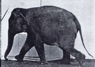
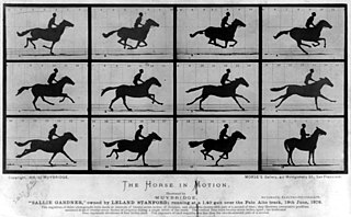
Gait is the pattern of movement of the limbs of animals, including humans, during locomotion over a solid substrate. Most animals use a variety of gaits, selecting gait based on speed, terrain, the need to maneuver, and energetic efficiency. Different animal species may use different gaits due to differences in anatomy that prevent use of certain gaits, or simply due to evolved innate preferences as a result of habitat differences. While various gaits are given specific names, the complexity of biological systems and interacting with the environment make these distinctions "fuzzy" at best. Gaits are typically classified according to footfall patterns, but recent studies often prefer definitions based on mechanics. The term typically does not refer to limb-based propulsion through fluid mediums such as water or air, but rather to propulsion across a solid substrate by generating reactive forces against it.

<span class="mw-page-title-main">Paw</span> Soft foot-like part of a mammal that has claws

A paw is the soft foot-like part of a mammal, generally a quadruped, that has claws.
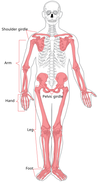
<span class="mw-page-title-main">Navicular bone</span> Small bone found in the feet of most mammals

The navicular bone is a small bone found in the feet of most mammals.

<span class="mw-page-title-main">Upper limb</span> Consists of the arm, forearm, and hand

The upper limbs or upper extremities are the forelimbs of an upright-postured tetrapod vertebrate, extending from the scapulae and clavicles down to and including the digits, including all the musculatures and ligaments involved with the shoulder, elbow, wrist and knuckle joints. In humans, each upper limb is divided into the arm, forearm and hand, and is primarily used for climbing, lifting and manipulating objects.

<span class="mw-page-title-main">Digitigrade</span> Standing or walking on digits/toes; animals which do so

In terrestrial vertebrates, digitigrade locomotion is walking or running on the toes. A digitigrade animal is one that stands or walks with its toes (phalanges) on the ground, and the rest of its foot lifted. Digitigrades include birds, cats, dogs, and many other mammals, but not plantigrades or unguligrades. Digitigrades generally move more quickly than other animals.

<span class="mw-page-title-main">Phalanx bone</span> Digital bone in the hands and feet of most vertebrates

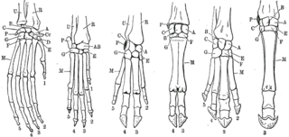
The phalanges are digital bones in the hands and feet of most vertebrates. In primates, the thumbs and big toes have two phalanges while the other digits have three phalanges. The phalanges are classed as long bones.

<span class="mw-page-title-main">Forelimb</span> One of the paired articulated appendages attached on the cranial end of a vertebrates torso

A forelimb or front limb is one of the paired articulated appendages (limbs) attached on the cranial (anterior) end of a terrestrial tetrapod vertebrate's torso. With reference to quadrupeds, the term foreleg or front leg is often used instead. In bipedal animals with an upright posture, the term upper limb is often used.

<span class="mw-page-title-main">Laminitis</span> Disease of the feet of hooved animals

Laminitis is a disease that affects the feet of ungulates and is found mostly in horses and cattle. Clinical signs include foot tenderness progressing to inability to walk, increased digital pulses, and increased temperature in the hooves. Severe cases with outwardly visible clinical signs are known by the colloquial term founder, and progression of the disease will lead to perforation of the coffin bone through the sole of the hoof or being unable to stand up, requiring euthanasia.

<span class="mw-page-title-main">Tarsus (skeleton)</span> Bones of the foot

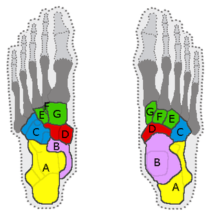
In the human body, the tarsus is a cluster of seven articulating bones in each foot situated between the lower end of the tibia and the fibula of the lower leg and the metatarsus. It is made up of the midfoot and hindfoot.

<span class="mw-page-title-main">Cursorial</span>

A cursorial organism is one that is adapted specifically to run. An animal can be considered cursorial if it has the ability to run fast or if it can keep a constant speed for a long distance. "Cursorial" is often used to categorize a certain locomotor mode, which is helpful for biologists who examine behaviors of different animals and the way they move in their environment. Cursorial adaptations can be identified by morphological characteristics, physiological characteristics, maximum speed, and how often running is used in life. There is much debate over how to define a cursorial animal specifically. The most accepted definitions include that a cursorial organism could be considered adapted to long-distance running at high speeds or has the ability to accelerate quickly over short distances. Among vertebrates, animals under 1 kg of mass are rarely considered cursorial, and cursorial behaviors and morphology are thought to only occur at relatively large body masses in mammals. There are a few mammals that have been termed "micro-cursors" that are less than 1 kg in mass and have the ability to run faster than other small animals of similar sizes.

Many vertebrates are limbless, limb-reduced, or apodous, with a body plan consisting of a head and vertebral column, but no adjoining limbs such as legs or fins. Jawless fish are limbless but may have preceded the evolution of vertebrate limbs, whereas numerous reptile and amphibian lineages – and some eels and eel-like fish – independently lost their limbs. Larval amphibians, tadpoles, are also often limbless. No mammals or birds are limbless, but some feature partial limb-loss or limb reduction.

<span class="mw-page-title-main">Terrestrial locomotion</span> Ability of animals to travel on land

Terrestrial locomotion has evolved as animals adapted from aquatic to terrestrial environments. Locomotion on land raises different problems than that in water, with reduced friction being replaced by the increased effects of gravity.

<span class="mw-page-title-main">Stifle joint</span>

The stifle joint is a complex joint in the hind limbs of quadruped mammals such as the sheep, horse or dog. It is the equivalent of the human knee and is often the largest synovial joint in the animal's body. The stifle joint joins three bones: the femur, patella, and tibia. The joint consists of three smaller ones: the femoropatellar joint, medial femorotibial joint, and lateral femorotibial joint.

A facultative biped is an animal that is capable of walking or running on two legs (bipedal), as a response to exceptional circumstances (facultative), while normally walking or running on four limbs or more. In contrast, obligate bipedalism is where walking or running on two legs is the primary method of locomotion. Facultative bipedalism has been observed in several families of lizards and multiple species of primates, including sifakas, capuchin monkeys, baboons, gibbons, gorillas, bonobos and chimpanzees. Different facultatively bipedal species employ different types of bipedalism corresponding to the varying reasons they have for engaging in facultative bipedalism. In primates, bipedalism is often associated with food gathering and transport. In lizards, it has been debated whether bipedal locomotion is an advantage for speed and energy conservation or whether it is governed solely by the mechanics of the acceleration and lizard's center of mass. Facultative bipedalism is often divided into high-speed (lizards) and low-speed (gibbons), but some species cannot be easily categorized into one of these two. Facultative bipedalism has also been observed in cockroaches and some desert rodents.

A limb is a jointed, muscled appendage of a tetrapod vertebrate animal used for weight-bearing, terrestrial locomotion and physical interaction with other objects. The distalmost portion of a limb is known as its extremity. The limbs' bony endoskeleton, known as the appendicular skeleton, is homologous among all tetrapods, who use their limbs for walking, running and jumping, swimming, climbing, grasping, touching and striking.

Lameness is an abnormal gait or stance of an animal that is the result of dysfunction of the locomotor system. In the horse, it is most commonly caused by pain, but can be due to neurologic or mechanical dysfunction. Lameness is a common veterinary problem in racehorses, sport horses, and pleasure horses. It is one of the most costly health problems for the equine industry, both monetarily for the cost of diagnosis and treatment, and for the cost of time off resulting in loss-of-use.

<i>Aardonyx</i> Extinct genus of dinosaur of the Jurassic from South Africa

Aardonyx is a genus of basal sauropodomorph dinosaur. It is known from the type species Aardonyx celestae found from the Early Jurassic Elliot Formation of South Africa. A. celestae was named after Celeste Yates, who prepared much of the first known fossil material of the species. It has arm features that are intermediate between prosauropods and sauropods.

<span class="mw-page-title-main">Limbs of the horse</span> Structures made of bones, joints, muscles, tendons, and ligaments

The limbs of the horse are structures made of dozens of bones, joints, muscles, tendons, and ligaments that support the weight of the equine body. They include two apparatuses: the suspensory apparatus, which carries much of the weight, prevents overextension of the joint and absorbs shock, and the stay apparatus, which locks major joints in the limbs, allowing horses to remain standing while relaxed or asleep. The limbs play a major part in the movement of the horse, with the legs performing the functions of absorbing impact, bearing weight, and providing thrust. In general, the majority of the weight is borne by the front legs, while the rear legs provide propulsion. The hooves are also important structures, providing support, traction and shock absorption, and containing structures that provide blood flow through the lower leg. As the horse developed as a cursorial animal, with a primary defense mechanism of running over hard ground, its legs evolved to the long, sturdy, light-weight, one-toed form seen today.

References

  1. 1 2 Lockley, M; Jackson, P (2008). "Morphodynamic perspectives on convergence between the feet and limbs of sauropods and humans: two cases of hypermorphosis". Ichnos. 15 (3–4): 140–157. doi:10.1080/10420940802467884. S2CID   86281623.
  2. Lee, DV; Stakebake, EF; Walter, RM; Carrier, DR (2004). "Effects of mass distribution on the mechanics of level trotting in dogs". J Exp Biol. 207 (10): 1715–1728. doi: 10.1242/jeb.00947 . PMID   15073204.
  3. Alexander, R McN; Maloiy, GMO; Hunter, B; Jayes, AS; j, Nturibi (1979). "Mechanical stresses in fast locomotion of buffalo (Syncerus caffer) and elephant (Loxodonta Africana)". J Zool. 189 (2): 135–144. doi:10.1111/j.1469-7998.1979.tb03956.x.
  4. DV, Lee; Sandord, GM (2005). "Directionally compliant legs influence the intrinsic pitch behavior of a trotting quadruped". Proc R Soc B (272): 567–572.
  5. 1 2 Griffin, TM; Main, RP; Farley, CT (2004). "Biomechanics of quadrupedal walking: how do four-legged animals achieve inverted pendulum-like movements?". J Exp Biol. 207 (20): 3545–3558. doi:10.1242/jeb.01177. PMID   15339951.
  6. 1 2 3 4 5 6 Weissengruber, GE; Forstenpointer, G (2004). "Shock absorbers and more: design principles of the lower hind limbs in African elephants (Loxodonta Africana)". J Morphol (260): 339.
  7. 1 2 Miller, CE; Basu, C; Fritsch, G; Hildebrandt, T; JR, Hutchinson (2008). "Ontogenetic scaling of foot musculoskeletal anatomy in elephants". J R Soc Interface. 5 (21): 465–475. doi:10.1098/rsif.2007.1220. PMC   2607390 . PMID   17974531.
  8. 1 2 Dutto, DJ; Hoyt, DF; Clayton, HM; Cogger, EA; SJ, Wickler (2006). "Joint Work and power for both the forelimb and hind limb during trotting in the horse". J Exp Biol. 209 (20): 3990–3999. doi:10.1242/jeb.02471. PMID   17023593.
  9. Hessert, MJ; Vyas, M; Leach, J; Hu, K; LA Lipsitz; V Novak (2005). "Foot pressure distribution during walking in young and old adults". BMC Geriatrics. 5 (1): 8–16. doi: 10.1186/1471-2318-5-8 . PMC   1173105 . PMID   15943881.
  10. Hutchinson, FR; Famini, D; Lair, R; Kram, R (2003). "Are fast moving elephants really running?". Nature. 422 (6931): 493–494. Bibcode:2003Natur.422..493H. doi:10.1038/422493a. PMID   12673241. S2CID   4403723.
  11. Beiwener, AA (2006). "Patterns of mechanical energy change in tetrapod gait: pendula, springs and work". Journal of Experimental Zoology . 305A (11): 899–911. doi:10.1002/jez.a.334. PMID   17029267.
  12. 1 2 McClure, RC (1999). "Functional Anatomy of the Horse Foot" (PDF). Agricultural MU Guide. Archived from the original (PDF) on January 30, 2023. Retrieved April 22, 2009.
  13. Howell, AB (1944). Speed in Animals: Their Specialization for Running and Leaping. Chicago: University of Chicago Press.
  14. Gambaryan, PP (1974). How Mammals Run. New York: John Wiley & Sons.
  15. Douglas, JE; Mittal, C; Thomason, JJ; Jofriet, JC (1996). "The modulus of elasticity of equine hoof wall: implications for the mechanical function of the hoof". J Exp Biol. 199 (8): 1829–1836. doi:10.1242/jeb.199.8.1829. PMID   8708582.
  16. Thompson, JJ; Biewener, AA; Bertram, JEA (1992). "Surface strain on the equine hoof wall in vivo: implications for the material design and functional morphology of the wall". J Exp Biol. 166 (166): 145–168. doi:10.1242/jeb.166.1.145.
  17. 1 2 Donatelli, R (1997). "The evolution and mechanics of the midfoot and hindfoot". Journal of Back and Musculoskeletal Rehabilitation. 8 (1): 57–64. doi:10.3233/BMR-1997-8108. PMID   24572715.
  18. 1 2 Gefen, A; Ravid, MM; Itzchak, Y (2001). "In vivo biomechanical behavior of the human heel pad during the stance phase of gait". J Biomech. 34 (12): 1661–1665. doi:10.1016/s0021-9290(01)00143-9. PMID   11716870.
  19. 1 2 Miller-Young, JE; Duncan, NA; Baroud, G (2002). "Material properties of the human calcaneal fat pad in compression: experiment and theory". J Biomech. 35 (12): 1523–1531. doi:10.1016/s0021-9290(02)00090-8. PMID   12445605.
  20. 1 2 3 4 5 Weissengruber, GE; Egger, GF; Hutchinson, JR; Groenewald, HB; L Elsasser; D Famini; G Forstenponter (2006). "The structure of the cushions in the feet of African elephants (Loxodonta Africana)". J Anat. 209 (6): 781–792. doi:10.1111/j.1469-7580.2006.00648.x. PMC   2048995 . PMID   17118065.
  21. 1 2 König, HE; Macher, R; Polsterer-Heindl, E; Sora, CM; C Hinterhofer; M Helmreich; P Böck (2003). "Stroßbrechende Einrichtungen am Zehenendorgan des Pferdes". Wiener Tierarztliche Monatsschrift (90): 267–273.
  22. McPoil TG, Brocato RS. The foot and ankle: biomechanical evaluation and treatment. In: Gould JA, Davies GJ, ed. Orthopaedic and Sports Physical Therapy. St. Louis: CV Mosby; 1985.
  23. 1 2 3 Perry, J (1992). Gait Analysis: Normal and Pathological Function. Thorofare, NJ: SLACK Inc.
  24. 1 2 3 Soderberg, GL (1997). Kinesiology Application to Pathological Motion (2nd ed.). Baltimore: Williams & Wilkins.
  25. Fisher, MS; Witte, H (2007). "Legs evolved only at the end!". Philosophical Transactions of the Royal Society A . 365 (1850): 185–198. Bibcode:2007RSPTA.365..185F. doi:10.1098/rsta.2006.1915. PMID   17148056. S2CID   20889363.
  26. Weissengruber, GE; Forstenpointer, G (2004). "Musculature of the crus and pes of the African elephant (Loxodonta Africana): insight into semiplantigrade limb architecture". Anat Embryol. 208 (6): 451–461. doi:10.1007/s00429-004-0406-1. PMID   15340844. S2CID   2142971.
  27. Smuts, MMS; Bezuidenhout, AJ (1994). "Osteology of the pelvic limb of the African elephant (Loxodonta Africana)". Onderstepoort J Vet Res. 61 (61): 51–66. PMID   7898898.
  28. Benz, A (2005). "The elephant's hoof: macroscopic and microscopic morphology of defined locations under consideration of pathological changes". Inaugural Dissertation. Vetsuisse-Fakultät Universität Zürich.
  29. 1 2 Ker, RF; Bennett, MB; Bibby, SR; Kester, RC; Alexander, RMcN (1987). "The spring in the arch of the human foot". Nature. 325 (6100): 147–149. Bibcode:1987Natur.325..147K. doi:10.1038/325147a0. PMID   3808070. S2CID   4358258.
  30. Tillmann, B. "Untere Extremität". In Leonhardt, H; Tillmann, B; Töndury, G; et al. (eds.). Anatomie des Menschen, Band I, Bewegungsapparat (3rd ed.). Stuttgart: Thieme. pp. 445–651.
  31. "Elephants Have a Sixth 'Toe'". ScienceMag.org. Archived from the original on 2012-01-13. Retrieved 2011-12-23.
  32. Lieberman, Daniel E; Venkadesan, Madhusudhan; Daoud, Adam I; Werbel, William A (August 2010). "Biomechanics of Foot Strikes & Applications to Running Barefoot or in Minimal Footwear" . Retrieved 3 July 2011.
  33. davejhavu (November 2007). "Favorite Runner 1". YouTube. Retrieved 3 July 2011.
  34. Fowler, ME. "An overview of foot conditions in Asian and African elephants". In Csuti, B; Sargent, EL; Bechert, US (eds.). The elephant's foot. Ames, IA: Iowa State University Press. pp. 3–7.