Cytauxzoonosis

Last updated

Cytauxzoonosis
Scientific classification OOjs UI icon edit-ltr.svg
Domain: Eukaryota
Clade: Diaphoretickes
Clade: SAR
Clade: Alveolata
Phylum: Apicomplexa
Class: Aconoidasida
Order: Piroplasmida
Family: Theileriidae
Genus: Cytauxzoon
Species:
C. felis
Binomial name
Cytauxzoon felis
(Kier)
Synonyms [1]
  • Theileria felis

Cytauxzoon felis is a protozoal organism transmitted to domestic cats by tick bites, and whose natural reservoir host is the bobcat. [2] [3] [4] [5] C. felis has been found in other wild felid species such as the cougar, as well as a white tiger in captivity. [6] [7] [8] C. felis infection is limited to the family Felidae which means that C. felis poses no zoonotic (transmission to humans) risk or agricultural (transmission to farm animals) risk. Until recently it was believed that after infection with C. felis, pet cats almost always died. As awareness of C. felis has increased it has been found that treatment is not always futile. More cats have been shown to survive the infection than was previously thought. [2] [5] [9] [10] New treatments offer as much as 60% survival rate. [11]

Contents

Terminology

Cytauxzoon felis belongs to the order Piroplasmida and the family Theileriidae. [4] C. felis is related to Theileria spp. of African ungulates. It is not a bacterium, not a virus, and not a fungus, but is instead a protozoan that infects the blood cells of cats.[ citation needed ]

Epidemiology

The first case of C. felis was documented in Missouri in 1976, and in the past it was believed to be limited to south central and southeastern United States. [4] There have been more cases popping up in the mid-Atlantic region in the United States, and even as far north as Pennsylvania and North Dakota in bobcats. [2] There has not been any age or sex predilection made, but intact animals have shown more prevalence in some studies; in particular intact males. In studies it is unclear if the sex predilection is due to the increased roaming of intact animals or another reason. [2] The most common seasons for the disease to become apparent are spring and summer and appear to affect outdoor cats the most often. The reason that outdoor cats get infected more is due to the increased exposure to ticks. Ticks are also active in spring and summer. [4]

Symptoms and signs

Most infected cats have been healthy before a very sudden onset of severe disease. The course of clinical disease is often swift with clinical signs of lethargy and inappetence within 5 to 20 days after the tick bite. [4] Cats develop a high fever, but the temperature may become low before death. Other clinical findings can be: dehydration, icterus (jaundice), enlarged liver and spleen, lymphadenopathy, pale mucus membranes, respiratory distress, tachycardia or bradycardia, and tick infestation (although ticks are not often found on infected cats since cats typically groom ticks off their fur). Signs of disease seen on blood work include hemolytic anemia, thrombocytopenia, increased or decreased white blood cell numbers, icterus, and elevated liver enzymes. Death usually follows the onset of clinical signs within a few days. [4] However, more recent studies show not all cats develop clinical signs after infection, and some cats survive the infection. [2] [5] [9] [10] [11]

Life cycle and transmission

The life cycle for Cytauxzoon felis has not been completely described. Bobcats become persistent carriers of the organism after they develop mild or subclinical infection and then serve as the reservoir host. [2] [3] [4] [5] C. felis is transmitted by the Lone star tick ( Amblyomma americanum ) that feeds on an infected bobcat then transmits the organism to another bobcat, or to a pet cat. [10] Many other types of ticks may feed on the cats, but the only other tick that has been shown to transmit the organism is the American dog tick ( Dermacentor variabilis ) but only in a research setting. [2] [5] [10] In the past, domestic cats were thought to always die from infection so they were considered terminal hosts or "dead end hosts". [4] Now there have been documented cases of cats that get sick and survive the infection and even those that never show clinical signs at all. [2] [5] [9] [10] Cats cannot catch the infection from another cat directly without being bitten by a tick.[ citation needed ]

The life cycle of the protozoa has a tissue phase and an erythrocytic (red blood cell) phase. The organism goes through asexual reproduction phase that is called schizogony which occurs in the mononuclear phagocytic cells. In the tissue phase, the macrophage cells that are infected often clog venules in organs such as the liver, spleen, lungs, and lymph nodes. It is this phase that is associated with clinical disease if the schizonts are numerous. Schizonts next form merozoites that break out of the macrophage cells and infect the red blood cells (erythrocytes). The erythrocytic disease is not as important for clinical disease, but is often used to diagnose C. felis because piroplasms (erythrocytes that have phagocytized merozoites) can be seen on a peripheral blood smear under the microscope. [4]

It is not clear why some cats become clinically affected with C. felis while others never show any signs. There are speculations about resistance in some cats, and speculations about different strains of C. felis with differing virulence. [2] [11] [12] There is not enough information to prove any of these theories, but cats in the same household or same regions can have drastically different clinical presentations. Some studies even show multi-cat households have had one cat die and another be persistently infected. [2] [5] When cats survive infection they have persistent parasitemia which shows up in the blood as piroplasms, but these cats do not have the tissue phase again and therefore do not again show the clinical illness. [3] [5] [10]

Diagnosis

One way to diagnose C. felis is by taking blood and performing a peripheral blood smear to look for the erythrocytic piroplasms. [4] The erythrocytic piroplasms are usually shaped like signet rings and are 1 to 1.5 µm. [4] Not all cats that are infected will have the piroplasms on their blood smear, especially if they are early in disease course. Another method of diagnosing infection in sick cats is to take needle aspirates of affected organs and find the schizonts inside mononuclear cells in the tissues; examination of tissue is also useful for the diagnosis after cats have died. Blood samples can be sent away for polymerase chain reaction (PCR) testing to confirm infection. Other diseases that might resemble cytauxzoonosis should be ruled out. A major rule-out for C. felis is Mycoplasma haemofelis (formerly known as Haemobartonella felis); clinical signs can be similar to cytauxzoonosis and the organism may be confused on the peripheral smear. [4] Because it causes similar signs in outdoor cats during the spring and summer, tularemia is another disease the veterinarian may want to rule out.[ citation needed ]

Other laboratory tests are often abnormal in sick cats. The CBC of an infected cat often shows a pancytopenia, or a decrease in red blood cells, white blood cells, and platelets; in some cases there is not a decrease in all three values. Clotting tests may be prolonged. Increased liver enzymes are common, and electrolyte disturbances, hyperglycemia, and acid-base disturbances can also be observed.[ citation needed ]

Treatment and prevention

The preventative measure of keeping cats inside in areas with high infection rates can prevent infection. [4] Approved tick treatments for cats can be used but have been shown not to fully prevent tick bites.[ citation needed ]

The most often used treatments for cytauxzoonosis are imidocarb dipropionate and a combination of atovaquone and azithromycin. Although imidocarb has been used for years, it is not particularly effective. [13] In a large study, [13] only 25% of cats treated with this drug and supportive care survived. 60% of sick cats treated with supportive care and the combination of the anti-malarial drug atovaquone and the antibiotic azithromycin survived infection. [13]

Quick referral to a veterinarian equipped to treat the disease may be beneficial. All infected cats require supportive care, including careful fluids, nutritional support, treatment for complications, and often blood transfusion. [11]

Cats that survive the infection should be kept indoors as they can be persistent carriers after surviving infection and might indirectly infect other cats after being themselves bitten by a vector tick. [3] [5]

Related Research Articles

Ehrlichiosis is a tick-borne disease of dogs usually caused by the rickettsial agent Ehrlichia canis. Ehrlichia canis is the pathogen of animals. Humans can become infected by E. canis and other species after tick exposure. German Shepherd Dogs are thought to be susceptible to a particularly severe form of the disease; other breeds generally have milder clinical signs. Cats can also be infected.

<span class="mw-page-title-main">Babesiosis</span> Malaria-like parasitic disease caused by infection with the alveoate Babesia or Theileria

Babesiosis or piroplasmosis is a malaria-like parasitic disease caused by infection with a eukaryotic parasite in the order Piroplasmida, typically a Babesia or Theileria, in the phylum Apicomplexa. Human babesiosis transmission via tick bite is most common in the Northeastern and Midwestern United States and parts of Europe, and sporadic throughout the rest of the world. It occurs in warm weather. People can get infected with Babesia parasites by the bite of an infected tick, by getting a blood transfusion from an infected donor of blood products, or by congenital transmission . Ticks transmit the human strain of babesiosis, so it often presents with other tick-borne illnesses such as Lyme disease. After trypanosomes, Babesia is thought to be the second-most common blood parasite of mammals. They can have major adverse effects on the health of domestic animals in areas without severe winters. In cattle the disease is known as Texas cattle fever or redwater.

<i>Plasmodium falciparum</i> Protozoan species of malaria parasite

Plasmodium falciparum is a unicellular protozoan parasite of humans, and the deadliest species of Plasmodium that causes malaria in humans. The parasite is transmitted through the bite of a female Anopheles mosquito and causes the disease's most dangerous form, falciparum malaria. It is responsible for around 50% of all malaria cases. P. falciparum is therefore regarded as the deadliest parasite in humans. It is also associated with the development of blood cancer and is classified as a Group 2A (probable) carcinogen.

<i>Feline leukemia virus</i> Species of virus

Feline leukemia virus (FeLV) is a retrovirus that infects cats. FeLV can be transmitted from infected cats when the transfer of saliva or nasal secretions is involved. If not defeated by the animal's immune system, the virus weakens the cat's immune system, which can lead to diseases which can be lethal. Because FeLV is cat-to-cat contagious, FeLV+ cats should only live with other FeLV+ cats.

Tick-borne diseases, which afflict humans and other animals, are caused by infectious agents transmitted by tick bites. They are caused by infection with a variety of pathogens, including rickettsia and other types of bacteria, viruses, and protozoa. The economic impact of tick-borne diseases is considered to be substantial in humans, and tick-borne diseases are estimated to affect ~80 % of cattle worldwide. Most of these pathogens require passage through vertebrate hosts as part of their life cycle. Tick-borne infections in humans, farm animals, and companion animals are primarily associated with wildlife animal reservoirs. many tick-borne infections in humans involve a complex cycle between wildlife animal reservoirs and tick vectors. The survival and transmission of these tick-borne viruses are closely linked to their interactions with tick vectors and host cells. These viruses are classified into different families, including Asfarviridae, Reoviridae, Rhabdoviridae, Orthomyxoviridae, Bunyaviridae, and Flaviviridae.

A trophozoite is the activated, feeding stage in the life cycle of certain protozoa such as malaria-causing Plasmodium falciparum and those of the Giardia group. The complementary form of the trophozoite state is the thick-walled cyst form. They are often different from the cyst stage, which is a protective, dormant form of the protozoa. Trophozoites are often found in the host's body fluids and tissues and in many cases, they are the form of the protozoan that causes disease in the host. In the protozoan, Entamoeba histolytica it invades the intestinal mucosa of its host, causing dysentery, which aid in the trophozoites traveling to the liver and leading to the production of hepatic abscesses.

<i>Plasmodium malariae</i> Species of single-celled organism

Plasmodium malariae is a parasitic protozoan that causes malaria in humans. It is one of several species of Plasmodium parasites that infect other organisms as pathogens, also including Plasmodium falciparum and Plasmodium vivax, responsible for most malarial infection. Found worldwide, it causes a so-called "benign malaria", not nearly as dangerous as that produced by P. falciparum or P. vivax. The signs include fevers that recur at approximately three-day intervals – a quartan fever or quartan malaria – longer than the two-day (tertian) intervals of the other malarial parasite.

<span class="mw-page-title-main">Atovaquone</span> Antimicrobial and antiprotozoan drug

Atovaquone, sold under the brand name Mepron, is an antimicrobial medication for the prevention and treatment of Pneumocystis jirovecii pneumonia (PCP).

Bartonellosis is an infectious disease produced by bacteria of the genus Bartonella. Bartonella species cause diseases such as Carrión's disease, trench fever, cat-scratch disease, bacillary angiomatosis, peliosis hepatis, chronic bacteremia, endocarditis, chronic lymphadenopathy, and neurological disorders.

Chlamydia felis is a Gram-negative, obligate intracellular bacterial pathogen that infects cats. It is endemic among domestic cats worldwide, primarily causing inflammation of feline conjunctiva, rhinitis and respiratory problems. C. felis can be recovered from the stomach and reproductive tract. Zoonotic infection of humans with C. felis has been reported. Strains FP Pring and FP Cello have an extrachromosomal plasmid, whereas the FP Baker strain does not. FP Cello produces lethal disease in mice, whereas the FP Baker does not. An attenuated FP Baker strain, and an attenuated 905 strain, are used as live vaccines for cats.

<i>Babesia</i> Genus of protozoan parasites

Babesia, also called Nuttallia, is an apicomplexan parasite that infects red blood cells and is transmitted by ticks. Originally discovered by the Romanian bacteriologist Victor Babeș in 1888, over 100 species of Babesia have since been identified.

<span class="mw-page-title-main">Anaplasmosis</span> Medical condition

Anaplasmosis is a tick-borne disease affecting ruminants, dogs, and horses, and is caused by Anaplasma bacteria. Anaplasmosis is an infectious but not contagious disease. Anaplasmosis can be transmitted through mechanical and biological vector processes. Anaplasmosis can also be referred to as "yellow bag" or "yellow fever" because the infected animal can develop a jaundiced look. Other signs of infection include weight loss, diarrhea, paleness of the skin, aggressive behavior, and high fever.

<i>Babesia microti</i> Species of parasitic protist in the Apicomplexa phylum

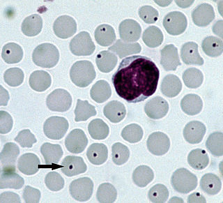
Babesia microti is a parasitic blood-borne piroplasm transmitted by deer ticks. B. microti is responsible for the disease babesiosis, a malaria-like disease which also causes fever and hemolysis.

<i>Theileria parva</i> Species of single-celled organism

Theileria parva is a species of parasites, named in honour of Arnold Theiler, that causes East Coast fever (theileriosis) in cattle, a costly disease in Africa. The main vector for T. parva is the tick Rhipicephalus appendiculatus. Theiler found that East Coast fever was not the same as redwater, but caused by a different protozoan.

<span class="mw-page-title-main">Cat-scratch disease</span> Human disease

Cat-scratch disease (CSD) or felinosis is an infectious disease that most often results from a scratch or bite of a cat. Symptoms typically include a non-painful bump or blister at the site of injury and painful and swollen lymph nodes. People may feel tired, have a headache, or a fever. Symptoms typically begin within 3–14 days following infection.

<i>Mycoplasma haemofelis</i> Parasitic bacterium

Mycoplasma haemofelis is a gram-negative epierythrocytic parasitic bacterium. It often appears in bloodsmears as small (0.6μm) coccoid bodies, sometimes forming short chains of three to eight organisms. It is usually the causative agent of feline infectious anemia (FIA) in the United States.

<span class="mw-page-title-main">Ticks of domestic animals</span>

Ticks of domestic animals directly cause poor health and loss of production to their hosts. Ticks also transmit numerous kinds of viruses, bacteria, and protozoa between domestic animals. These microbes cause diseases which can be severely debilitating or fatal to domestic animals, and may also affect humans. Ticks are especially important to domestic animals in tropical and subtropical countries, where the warm climate enables many species to flourish. Also, the large populations of wild animals in warm countries provide a reservoir of ticks and infective microbes that spread to domestic animals. Farmers of livestock animals use many methods to control ticks, and related treatments are used to reduce infestation of companion animals.

Cytauxzoon is a genus of parasitic alveolates in the phylum Apicomplexa. The name is derived from the Greek meaning an increase in the number of cells in an animal.

Anaplasma bovis is gram negative, obligate intracellular organism, which can be found in wild and domestic ruminants, and potentially a wide variety of other species. It is one of the last species of the Family Anaplasmaceae to be formally described. It preferentially infects host monocytes, and is often diagnosed via blood smears, PCR, and ELISA. A. bovis is not currently considered zoonotic, and does not frequently cause serious clinical disease in its host. This organism is transmitted by tick vectors, so tick bite prevention is the mainstay of A. bovis control, although clinical infections can be treated with tetracyclines. This organism has a global distribution, with infections noted in many areas, including Korea, Japan, Europe, Brazil, Africa, and North America.

<i>Babesia canis</i> Species of single-celled organism

Babesia canis is a parasite that infects red blood cells and can lead to anemia. This is a species that falls under the overarching genus Babesia. It is transmitted by the brown dog tick and is one of the most common piroplasm infections. The brown dog tick is adapted to warmer climates and is found in both Europe and the United States, especially in shelters and greyhound kennels. In Europe, it is also transmitted by Dermacentor ticks with an increase in infections reported due to people traveling with their pets.

References

  1. "Cytauxzoon felis". CABI. Retrieved 14 January 2019.
  2. 1 2 3 4 5 6 7 8 9 10 Birkenheuer, A.J.; Le, J.A.; Valenzisi, A.M.; Tucker, M.D.; Levy, M.G.; Breitschwerdt, E.B. (2006). "Cytauxzoon felis in cats in the mid-Atlantic states: 34 cases (1998-2004)". Journal of the American Veterinary Medical Association. 228 (4): 568–571. doi:10.2460/javma.228.4.568. PMID   16478435.
  3. 1 2 3 4 Brown, H.M.; Latimer, K.S.; Erikson, L.E.; Cashwell, M.E.; Britt, J.O.; Peterson, D.S. (2008). "Detection of Persistent Cytauxzoon Felis Infection by Polymerase Chain Reaction in Three Asymptomatic Domestic Cats". Journal of Veterinary Diagnostic Investigation. 20 (4): 485–488. doi: 10.1177/104063870802000411 . PMID   18599854.
  4. 1 2 3 4 5 6 7 8 9 10 11 12 13 Cohn, L.A. (2005). "Feline Cytauxzoonosis". American Association of Feline Practitioners Rounds. 27: 69–75.
  5. 1 2 3 4 5 6 7 8 9 Habera, M.D.; Tuckera, M.D.; Marra, H.S.; Levyb, J.K.; Burgessc, J.; Lappind, M.R.; Birkenheuera, A.J. (2007). "The detection of Ctyauxzoon felis in apparently healthy free-roaming cats in the US". Veterinary Parasitology. 146 (3–4): 316–320. doi:10.1016/j.vetpar.2007.02.029. PMID   17391852.
  6. Garner, M.M.; Lung, N.P.; Citino, S.; Greiner, E.C.; Harvey, J.W.; Homer, B.L. (1996). "Fatal Cytauxzoonosis in a Captive reared White Tiger (Panthera tigris)". Veterinary Pathology. 33 (1): 82–86. doi: 10.1177/030098589603300111 . PMID   8826011. S2CID   10450447.
  7. Kier, A.B.; Wightman, S.R.; Wagner, J.E. (1982). "Interspecies transmission of Cytauxzoon felis". Am J Vet Res. 43 (1): 102–105. PMID   6807139.
  8. Rotstein, D.S.; Taylor, S.K.; Harvey, J.W.; Bean, J. (1999). "Hematologic effects of cytauxzoonosis in Florida panthers and Texas cougars in Florida". Journal of Wildlife Diseases. 35 (3): 613–617. doi: 10.7589/0090-3558-35.3.613 . PMID   10479103. S2CID   30210414.
  9. 1 2 3 Meinkoth, J.; Kocan, A.A.; Whitworth, L.; Murphy, G.; Fox, C.; Woods, J.P. (2000). "Cats Surviving Natural Infection with Cytauxzoon felis: 18 Cases (1997-1998)". J Vet Med. 14 (5): 521–525. doi:10.1892/0891-6640(2000)014<0521:csniwf>2.3.co;2. PMID   11012116.
  10. 1 2 3 4 5 6 Reichard, M.V.; Meinkoth, J.H.; Edwards, A.C.; Snider, T.A.; Kocan, K.M.; Blouin, E.F.; Little, S.E. (2009). "Transmission of Cytauxzoon felis to a domestic cat by Amblyomma americanum". Veterinary Parasitology. 161 (1–2): 110–115. doi:10.1016/j.vetpar.2008.12.016. PMID   19168288.
  11. 1 2 3 4 Cohn, L.A.; Birkenheuer, A.J.; Brunker J.D.; Ratcliff E.R.; Craig, A.W. (2011). "Efficacy of Atovaquone and Azithromycin or Imidocarb Dipropionate in Cats with Acute Cytauxzoonosis". J Vet Intern Med. 25 (1): 55–60. doi: 10.1111/j.1939-1676.2010.0646.x . PMID   21143646.
  12. Brown, H.M.; Berghaus, R.D.; Latimer, K.S.; Britt, J.O.; Rakich, P.M.; Peterson, D.S. (2009). "Genetic Variability of Cytauxzoon felis from 88 Infected Domestic Cats in Arkansas and Georgia". Journal of Veterinary Diagnostic Investigation. 21 (1): 59–63. doi: 10.1177/104063870902100109 . PMID   19139502.
  13. 1 2 3 Cohn, L.A.; Birkenheuer, A.J.; Brunker, J.D.; Ratcliff, E.R.; Craig, A.W. (2011). "Efficacy of Atovaquone and Azithromycin or Imidocarb Dipropionate in Cats with Acute Cytauxzoonosis". J Vet Intern Med. 25 (1): 55–60. doi: 10.1111/j.1939-1676.2010.0646.x . PMID   21143646.