DNA-functionalized quantum dots

Last updated

DNA-functionalization of quantum dots is the attachment of strands of DNA to the surface of a quantum dot. Although quantum dots with cadmium (Cd) have some cytotoxic release, researchers have functionalized quantum dots for biocompatibility and bound them to DNA in order to combine the advantages of both materials. Quantum dots are commonly used for imaging biological systems in vitro and in vivo in animal studies due to their excellent optical properties when excited by light, while DNA has numerous bioengineering applications, including: genetic engineering, self-assembling nanostructures, protein binding, and biomarkers. The ability to visualize the chemical and biological processes of DNA allows feedback to optimize and learn about these small scale behaviors. [1] [2]

Contents

Figure 1: Quantum dot solutions emitting yellow-orange and light-blue light, excited by UV light (manufactured at California Polytechnic San Luis Obispo) Quantum Dots.jpg
Figure 1: Quantum dot solutions emitting yellow-orange and light-blue light, excited by UV light (manufactured at California Polytechnic San Luis Obispo)

Background

Quantum dots are inorganic nanocrystal semiconductors that behave exceptionally well as fluorophores. In the field of biology, fluorophores are one of the few tools that allow us to peer inside of a live biological system at a cellular level. As a fluorophore, the size of a quantum dot directly reflects the wavelength of light emitted, allowing for a highly tunable color spectrum. Since the size of quantum dots are controllable and an increased size produces an increased wavelength range of emission, researchers are able to paint pictures on the cellular and sub-cellular levels with this technology. The current problem with common CdSe-ZnS quantum dots is that Cd is toxic to cells. [3]

Figure 2: Gas mask used in clean room Face Mask.jpg
Figure 2: Gas mask used in clean room

To prevent this problem researches are developing ways to modify the quantum dot surfaces for biocompatibility, in addition to the development of Cd-free quantum dots ("CFQDs"). After a surface modification has been made to limit toxicity, the particle can be further coated with a hydrogel or bioconjugate layer to selectively bind to DNA, which may then be used for cellular or molecular level detection. [2]

Figure 3: Schematic of a quantum dot coated with an organic protein bound to DNA DNA-functionalized Quantum Dot.png
Figure 3: Schematic of a quantum dot coated with an organic protein bound to DNA

Surface Modification Methods

Hydrogel encapsulation of quantum dots

In order to coat the toxic cadmium ions of the CdSe core, hydrogel layers may be used to coat quantum dots for biocompatibility. The purpose of the outer ZnS shell in this case is to interact with dangling bonds, in addition to maintaining the fluorescent strength of a functional quantum dot fluorophore. Within a hydrogel encapsulation, the ZnSe shell surface may be charged to bind to the hydrophobic interior of a micelle, which then allows the hydrophilic exterior to remain in contact with an aqueous solution (i.e. the human body and most other biological systems). The hydrogel layer works as a simplified intermediary bond for DNA or other organic materials.

Bioconjugation of Quantum Dots

Another surface modification type is bioconjugation. This method uses two biomolecules which are covalently bonded to one another to form a protective shell around the quantum dot. Hydrophobic bioconjugation inhibits the breakdown of the quantum dot structure by sources within the body that may cause degradation. The bioconjugates can be further customized by attaching affinity ligands to the surface of the structure. These ligands allow the quantum dot to bind to various antigens and can be used to specifically target certain cells. This is the driving mechanism for tumor targeting.

Core-shell CdSe-ZnS quantum dots can be protected through bioconjugation, using a coordinating ligand and an amphiphilic polymer. One study used tri-n-octylphosphine oxide (TOPO) as a ligand, and a triblock polymer structure consisting of two hydrophobic segments, and one hydrophilic segment, all with hydrophobic hydrocarbon side chains. The strong hydrophobic interactions between the TOPO and polymer hydrocarbon allow the two layers to "bond" to one another, forming a hydrophobic protection structure. This structure resists degradation via hydrolysis and enzymes, which are common methods of degradation in vivo. This bioconjugation layer protects quantum dot optical properties in a wide range of pH (1-14), salt conditions (0.01-1.0M), and even after 1.0M hydrochloric acid treatment. [4]

Carboxyl attachments

Figure 4: Quantum dot coated with zinc oxide, carboxyl groups, and oligonucleotides to allow DNA binding. Quantum dot coated with ZnS, Carboxyl groups, and Oligionucleotides.svg
Figure 4: Quantum dot coated with zinc oxide, carboxyl groups, and oligonucleotides to allow DNA binding.

Carboxyl groups can be immobilized on the surface of a quantum dot coated in zinc oxide . Single strands of DNA can then be modified with an added amino group in order to covalently bond to the carboxyl group, due to an amide bond formed between the carboxyl and amino groups in the presence of 1-ethyl-3-(3-dimethylaminopropyl)-carbodiimide (EDC). [5] Factors that can influence the binding of single stranded DNA to the carboxyl group are pH and ionic strength. The pH determines how many protons are available to form covalent bonds, with less being present as the pH gets higher. This results in fewer strands of DNA binding to each quantum dot. Lower ionic strength results in more stable quantum dots, but also causes DNA strands to repel each other. Optimum coupling conditions for over 10 DNA strands per quantum dot are at a pH of 7 and an ionic strength of 0M. [6] A neutral pH of 7 allows enough protons from the carboxyl group to facilitate covalent bonding of amino modified DNA, but not enough protons to destabilize the colloids.

Intermolecular Forces

The addition of DNA to the surface of a quantum dot changes the intermolecular forces that occur between un-conjugated quantum dots. Changing the intermolecular forces between the quantum dots can alter many characteristics important for the use of quantum dots in aqueous conditions. As the surface of quantum dots are conjugated with DNA, the colloidal stability and solubility are affected.

Colloidal stability

Quantum dots conjugated with DNA are subject to electrostatic repulsion and Van der Waals forces which affect the colloidal stability of the quantum dot-DNA conjugates. Binding DNA to the surface of a quantum dot increases the stability of the quantum dots. The DNA chains provide more electrostatic repulsion than the surface of the quantum dots, which prevents them from aggregating and falling out of solution. The colloidal stability is estimated from the total interaction energy between two particles which calculated by the DLVO equation [7]

Ves is the electrostatic repulsion forces between two identical spherical particles from the electric double layer of each particle. It is calculated with the equation [6]

Where:

VvdW is the attractive force between all particles. The Van der Waals forces are calculated with the equation [6]

Where

The colloidal stability of the quantum dots can differ with changing pH and ionic strength. Overall, the DNA conjugation increases the stability of the quantum dots by providing electrostatic and steric repulsion, which prevents the particles from aggregating due to van der Waals forces. [6]

Solubility

In order to use quantum dots in many biology related applications, the quantum dots must be soluble in aqueous environments. For quantum dots to be solubilized in water, the amphiphilic ligands must be on the surface of quantum dots. DNA can be used as a solubilization ligand due to its amphiphilic nature. [1] This allows quantum dots functionalized with DNA to be used in the aqueous conditions often found in biology and medical research. The increased solubility is necessary in order to allow quantum dots to be used as a DNA imaging probe in a biological system.

Applications

Quantum dots have become powerful imaging tools and are continually evolving for biocompatibility in the hopes of successfully imaging humans and other live biological systems. By reducing the amount of Cd released around cells, researchers have been striving to create in vitro and in vivo testing methods for imaging nano- and micro-scaled structures. The high resolution within the nanometer range demonstrates the usefulness for imaging DNA behavior for both bioengineering feedback and biological and chemical observation and analysis. The ability to control the emission spectra by altering the size of the quantum dots allows researchers to code many different targets by color. [8]

Size (nm) Emission Peak (nm) Color
2.2 [9] 495blue
2.9 [9] 550green
3.1 [10] 576yellow
4.1 [9] 595orange
4.4 [11] 611orange
4.8 [10] 644red
7.3 [9] 655dark red

Quantifying and imaging gene expression

Since quantum dots have high photostability and luminescence, researchers are using them to light up mRNA within cells to image gene expression. Amine-modified oligonucleotide probes attached to carboxyl groups on quantum dots show sequence-specific hybridization. These probes can also detect low expressing genes. [12] This potentially allows researchers to understand when and where certain proteins are made.

Self-assembling nanostructures

Self-assembled quantum dots form spontaneously under specific conditions during molecular beam epitaxy or another form of atomic deposition. This spontaneous formation is a consequence of the lattice-mismatch between the deposited semiconductor material and the underlying substrate. The resulting structure formed on the substrate surface is a three-dimensional "island" nanostructure. The islands are formed into quantum dots by covering them with another semiconductor material, in a process called quantum confinement. [13] [14] Self-assembled quantum dots provide opportunities in technological applications such as quantum cryptography, quantum computing, optics and optoelectronics. [13]

Single-molecule imaging

In the past, green fluorescent protein (GFP) was used to track movement inside cells. However, GFP does not light up well and is unstable after application. Thus, GFP prevented long term studies of protein movement. By using quantum dots, which are more stable, researchers can now track proteins through cells undergoing different pathways. [15] In order to overcome the inability for the cameras to capture depth, researchers have developed a 3D tracking apparatus that can accurately map the path of proteins inside cells. [16]

Live-time protein tracking

Because quantum dots have fine-tuned wavelength spectrums along with high emission intensities and small size, quantum dots have become the norm for molecule tracking. However, quantum dots have two levels: bright and dark. For low quantities, this is a problem as researchers need to retrace where the molecule went during the dark stage, which can vary from several milliseconds to hours. The blinking phenomenon is not a problem when imaging larger objects (i.e. tumors) as there will be enough quantum dots in the bright state to image, even while several may remain in their dark stage. [17]

See also

Related Research Articles

<span class="mw-page-title-main">Quantum dot</span> Zero-dimensional, nano-scale semiconductor particles with novel optical and electronic properties

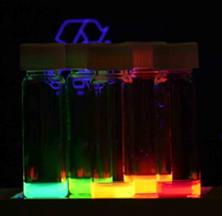
Quantum dots (QDs) or semiconductor nanocrystals are semiconductor particles a few nanometres in size with optical and electronic properties that differ from those of larger particles via quantum mechanical effects. They are a central topic in nanotechnology and materials science. When a quantum dot is illuminated by UV light, an electron in the quantum dot can be excited to a state of higher energy. In the case of a semiconducting quantum dot, this process corresponds to the transition of an electron from the valence band to the conductance band. The excited electron can drop back into the valence band releasing its energy as light. This light emission (photoluminescence) is illustrated in the figure on the right. The color of that light depends on the energy difference between the conductance band and the valence band, or the transition between discrete energy states when the band structure is no longer well-defined in QDs.

<span class="mw-page-title-main">Fluorophore</span> Agents that emit light after excitation by light

A fluorophore is a fluorescent chemical compound that can re-emit light upon light excitation. Fluorophores typically contain several combined aromatic groups, or planar or cyclic molecules with several π bonds.

<span class="mw-page-title-main">Colloidal gold</span> Suspension of gold nanoparticles in a liquid

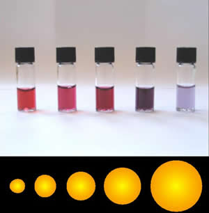
Colloidal gold is a sol or colloidal suspension of nanoparticles of gold in a fluid, usually water. The colloid is coloured usually either wine red or blue-purple . Due to their optical, electronic, and molecular-recognition properties, gold nanoparticles are the subject of substantial research, with many potential or promised applications in a wide variety of areas, including electron microscopy, electronics, nanotechnology, materials science, and biomedicine.

<span class="mw-page-title-main">Förster resonance energy transfer</span> Photochemical energy transfer mechanism

Förster resonance energy transfer (FRET), fluorescence resonance energy transfer, resonance energy transfer (RET) or electronic energy transfer (EET) is a mechanism describing energy transfer between two light-sensitive molecules (chromophores). A donor chromophore, initially in its electronic excited state, may transfer energy to an acceptor chromophore through nonradiative dipole–dipole coupling. The efficiency of this energy transfer is inversely proportional to the sixth power of the distance between donor and acceptor, making FRET extremely sensitive to small changes in distance.

Cell-penetrating peptides (CPPs) are short peptides that facilitate cellular intake and uptake of molecules ranging from nanosize particles to small chemical compounds to large fragments of DNA. The "cargo" is associated with the peptides either through chemical linkage via covalent bonds or through non-covalent interactions.

<span class="mw-page-title-main">Janus particles</span> Type of nanoparticle or microparticle

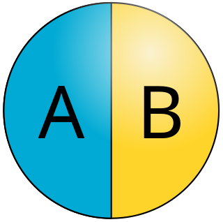
Janus particles are special types of nanoparticles or microparticles whose surfaces have two or more distinct physical properties. This unique surface of Janus particles allows two different types of chemistry to occur on the same particle. The simplest case of a Janus particle is achieved by dividing the particle into two distinct parts, each of them either made of a different material, or bearing different functional groups. For example, a Janus particle may have one half of its surface composed of hydrophilic groups and the other half hydrophobic groups, the particles might have two surfaces of different color, fluorescence, or magnetic properties. This gives these particles unique properties related to their asymmetric structure and/or functionalization.

<span class="mw-page-title-main">Platinum nanoparticle</span>

Platinum nanoparticles are usually in the form of a suspension or colloid of nanoparticles of platinum in a fluid, usually water. A colloid is technically defined as a stable dispersion of particles in a fluid medium.

<span class="mw-page-title-main">Fluorescence in the life sciences</span> Scientific investigative technique

Fluorescence is used in the life sciences generally as a non-destructive way of tracking or analysing biological molecules. Some proteins or small molecules in cells are naturally fluorescent, which is called intrinsic fluorescence or autofluorescence. Alternatively, specific or general proteins, nucleic acids, lipids or small molecules can be "labelled" with an extrinsic fluorophore, a fluorescent dye which can be a small molecule, protein or quantum dot. Several techniques exist to exploit additional properties of fluorophores, such as fluorescence resonance energy transfer, where the energy is passed non-radiatively to a particular neighbouring dye, allowing proximity or protein activation to be detected; another is the change in properties, such as intensity, of certain dyes depending on their environment allowing their use in structural studies.

<span class="mw-page-title-main">Nanoparticle–biomolecule conjugate</span> Tailored macromolecule with covalently-bonded bio-active substances targeting specific tissues

A nanoparticle–biomolecule conjugate is a nanoparticle with biomolecules attached to its surface. Nanoparticles are minuscule particles, typically measured in nanometers (nm), that are used in nanobiotechnology to explore the functions of biomolecules. Properties of the ultrafine particles are characterized by the components on their surfaces more so than larger structures, such as cells, due to large surface area-to-volume ratios. Large surface area-to-volume-ratios of nanoparticles optimize the potential for interactions with biomolecules.

<span class="mw-page-title-main">Core–shell semiconductor nanocrystal</span>

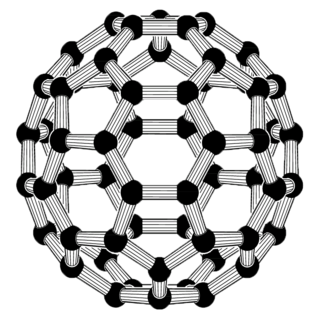
Core–shell semiconducting nanocrystals (CSSNCs) are a class of materials which have properties intermediate between those of small, individual molecules and those of bulk, crystalline semiconductors. They are unique because of their easily modular properties, which are a result of their size. These nanocrystals are composed of a quantum dot semiconducting core material and a shell of a distinct semiconducting material. The core and the shell are typically composed of type II–VI, IV–VI, and III–V semiconductors, with configurations such as CdS/ZnS, CdSe/ZnS, CdSe/CdS, and InAs/CdSe Organically passivated quantum dots have low fluorescence quantum yield due to surface related trap states. CSSNCs address this problem because the shell increases quantum yield by passivating the surface trap states. In addition, the shell provides protection against environmental changes, photo-oxidative degradation, and provides another route for modularity. Precise control of the size, shape, and composition of both the core and the shell enable the emission wavelength to be tuned over a wider range of wavelengths than with either individual semiconductor. These materials have found applications in biological systems and optics.

The behavior of quantum dots (QDs) in solution and their interaction with other surfaces is of great importance to biological and industrial applications, such as optical displays, animal tagging, anti-counterfeiting dyes and paints, chemical sensing, and fluorescent tagging. However, unmodified quantum dots tend to be hydrophobic, which precludes their use in stable, water-based colloids. Furthermore, because the ratio of surface area to volume in a quantum dot is much higher than for larger particles, the thermodynamic free energy associated with dangling bonds on the surface is sufficient to impede the quantum confinement of excitons. Once solubilized by encapsulation in either a hydrophobic interior micelle or a hydrophilic exterior micelle, the QDs can be successfully introduced into an aqueous medium, in which they form an extended hydrogel network. In this form, quantum dots can be utilized in several applications that benefit from their unique properties, such as medical imaging and thermal destruction of malignant cancers.

<span class="mw-page-title-main">Surface chemistry of neural implants</span>

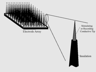
As with any material implanted in the body, it is important to minimize or eliminate foreign body response and maximize effectual integration. Neural implants have the potential to increase the quality of life for patients with such disabilities as Alzheimer's, Parkinson's, epilepsy, depression, and migraines. With the complexity of interfaces between a neural implant and brain tissue, adverse reactions such as fibrous tissue encapsulation that hinder the functionality, occur. Surface modifications to these implants can help improve the tissue-implant interface, increasing the lifetime and effectiveness of the implant.

<span class="mw-page-title-main">Self-healing hydrogels</span> Type of hydrogel

Self-healing hydrogels are a specialized type of polymer hydrogel. A hydrogel is a macromolecular polymer gel constructed of a network of crosslinked polymer chains. Hydrogels are synthesized from hydrophilic monomers by either chain or step growth, along with a functional crosslinker to promote network formation. A net-like structure along with void imperfections enhance the hydrogel's ability to absorb large amounts of water via hydrogen bonding. As a result, hydrogels, self-healing alike, develop characteristic firm yet elastic mechanical properties. Self-healing refers to the spontaneous formation of new bonds when old bonds are broken within a material. The structure of the hydrogel along with electrostatic attraction forces drive new bond formation through reconstructive covalent dangling side chain or non-covalent hydrogen bonding. These flesh-like properties have motivated the research and development of self-healing hydrogels in fields such as reconstructive tissue engineering as scaffolding, as well as use in passive and preventive applications.

Quantum dots (QDs) are semiconductor nanoparticles with a size less than 10 nm. They exhibited size-dependent properties especially in the optical absorption and the photoluminescence (PL). Typically, the fluorescence emission peak of the QDs can be tuned by changing their diameters. So far, QDs were consisted of different group elements such as CdTe, CdSe, CdS in the II-VI category, InP or InAs in the III-V category, CuInS2 or AgInS2 in the I–III–VI2 category, and PbSe/PbS in the IV-VI category. These QDs are promising candidates as fluorescent labels in various biological applications such as bioimaging, biosensing and drug delivery.

Nanoparticle drug delivery systems are engineered technologies that use nanoparticles for the targeted delivery and controlled release of therapeutic agents. The modern form of a drug delivery system should minimize side-effects and reduce both dosage and dosage frequency. Recently, nanoparticles have aroused attention due to their potential application for effective drug delivery.

Hedi Mattoussi is a Tunisian-American materials scientist and professor at Florida State University. His research considers colloidal inorganic nanocrystals for biological imaging and sensing. He is a Fellow of the American Physical Society, American Chemical Society and Materials Research Society.

A protein corona is a dynamic coating of biomolecules, usually proteins, around the surface of a nanoparticle that forms spontaneously in colloidal nanomaterials upon exposure to biological mediums. Protein coronas can form in many different patterns depending on their size, shape, composition, charge, and surface functional groups, and have properties that vary in different environmental factors like temperature, pH, shearing stress, immersed media composition, and exposing time. These coatings are also changeable according to the conditions of the biochemical and physiochemical surface interactions. Types of protein coronas are known to be divided into two categories: “hard” and “soft”. “Hard” coronas have higher-affinity proteins that are irreversibly bonded to the nanoparticle surface, while “soft” coronas have lower-affinity proteins on the nanoparticle surface that are reversibly bound. These reversibly-bound proteins allow for the biomolecules in “soft” protein coronas to be exchanged or detached over time for various applications. This process is governed by the intermolecular protein-nanoparticle and protein-protein interactions that exist within a solution. In "soft" protein coronas, it is common to observe an exchange of proteins at the surface; larger proteins with lower affinities will often aggregate to the surface of the nanoparticle first, and over time, smaller proteins with higher affinities will replace them, "hardening" the corona, known as the Vroman effect.

Silicon quantum dots are metal-free biologically compatible quantum dots with photoluminescence emission maxima that are tunable through the visible to near-infrared spectral regions. These quantum dots have unique properties arising from their indirect band gap, including long-lived luminescent excited-states and large Stokes shifts. A variety of disproportionation, pyrolysis, and solution protocols have been used to prepare silicon quantum dots, however it is important to note that some solution-based protocols for preparing luminescent silicon quantum dots actually yield carbon quantum dots instead of the reported silicon. The unique properties of silicon quantum dots lend themselves to an array of potential applications: biological imaging, luminescent solar concentrators, light emitting diodes, sensors, and lithium-ion battery anodes.

<span class="mw-page-title-main">Dextran drug delivery systems</span> Polymeric drug carrier

Dextran drug delivery systems involve the use of the natural glucose polymer dextran in applications as a prodrug, nanoparticle, microsphere, micelle, and hydrogel drug carrier in the field of targeted and controlled drug delivery. According to several in vitro and animal research studies, dextran carriers reduce off-site toxicity and improve local drug concentration at the target tissue site. This technology has significant implications as a potential strategy for delivering therapeutics to treat cancer, cardiovascular diseases, pulmonary diseases, bone diseases, liver diseases, colonic diseases, infections, and HIV.

Protein nanotechnology is a burgeoning field of research that integrates the diverse physicochemical properties of proteins with nanoscale technology. This field assimilated into pharmaceutical research to give rise to a new classification of nanoparticles termed protein nanoparticles (PNPs). PNPs garnered significant interest due to their favorable pharmacokinetic properties such as high biocompatibility, biodegradability, and low toxicity Together, these characteristics have the potential to overcome the challenges encountered with synthetic NPs drug delivery strategies. These existing challenges including low bioavailability, a slow excretion rate, high toxicity, and a costly manufacturing process, will open the door to considerable therapeutic advancements within oncology, theranostics, and clinical translational research.

References

  1. 1 2 X. Michalet; F. F. Pinaud; L. A. Bentolila; et al. (2005). "Quantum Dots for Live Cells, in Vivo Imaging, and Diagnostics". Science. 307 (5709): 538–544. Bibcode:2005Sci...307..538M. doi:10.1126/science.1104274. PMC   1201471 . PMID   15681376.
  2. 1 2 Catherine J. Murphy; Eric B. Brauns; Latha Gearheart (1996). "Quantum Dots as Inorganic DNA-Binding Proteins". MRS Proceedings. 452: 452–597. doi:10.1557/PROC-452-597.
  3. Santos, Ana R.; Miguel, Ana S.; Macovei, Anca. (2013). "CdSe/ZnS Quantum Dots trigger DNA repair and antioxidant enzyme systems in Medicago sativa cells in suspension culture". BMC Biotechnology. 13: 111. doi: 10.1186/1472-6750-13-111 . PMC   3901376 . PMID   24359290.
  4. Gao, Xiaohu (2004). "In Vivo Cancer Targeting and Imaging with Semiconductor Quantum Dots". Nature Biotechnology. 22 (8): 969–976. doi:10.1038/nbt994. PMID   15258594. S2CID   41561027.
  5. Pong, Boon Kin; Trout, Bernhardt L.; Lee, Jim Yang (2007). "Preparation of DNA-functionalized CdSe/ZnS Quantum Dots". Chemical and Pharmaceutical Engineering. 1.
  6. 1 2 3 4 Dazhi Sun; Oleg Gang (2013). "DNA-Functionalized Quantum Dots: Fabrication, Structural, and Physicochemical Properties". Langmuir. 29 (23): 7038–7046. doi:10.1021/la4000186. PMID   23706124.
  7. Russel, W. B. (1989). Colloidal Dispersions. Cambridge University Press. ISBN   9780511608810.
  8. Mingyong Han; Xiaohu Gao; Jack Z. Su (2001). "Quantum-dot-tagged microbeads for multiplexed optical coding of biomolecules". Nature Biotechnology. 19 (7): 631–635. doi:10.1038/90228. PMID   11433273. S2CID   3118240.
  9. 1 2 3 4 Andrew M. Smith; Hongwei Duan; Aaron M. Mohs (2008). "Bioconjugated quantum dots for in vivo molecular and cellular imaging". Drug Delivery. 60 (11): 1226–1240. doi:10.1016/j.addr.2008.03.015. PMC   2649798 . PMID   18495291.
  10. 1 2 Daniele Gerion; Fabien Pinaud; Shara C. Williams (2001). "Synthesis and Properties of Biocompatible Water-Soluble Silica-Coated CdSe/ZnS Semiconductor Quantum Dots". J. Phys. Chem. B. 105 (37): 8861–8871. doi:10.1021/jp0105488. S2CID   8818995.
  11. Warren CW Chan; Dustin J Maxwell; Xiaohu Gao (2002). "Luminescent quantum dots for multiplexed biological detection and imaging". Current Opinion in Biotechnology. 13 (1): 40–46. doi:10.1016/S0958-1669(02)00282-3. PMID   11849956.
  12. Choi, Youngseon; Kim, Hwa Pyung; Hong, Suk Min; Ryu, Ji Young; Han, Sung Jun; Song, Rita (18 September 2009). "In situ Visualization of Gene Expression Using Polymer-Coated Quantum-Dot-DNA Conjugates". Small. 5 (18): 2085–2091. doi:10.1002/smll.200900116. PMID   19517489.
  13. 1 2 Ryman-Rasmussen, Jessica P; Riviere, Jim E; Monteiro-Riviere, Nancy A (10 August 2006). "Surface Coatings Determine Cytotoxicity and Irritation Potential of Quantum Dot Nanoparticles in Epidermal Keratinocytes". Journal of Investigative Dermatology. 127 (1): 143–153. doi: 10.1038/sj.jid.5700508 . PMID   16902417.
  14. Petroff, Pierre M.; Lorke, Axel; Imamoglu, Atac (May 2001). "Epitaxially Self-Assembled Quantum Dots". Physics Today. 54 (5). Scitation: 46–52. doi: 10.1063/1.1381102 .
  15. Baba, Koichi; Nishida, Kohji (2012). "Single-Molecule Tracking in Living Cells Using Single Quantum Dot Applications". Theranostics. 2 (7): 655–667. doi:10.7150/thno.3890. PMC   3418928 . PMID   22896768.
  16. Wells, Nathan P.; Lessard, Guillaume A.; Goodwin, Peter M.; Phipps, Mary E.; Cutler, Patrick J.; Lidke, Diane S.; Wilson, Bridget S.; Werner, James H. (10 November 2010). "Time-Resolved Three-Dimensional Molecular Tracking in Live Cells". Nano Letters. 10 (11): 4732–4737. Bibcode:2010NanoL..10.4732W. doi:10.1021/nl103247v. PMC   3061257 . PMID   20957984.
  17. Pinaud, Fabien; Clarke, Samuel; Sittner, Assa; Dahan, Maxime (30 March 2010). "Probing cellular events, one quantum dot at a time". Nature Methods. 7 (4): 275–285. doi:10.1038/NMETH.1444. PMID   20354518. S2CID   205419295.