Ascomycota is a phylum of the kingdom Fungi that, together with the Basidiomycota, forms the subkingdom Dikarya. Its members are commonly known as the sac fungi or ascomycetes. It is the largest phylum of Fungi, with over 64,000 species. The defining feature of this fungal group is the "ascus", a microscopic sexual structure in which nonmotile spores, called ascospores, are formed. However, some species of the Ascomycota are asexual, meaning that they do not have a sexual cycle and thus do not form asci or ascospores. Familiar examples of sac fungi include morels, truffles, brewers' and bakers' yeast, dead man's fingers, and cup fungi. The fungal symbionts in the majority of lichens such as Cladonia belong to the Ascomycota.

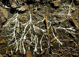
A hypha is a long, branching, filamentous structure of a fungus, oomycete, or actinobacterium. In most fungi, hyphae are the main mode of vegetative growth, and are collectively called a mycelium.
A mycorrhiza is a symbiotic association between a fungus and a plant. The term mycorrhiza refers to the role of the fungus in the plant's rhizosphere, its root system. Mycorrhizae play important roles in plant nutrition, soil biology, and soil chemistry.

Rusts are fungal plant pathogens of the order Pucciniales causing plant fungal diseases.

Mistletoe is the common name for obligate hemiparasitic plants in the order Santalales. They are attached to their host tree or shrub by a structure called the haustorium, through which they extract water and nutrients from the host plant.

A parasitic plant is a plant that derives some or all of its nutritional requirements from another living plant. They make up about 1% of angiosperms and are found in almost every biome. All parasitic plants develop a specialized organ called the haustorium, which penetrates the host plant, connecting them to the host vasculature – either the xylem, phloem, or both. For example, plants like Striga or Rhinanthus connect only to the xylem, via xylem bridges (xylem-feeding). Alternately, plants like Cuscuta and some members of Orobanche connect to both the xylem and phloem of the host. This provides them with the ability to extract resources from the host. These resources can include water, nitrogen, carbon and/or sugars. Parasitic plants are classified depending on the location where the parasitic plant latches onto the host, the amount of nutrients it requires, and their photosynthetic capability. Some parasitic plants can locate their host plants by detecting volatile chemicals in the air or soil given off by host shoots or roots, respectively. About 4,500 species of parasitic plants in approximately 20 families of flowering plants are known.
Glomus is a genus of arbuscular mycorrhizal (AM) fungi, and all species form symbiotic relationships (mycorrhizae) with plant roots. Glomus is the largest genus of AM fungi, with ca. 85 species described, but is currently defined as non-monophyletic.
Suspensors are anatomical structures found in certain fungi and plants.

Geosiphon is a genus of fungus in the family Geosiphonaceae. The genus is monotypic, containing the single species Geosiphon pyriformis, first described by Kützing in 1849 as Botrydium pyriforme. In 1915, Von Wettstein characterized Geosiphon pyriforme as a multinucleate alga containing endosymbiotic cyanobacteria, although he also noted the presence of chitin, a component of fungal cell walls. In 1933, Knapp was the first to suggest the fungal origin of the species and described it as a lichen with endosymbiotic cyanobacteria. It is the only member of the Glomeromycota known to not form a symbiosis with terrestrial plants in the form of arbuscular mycorrhiza.
Saprotrophic nutrition or lysotrophic nutrition is a process of chemoheterotrophic extracellular digestion involved in the processing of decayed organic matter. It occurs in saprotrophs, and is most often associated with fungi and soil bacteria. Saprotrophic microscopic fungi are sometimes called saprobes. Saprotrophic plants or bacterial flora are called saprophytes, although it is now believed that all plants previously thought to be saprotrophic are in fact parasites of microscopic fungi or other plants. The process is most often facilitated through the active transport of such materials through endocytosis within the internal mycelium and its constituent hyphae.
This is a glossary of some of the terms used in phytopathology.
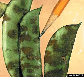
Colletotrichum lindemuthianum is a fungus which causes anthracnose, or black spot disease, of the common bean plant. It is considered a hemibiotrophic pathogen because it spends part of its infection cycle as a biotroph, living off of the host but not harming it, and the other part as a necrotroph, killing and obtaining nutrients from the host tissues.
Entropezites is an extinct monotypic genus of [hypermycoparasitic] fungus in the order Hypocreales. At present it contains the single species Entropezites patricii.

Phoradendron tomentosum, the leafy mistletoe, hairy mistletoe or Christmas mistletoe, is a plant parasite. It is characterized by its larger leaves and smaller berries than dwarf mistletoe. Leafy mistletoe seldom kill but they do rob their hosts of moisture and some minerals, causing stress during drought and reducing crop productions on fruit and nut trees. Leafy mistletoe has the ability to photosynthesize on its own but it relies on other plants in order to obtain its nutrients. It attaches itself to a tree and then grows haustoria, in order to get the food and water it needs.

Purpureocillium lilacinum is a species of filamentous fungus in the family Ophiocordycipitaceae. It has been isolated from a wide range of habitats, including cultivated and uncultivated soils, forests, grassland, deserts, estuarine sediments and sewage sludge, and insects. It has also been found in nematode eggs, and occasionally from females of root-knot and cyst nematodes. In addition, it has frequently been detected in the rhizosphere of many crops. The species can grow at a wide range of temperatures – from 8 to 38 °C for a few isolates, with optimal growth in the range 26 to 30 °C. It also has a wide pH tolerance and can grow on a variety of substrates. P. lilacinum has shown promising results for use as a biocontrol agent to control the growth of destructive root-knot nematodes.

An ectomycorrhiza is a form of symbiotic relationship that occurs between a fungal symbiont, or mycobiont, and the roots of various plant species. The mycobiont is often from the phyla Basidiomycota and Ascomycota, and more rarely from the Zygomycota. Ectomycorrhizas form on the roots of around 2% of plant species, usually woody plants, including species from the birch, dipterocarp, myrtle, beech, willow, pine and rose families. Research on ectomycorrhizas is increasingly important in areas such as ecosystem management and restoration, forestry and agriculture.
Orchid mycorrhizae are endomycorrhizal fungi which develop symbiotic relationships with the roots and seeds of plants of the family Orchidaceae. Nearly all orchids are myco-heterotrophic at some point in their life cycle. Orchid mycorrhizae are critically important during orchid germination, as an orchid seed has virtually no energy reserve and obtains its carbon from the fungal symbiont.
Hemibiotrophs are the spectrum of plant pathogens, including bacteria, oomycete and a group of plant pathogenic fungi that keep its host alive while establishing itself within the host tissue, taking up the nutrients with brief biotrophic-like phase. It then, in later stages of infection switches to a necrotrophic life-style, where it rampantly kills the host cells, deriving its nutrients from the dead tissues.

Exobasidium camelliae is a phytopathagenic fungus that infects ornamental shrubs of the Camellia genus. It absorbs nutrients from the host through its haustoria and causes the leaves of the host plant to be thicker and lighter green than usual. It forms a hymenium between cells four to six layers above the lower epidermis which is subsequently sloughed off to reveal its basidia.
The Melaniellaceae are a family of fungi in the division Basidiomycota and order of Doassansiales. The family contains 1 genera and 2 species. They have a distribution in south and south-east Asia.