Heme A

Last updated
Heme A
Heme a.svg
Haem-a-in-cyctochrome-c-oxidase-PDB-1OCR-3D-SF-A.png
Names
Other names
Iron cytoporphyrin IX, formilporphyrin
Identifiers
3D model (JSmol)
ChemSpider
MeSH Heme+a
PubChem CID
  • InChI=1S/C49H59N4O6.Fe/c1-9-34-31(6)39-25-45-49(46(55)18-12-17-30(5)16-11-15-29(4)14-10-13-28(2)3)33(8)40(52-45)24-44-37(27-54)36(20-22-48(58)59)43(53-44)26-42-35(19-21-47(56)57)32(7)38(51-42)23-41(34)50-39;/h9,13,15,17,23-27,43,46,55H,1,10-12,14,16,18-22H2,2-8H3,(H4-,50,51,52,53,56,57,58,59);/q-1;+2/p-2/b29-15+,30-17+,42-26-; Yes check.svgY
    Key: RRRJRRNGYOECDS-ZHOBENDVSA-L Yes check.svgY
  • InChI=1/C49H59N4O6.Fe/c1-9-34-31(6)39-25-45-49(46(55)18-12-17-30(5)16-11-15-29(4)14-10-13-28(2)3)33(8)40(52-45)24-44-37(27-54)36(20-22-48(58)59)43(53-44)26-42-35(19-21-47(56)57)32(7)38(51-42)23-41(34)50-39;/h9,13,15,17,23-27,43,46,55H,1,10-12,14,16,18-22H2,2-8H3,(H4-,50,51,52,53,56,57,58,59);/q-1;+2/p-2/b29-15+,30-17+,42-26-;/rC49H57FeN4O6/c1-9-34-31(6)39-25-45-49(46(56)18-12-17-30(5)16-11-15-29(4)14-10-13-28(2)3)33(8)40-24-44-37(27-55)36(20-22-48(59)60)43-26-42-35(19-21-47(57)58)32(7)38-23-41(34)51(39)50(52(38)42,53(40)45)54(43)44/h9,13,15,17,23-27,43,46,56H,1,10-12,14,16,18-22H2,2-8H3,(H,57,58)(H,59,60)/q-1/b29-15+,30-17+
    Key: RRRJRRNGYOECDS-DRKRPJRXBB
  • OC(=O)CC/c6c(\C)c3n7c6cc2c(/CCC(O)=O)c(/C=O)c1cc5n8c(cc4n([Fe]78n12)c(c=3)c(C=C)c4c)c(\C(O)CC\C=C(/C)CC\C=C(/C)CC\C=C(C)/C)c5\C
Properties
C49H56O6N4Fe
Molar mass 852.837
Except where otherwise noted, data are given for materials in their standard state (at 25 °C [77 °F], 100 kPa).
Yes check.svgY  verify  (what is  Yes check.svgYX mark.svgN ?)

Heme A (or haem A) is a heme, a coordination complex consisting of a macrocyclic ligand called a porphyrin, chelating an iron atom. Heme A is a biomolecule and is produced naturally by many organisms. Heme A, often appears a dichroic green/red when in solution, is a structural relative of heme B, a component of hemoglobin, the red pigment in blood.

Contents

Relationship to other hemes

Heme A differs from heme B in that a methyl side chain at ring position 8 is oxidized to a formyl group and a hydroxyethylfarnesyl group, an isoprenoid chain, has been attached to the vinyl side chain at ring position 2 of the iron tetrapyrrole heme. Heme A is similar to heme o, in that both have this farnesyl addition at position 2 but heme O does not have the formyl group at position 8, still containing the methyl group. The correct structure of heme A, based upon NMR and IR experiments of the reduced, Fe(II) form of the heme, was published in 1975. [1] The structure was confirmed by synthesis of the dimethyl ester of the iron-free form. [2]

History

Heme A was first isolated by the German biochemist Otto Warburg in 1951 and shown by him to be the active component of the integral membrane metalloprotein cytochrome c oxidase. [3]

Stereochemistry

The final structural question of the exact geometric configuration about the first carbon at ring position 3 of ring I, the carbon bound to the hydroxyl group, has been shown to be the chiral S configuration. [4]

Like heme B, heme A is often attached to the apoprotein through a coordinate bond between the heme iron and a conserved amino acid side-chain. In the important respiratory protein cytochrome c oxidase (CCO) this ligand 5 for the heme A at the oxygen reaction center is a histidyl group. [5] Histidine is a common ligand for many hemeproteins including hemoglobin and myoglobin. Haem-a-in-cyctochrome-c-oxidase-PDB-1OCR-3D-balls-C.png
Heme A in the cytochrome a portion of cytochrome c oxidase, bound by two histidine residues (shown in pink) [6]

An example of a metalloprotein that contains heme A is cytochrome c oxidase. This very complicated protein contains heme A at two different sites, each with a different function. The iron of the heme A of cytochrome a is hexacoordinated, that is bound with 6 other atoms. The iron of the heme A of cytochrome a3 is sometimes bound by 5 other atoms leaving the sixth site available to bind dioxygen (molecular oxygen). [6] In addition, this enzyme binds 3 copper, magnesium, zinc, and several potassium and sodium ions. The two heme A groups in CCO are thought to readily exchange electrons between each other, the copper ions and the closely associated protein cytochrome c.

Both the formyl group and the isoprenoid side chain are thought to play important roles in conservation of the energy of oxygen reduction by cytochrome c oxidase. CCO is thought to be responsible for conserving the energy of dioxygen reduction by pumping protons into the inter-membrane mitochondrial space. Both the formyl and hydroxyethylfarnesyl groups of heme A are thought to play important roles in this critical process, as published by the influential group of S. Yoshikawa. [7]

See also

Related Research Articles

<span class="mw-page-title-main">Cytochrome</span> Redox-active proteins containing a heme with a Fe atom as a cofactor

Cytochromes are redox-active proteins containing a heme, with a central iron (Fe) atom at its core, as a cofactor. They are involved in electron transport chain and redox catalysis. They are classified according to the type of heme and its mode of binding. Four varieties are recognized by the International Union of Biochemistry and Molecular Biology (IUBMB), cytochromes a, cytochromes b, cytochromes c and cytochrome d.

<span class="mw-page-title-main">Hemoglobin</span> Oxygen-transport metalloprotein in red blood cells of most vertebrates

Hemoglobin, is the iron-containing oxygen-transport protein present in red blood cells (erythrocytes) of almost all vertebrates as well as the tissues of some invertebrate animals. Hemoglobin in blood carries oxygen from the respiratory organs to the other tissues of the body, where it releases the oxygen to enable aerobic respiration which powers the animal's metabolism. A healthy human has 12 to 20 grams of hemoglobin in every 100 mL of blood. Hemoglobin is a metalloprotein and chromoprotein.

<span class="mw-page-title-main">Oxidative phosphorylation</span> Metabolic pathway

Oxidative phosphorylation or electron transport-linked phosphorylation or terminal oxidation is the metabolic pathway in which cells use enzymes to oxidize nutrients, thereby releasing chemical energy in order to produce adenosine triphosphate (ATP). In eukaryotes, this takes place inside mitochondria. Almost all aerobic organisms carry out oxidative phosphorylation. This pathway is so pervasive because it releases more energy than alternative fermentation processes such as anaerobic glycolysis.

<span class="mw-page-title-main">Myoglobin</span> Iron and oxygen-binding protein

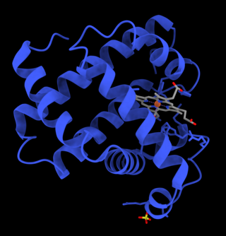
Myoglobin is an iron- and oxygen-binding protein found in the cardiac and skeletal muscle tissue of vertebrates in general and in almost all mammals. Myoglobin is distantly related to hemoglobin. Compared to hemoglobin, myoglobin has a higher affinity for oxygen and does not have cooperative binding with oxygen like hemoglobin does. Myoglobin consists of non-polar amino acids at the core of the globulin, where the heme group is non-covalently bounded with the surrounding polypeptide of myoglobin. In humans, myoglobin is only found in the bloodstream after muscle injury.

<span class="mw-page-title-main">Cytochrome c</span> Protein-coding gene in the species Homo sapiens

The cytochrome complex, or cyt c, is a small hemeprotein found loosely associated with the inner membrane of the mitochondrion where it plays a critical role in cellular respiration. It transfers electrons between Complexes III and IV. Cytochrome c is highly water-soluble, unlike other cytochromes. It is capable of undergoing oxidation and reduction as its iron atom converts between the ferrous and ferric forms, but does not bind oxygen. It also plays a major role in cell apoptosis. In humans, cytochrome c is encoded by the CYCS gene.

<span class="mw-page-title-main">Hemoprotein</span> Protein containing a heme prosthetic group

A hemeprotein, or heme protein, is a protein that contains a heme prosthetic group. They are a very large class of metalloproteins. The heme group confers functionality, which can include oxygen carrying, oxygen reduction, electron transfer, and other processes. Heme is bound to the protein either covalently or noncovalently or both.

<span class="mw-page-title-main">Cytochrome c oxidase</span> Complex enzyme found in bacteria, archaea, and mitochondria of eukaryotes

The enzyme cytochrome c oxidase or Complex IV, is a large transmembrane protein complex found in bacteria, archaea, and mitochondria of eukaryotes.

<span class="mw-page-title-main">Heme</span> Chemical coordination complex of an iron ion chelated to a porphyrin

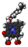
Heme, or haem, is a precursor to hemoglobin, which is necessary to bind oxygen in the bloodstream. Heme is biosynthesized in both the bone marrow and the liver.

<span class="mw-page-title-main">Porphyrin</span> Heterocyclic organic compound with four modified pyrrole subunits

Porphyrins are a group of heterocyclic macrocycle organic compounds, composed of four modified pyrrole subunits interconnected at their α carbon atoms via methine bridges (=CH−). In vertebrates, an essential member of the porphyrin group is heme, which is a component of hemoproteins, whose functions include carrying oxygen in the bloodstream. In plants, an essential porphyrin derivative is chlorophyll, which is involved in light-harvesting and electron transfer in photosynthesis.

<span class="mw-page-title-main">Metalloprotein</span> Protein that contains a metal ion cofactor

Metalloprotein is a generic term for a protein that contains a metal ion cofactor. A large proportion of all proteins are part of this category. For instance, at least 1000 human proteins contain zinc-binding protein domains although there may be up to 3000 human zinc metalloproteins.

<span class="mw-page-title-main">Rieske protein</span> Protein family with an iron–sulfur center transferring electrons

Rieske proteins are iron–sulfur protein (ISP) components of cytochrome bc1 complexes and cytochrome b6f complexes and are responsible for electron transfer in some biological systems. John S. Rieske and co-workers first discovered the protein and in 1964 isolated an acetylated form of the bovine mitochondrial protein. In 1979 Trumpower's lab isolated the "oxidation factor" from bovine mitochondria and showed it was a reconstitutively-active form of the Rieske iron-sulfur protein
It is a unique [2Fe-2S] cluster in that one of the two Fe atoms is coordinated by two histidine residues rather than two cysteine residues. They have since been found in plants, animals, and bacteria with widely ranging electron reduction potentials from -150 to +400 mV.

<span class="mw-page-title-main">Sulfite oxidase</span>

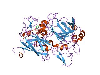
Sulfite oxidase is an enzyme in the mitochondria of all eukaryotes, with exception of the yeasts. It oxidizes sulfite to sulfate and, via cytochrome c, transfers the electrons produced to the electron transport chain, allowing generation of ATP in oxidative phosphorylation. This is the last step in the metabolism of sulfur-containing compounds and the sulfate is excreted.

Nitrite reductase refers to any of several classes of enzymes that catalyze the reduction of nitrite. There are two classes of NIR's. A multi haem enzyme reduces NO2 to a variety of products. Copper containing enzymes carry out a single electron transfer to produce nitric oxide.

<span class="mw-page-title-main">Ferrochelatase</span>

Protoporphyrin ferrochelatase (EC 4.98.1.1, formerly EC 4.99.1.1, or ferrochelatase; systematic name protoheme ferro-lyase (protoporphyrin-forming)) is an enzyme encoded by the FECH gene in humans. Ferrochelatase catalyses the eighth and terminal step in the biosynthesis of heme, converting protoporphyrin IX into heme B. It catalyses the reaction:

Iron-binding proteins are carrier proteins and metalloproteins that are important in iron metabolism and the immune response. Iron is required for life.

<span class="mw-page-title-main">Protoporphyrin IX</span> Chemical compound

Protoporphyrin IX is an organic compound, classified as a porphyrin, that plays an important role in living organisms as a precursor to other critical compounds like heme (hemoglobin) and chlorophyll. It is a deeply colored solid that is not soluble in water. The name is often abbreviated as PPIX.

<span class="mw-page-title-main">Heme O</span> Chemical compound

Heme O differs from the closely related heme A by having a methyl group at ring position 8 instead of the formyl group. The isoprenoid chain at position 2 is the same.

<span class="mw-page-title-main">Dioxygenase</span> Class of enzymes

Dioxygenases are oxidoreductase enzymes. Aerobic life, from simple single-celled bacteria species to complex eukaryotic organisms, has evolved to depend on the oxidizing power of dioxygen in various metabolic pathways. From energetic adenosine triphosphate (ATP) generation to xenobiotic degradation, the use of dioxygen as a biological oxidant is widespread and varied in the exact mechanism of its use. Enzymes employ many different schemes to use dioxygen, and this largely depends on the substrate and reaction at hand.

A transition metal oxo complex is a coordination complex containing an oxo ligand. Formally O2-, an oxo ligand can be bound to one or more metal centers, i.e. it can exist as a terminal or (most commonly) as bridging ligands (Fig. 1). Oxo ligands stabilize high oxidation states of a metal. They are also found in several metalloproteins, for example in molybdenum cofactors and in many iron-containing enzymes. One of the earliest synthetic compounds to incorporate an oxo ligand is potassium ferrate (K2FeO4), which was likely prepared by Georg E. Stahl in 1702.

<span class="mw-page-title-main">Galactose oxidase</span>

Galactose oxidase is an enzyme that catalyzes the oxidation of D-galactose in some species of fungi.

References

  1. Caughey, W.S.; Smythe, G.A.; O'Keefe, D.H.; Maskasky, J.E.; Smith, M.L. (1975). "Heme A of Cytochrome c Oxidase". Journal of Biological Chemistry . 250 (19): 7602–7622. doi: 10.1016/S0021-9258(19)40860-0 . PMID   170266.
  2. Battersby, Alan R.; McDonald, Edward; Thompson, Mervyn; Chaudhry, Irshad A.; Clezy, Peter S.; Fookes, Christopher J. R.; Hai, Ton That (1985). "Isolation, crystallisation, and synthesis of the dimethyl ester of porphyrin a, the iron-free prosthetic group of cytochrome c oxidase". Journal of the Chemical Society, Perkin Transactions 1: 135. doi:10.1039/P19850000135.
  3. Warburg, O; Gewitz H S. (1951). "Cytohämin aus Herzmuskel". Zeitschrift für Physiologische Chemie . 288 (1): 1–4. doi:10.1515/bchm2.1951.288.1.1. PMID   14860765.
  4. Yamashita E, Aoyama H, Yao M, et al. (2005). "Absolute configuration of the hydroxyfarnesylethyl group of heme A, determined by X-ray structural analysis of bovine heart cytochrome c oxidase using methods applicable at 2.8 Angstrom resolution". Acta Crystallographica D . 61 (10): 1373–1377. doi: 10.1107/S0907444905023358 . PMID   16204889.
  5. Tsukihara T, Shimokata K, Katayama Y, et al. (2003). "The low-spin heme of cytochrome c oxidase as the driving element of the proton-pumping process". PNAS . 100 (26): 15304–15309. Bibcode:2003PNAS..10015304T. doi: 10.1073/pnas.2635097100 . PMC   307562 . PMID   14673090.
  6. 1 2 Yoshikawa, S.; Shinzawa-Itoh, K.; Nakashima, R.; et al. (1998). "Redox-Coupled Crystal Structural Changes in Bovine Heart Cytochrome c Oxidase". Science . 280 (5370): 1723–1729. doi:10.1126/science.280.5370.1723. PMID   9624044. S2CID   37147458.
  7. Shimokata K, Katayama Y, Murayama H, et al. (2007). "The proton pumping pathway of bovine heart cytochrome c oxidase". PNAS . 104 (10): 4200–4205. Bibcode:2007PNAS..104.4200S. doi: 10.1073/pnas.0611627104 . PMC   1820732 . PMID   17360500.