Intermammary cleft

Last updated
Intermammary cleft
Bringuhar.jpg
Intermammary cleft dividing the breasts
Details
Identifiers
Latin sulcus intermammarius
TA98 A16.0.02.002
TA2 7110
FMA 55264
Anatomical terminology

The intermammary cleft, intermammary sulcus, or sulcus intermammarius is a surface feature of males and females that marks the division of the two breasts with the sternum (breastbone) in the middle. [1] [2] [3] [4] [5] The International Federation of Associations of Anatomists (IFAA) uses the terms "sulcus intermammarius" or "intermammary cleft" when referring to the area between the breasts. [6] [7] [8]

Contents

Etymology

"Intermammary" ("inter", between + "mamma", breasts + "ry", place [9] [10] [11] ) means something that is located or performed between the breasts (example: intermammary intercourse). [12] "Sulcus" is a Latin word that means a furrow or groove, commonly used to mean a fold, fissure or furrow of the brain (example: lateral sulcus). [13] [14] In popular usage the area is commonly referred to as a cleavage of breasts. In surgical parlance, the cleavage or intermammary cleft is also known as the "medial definition" or "medial fold" of breasts. [15] [16] An imaginary line between the nipples that crosses the intermammary cleft, serving as a landmark for some CPR procedures, is known as the "intermammary line". [17]

Anatomy

Skin

At the midline of the breast—the intermammary cleft—a deep layer of superficial fascia (lowermost layer of the skin) is firmly attached to the pectoral fascia (outer side of chest muscles) and the periosteum (bone membrane) of the sternum. It forms a shelving edge that supports the breasts. [18]

Muscles

The sternalis muscle is an anatomical variation that lies in front of the sternal end of the pectoralis major runs along the anterior aspect of the body of the sternum. The sternalis muscle often originates from the upper part of the sternum and can display varying insertions such as the pectoral fascia, lower ribs, costal cartilages, rectus sheath, aponeurosis of the abdominal external oblique muscle. [19] [20] [21] There is still a great deal of disagreement about its innervation and its embryonic origin. [19] The sternal side (towards the breastbone) of the pectoralis major is distinct from the clavicular side (towards the collarbone), and the two are separated by a fascial interval. [22] The sternal side is usually more robust and has a fiber orientation that best emulates the downward pull of the subscapularis muscle (armpit muscle). [23]

Ligaments

A dense structure of Cooper's ligaments is medially inserted into the skin overlying the sternum, determining the shape of the breasts and intermammary sulcus. [24] These ligaments helpful in supporting the breasts in an youthful disposition; loss of elasticity or attenuation of these ligaments results in ptosis (sagging) of breasts. [25] [26]

Anatomy of the cleavage, the red outlined area being the intermammary cleft Intermammary sulcus anatomy.png
Anatomy of the cleavage, the red outlined area being the intermammary cleft

Arteries

Major blood supply in the area happens through the intermammary artery (also known as the internal thoracic artery) in the thoracic cavity. [27] Among women, this artery is larger. [28] From which anterior perforating branches carry blood into the circummammary arterial plexus, a vascular network formed by branches between minute arteries around the breasts, with lateral mammary branches curving around the lateral border of the pectoralis major. [27] [28] [29]

Lymphs

Lymph vessels originating at the base of the nipples can ventrally extend as far as the intermammary cleft [30] and to the opposite breast. [1] The intermammary lymphatics begin as a bunch of small channels consisting of a single layer of epithelium that is supported by stroma tissues. Each mesh of this network surrounds one or more of the ultimate lobules of the glands and receives its lymph from the interacinous spaces between the acini of glands. [31]

Nerves

The first intercostal nerve, a tiny branch ( ramus ) of the anterior division of the first thoracic nerve, runs along the first intercostal space (the lower margin of the first rib), travels towards the sternum to innervate the skin near the midline, and, as the first anterior cutaneous (skin) branch of the thorax, ends on the front of the chest. [32] [33] [34] The nerve supply to sternalis muscles comes from the right third intercostal nerve through its anterior cutaneous branch. [35]

Clinical conditions

Chest Wall Keloid.JPG
Painful, inflammatory keloid of the chest wall
Tinea versicolor1.jpg
Pale coin sized eruptions of tinea versicolor on torso
Psoriasis en gouttes enfant 4.jpg
Psoriasis across chest

Poikiloderma of Civatte

Poikiloderma of Civatte, a condition of dilated blood vessels and red to red-brown spots, is common to upper part of the cleavage, especially for those who wear sports bras or push-up bras for prolonged periods, and commonly affects fair skinned middle-aged to elderly women. [36] [37] It is characterized by hypopigmentation, hyperpigmentation, telangiectasias and superficial skin atrophy (occasional itching is reported), is another condition caused by long exposure to sunlight. [38] [39] Poikiloderma and dermatoheliosis are treated by desquamation (skin peeling). [36] [38]

Hidradenitis suppurativa

Hidradenitis suppurativa (HS) is a non-contagious chronic skin disease affects intertriginous skin of apocrine sweat gland bearing areas like inframammary fold, and intermammary sulcus. It is characterized by clusters of abscesses, epidermoid cysts, sebaceous cysts, pilonidal cysts. [40] [41] There is no single effective treatment for HS. The recommended treatments include antibiotics, antiandrogens, corticosteroids, ciclosporins, and TNF inhibitors. [41]

Tinea versicolor

Tinea versicolor is a condition characterized by pinhead to coin sized eruptions on the body trunk and proximal extremities, often affecting the breast cleavage as a narrow band of lesions. [42] [43] The majority of tinea versicolor is caused by the fungus Malassezia globosa , although Malassezia furfur is responsible for a small number of cases. [44] [45] Topical antifungal medications containing selenium sulfide are often recommended to treat tinea versicolor. [46]

Psoriasis

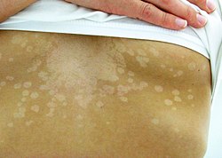
Intermammary cleft can get attacked by plaque type psoriasis, which can in turn can cause erythematosus. [47] Prurigo pigmentosa is a rare skin condition of unknown cause that affects depressed places on chest and back like the intermammary area. It is characterized by the sudden onset of erythematous papules that leave a reticulated hyperpigmentation when they heal. [48] [49] Confluent and reticulated papillomatosis, characterized by asymptomatic, small, red to brown, slightly verrucous papules occurs on upper torso, the cleavage area and back. [50] Granular parakeratosis, though mostly an ailment of the armpit area, is also found on the cleavage. [51]

Hirsutism

Most women have an increase of hair as they grow older, but some gets more hair on their cleavage, face and elsewhere because of hirsutism, often as a result of polycystic ovary syndrome. The hair on the cleavage is upsetting for many women. There are two ways to remove the hair — temporary (i.e. shaving, waxing, plucking, hair removal creams or bleaching) and permanent (electrolysis or laser hair removal. Contraceptive pills also help. [52] [53]

Symmastia

Symmastia is a condition defined as a confluence of the breast tissue of both breasts across the intermammary cleft that normally divides them. It can be surgically corrected by a plastic surgeon through symmastia revision. [54] Symmastia can either be a congenital anomaly or iatrogenic. [55] Congenital symmastia is a rare condition with few published cases. Iatrogenic symmastia may occur following breast augmentation, forming what is also colloquially referred to as a "uniboob" or "breadloafing" as a result of the release of skin and muscle tissue around the sternum due to over-dissection. [56]

In alternative medicine

The mandala (diagram) for anahata chakra, the yogic heart that lies at the depression on the sternum between the nipples Chakra4.svg
The mandala (diagram) for anahata chakra , the yogic heart that lies at the depression on the sternum between the nipples

The cleavage area is special in Ayurvedic and Yogic philosophy as the fourth chakra' or anahata chakra (अनाहत meaning "unstruck" in Sanskrit, the heart chakra) supposedly lies at the level of the depression in the sternum. [58] [57] The astral anahata chakra supposedly lies between the breasts, just inside the front of the chest level with the nipples. [59] [60] [61] According to Yogashikha Upanishad , the sacred text on yoga, 101 nadis (energy channels) connect the anahata chakra with the rest of the body, including ida, pingala and shushumna, the three major nadis. [62]

According to yoga philosophy, the kundalini shakti (the feminine energy) rises from the muladhara chakra (root chakra) in the pelvic area to reach the cleavage area, a fundamental center for growth of a human being, [63] where at the anahata chakra it is expressed as love, hate and fear. [64] In the cleavage area, according to yoga philosophy, lies the yogic heart of a person, not the heart of flesh, [57] that serves as the bridge between the three lower chakras and the three higher chakras, [65] and when the kundalini remains in the Anahata Chakra, a person is inclined to good and noble desires, thoughts and acts. [66]

According to Traditional Chinese medicine (TCM), shan zhong (Ren-17, 膻中; dan jung, 단중 in Korean) is the acupoint that lies at the intersection of the mid sternal line and a line connecting the nipples. The name shan zhong refers to its location at the center of the chest, seated on an "altar" (i.e. the sternum ) or a "place of worship". [67] It was described by Lingshu Jing, the Divine Pivot as the location of the pericardium. It also is the focal point for regulate the flow of qi , the vital force of any living entity, in the entire body, especially in the chest and breasts. [68] [69] It also helps to provide emotional relief and calm the spirit. [69]

See also

Related Research Articles

<span class="mw-page-title-main">Rib cage</span> Bone structure that protects the vital organs and major blood vessels

The rib cage is an endoskeletal enclosure in the thorax of most vertebrate animals that comprises the ribs, vertebral column and sternum, which protects vital organs such as the heart, lungs and great vessels. The circumferential enclosure formed by left and right rib cages, together known as the thoracic cage, is a semi-rigid bony and cartilaginous structure which surrounds the thoracic cavity and supports the shoulder girdles to form the core part of the axial skeleton.

<span class="mw-page-title-main">Nipple</span> Part of the breast

The nipple is a raised region of tissue on the surface of the breast from which, in females, milk leaves the breast through the lactiferous ducts to breastfeed an infant. The milk can flow through the nipple passively or it can be ejected by smooth muscle contractions that occur along with the ductal system. Male mammals also have nipples but without the same level of function, and often surrounded by body hair.

<span class="mw-page-title-main">Anahata</span> Heart chakra

Anahata or heart chakra is the fourth primary chakra, according to Hindu Yogic, Shakta and Buddhist Tantric traditions. In Sanskrit, anahata means "unhurt, unstruck, and unbeaten". Anahata Nad refers to the Vedic concept of unstruck sound. Anahata is associated with balance, calmness, and serenity.

<span class="mw-page-title-main">Tinea versicolor</span> Skin disease

Tinea versicolor is a condition characterized by a skin eruption on the trunk and proximal extremities. The majority of tinea versicolor is caused by the fungus Malassezia globosa, although Malassezia furfur is responsible for a small number of cases. These yeasts are normally found on the human skin and become troublesome only under certain conditions, such as a warm and humid environment, although the exact conditions that cause initiation of the disease process are poorly understood.

<span class="mw-page-title-main">Breast reduction</span> Plastic surgery procedure

Reduction mammoplasty is the plastic surgery procedure for reducing the size of large breasts. In a breast reduction surgery for re-establishing a functional bust that is proportionate to the patient's body, the critical corrective consideration is the tissue viability of the nipple–areola complex (NAC), to ensure the functional sensitivity and lactational capability of the breasts. The indications for breast reduction surgery are three-fold – physical, aesthetic, and psychological – the restoration of the bust, of the patient's self-image, and of the patient's mental health.

<span class="mw-page-title-main">Pectoralis major</span> Main human chest muscle

The pectoralis major is a thick, fan-shaped or triangular convergent muscle of the human chest. It makes up the bulk of the chest muscles and lies under the breast. Beneath the pectoralis major is the pectoralis minor muscle.

<span class="mw-page-title-main">Rectus abdominis muscle</span> Paired straight muscle

The rectus abdominis muscle, also known as the "abdominal muscle" or simply the "abs", is a pair of segmented skeletal muscle on the ventral aspect of a person's abdomen. The paired muscle is separated at the midline by a band of dense connective tissue called the linea alba, and the connective tissue defining each lateral margin of the rectus abdominus is the linea semilunaris. The muscle extends from the pubic symphysis, pubic crest and pubic tubercle inferiorly, to the xiphoid process and costal cartilages of the 5th–7th ribs superiorly.

<span class="mw-page-title-main">Cleavage (breasts)</span> Separation between human breasts

Cleavage is the narrow depression or hollow between the breasts of a woman. The superior portion of cleavage may be accentuated by clothing such as a low-cut neckline that exposes the division, and often the term is used to describe the low neckline itself, instead of the term décolletage. Joseph Breen, head of the U.S. film industry's Production Code Administration, coined the term in its current meaning when evaluating the 1943 film The Outlaw, starring Jane Russell. The term was explained in Time magazine on August 5, 1946. It is most commonly used in the parlance of Western female fashion to refer to necklines that reveal or emphasize décolletage.

<span class="mw-page-title-main">Sulcus (morphology)</span> Groove in an organ surface

In biological morphology and anatomy, a sulcus is a furrow or fissure. It may be a groove, natural division, deep furrow, elongated cleft, or tear in the surface of a limb or an organ, most notably on the surface of the brain, but also in the lungs, certain muscles, as well as in bones, and elsewhere. Many sulci are the product of a surface fold or junction, such as in the gums, where they fold around the neck of the tooth.

<span class="mw-page-title-main">Internal thoracic artery</span> Artery of the thorax

In human anatomy, the internal thoracic artery (ITA), also known as the internal mammary artery, is an artery that supplies the anterior chest wall and the breasts. It is a paired artery, with one running along each side of the sternum, to continue after its bifurcation as the superior epigastric and musculophrenic arteries.

<span class="mw-page-title-main">Lateral thoracic artery</span> Large blood vessel

In human anatomy, the lateral thoracic artery is a blood vessel that supplies oxygenated blood to the lateral structures of the thorax and breast.

<span class="mw-page-title-main">Intercostal nerves</span> Nerves in the thorax and abdomen

The intercostal nerves are part of the somatic nervous system, and arise from the anterior rami of the thoracic spinal nerves from T1 to T11. The intercostal nerves are distributed chiefly to the thoracic pleura and abdominal peritoneum, and differ from the anterior rami of the other spinal nerves in that each pursues an independent course without plexus formation.

<span class="mw-page-title-main">Costal cartilage</span> Resilient, smooth, glass-like tissue at the front ends of ribs in verterbrates

The costal cartilages are bars of hyaline cartilage that serve to prolong the ribs forward and contribute to the elasticity of the walls of the thorax. Costal cartilage is only found at the anterior ends of the ribs, providing medial extension.

<span class="mw-page-title-main">Intercostobrachial nerve</span>

The intercostobrachial nerve is the name applied to the lateral cutaneous branch of the second intercostal nerve. It arises anterior to the long thoracic nerve. It provides sensory innervation to the skin of the axilla, and a variable region of the medial side of the upper arm.

Mastopexy is the plastic surgery mammoplasty procedure for raising sagging breasts upon the chest of the woman, by changing and modifying the size, contour, and elevation of the breasts. In a breast-lift surgery to re-establish an aesthetically proportionate bust for the woman, the critical corrective consideration is the tissue viability of the nipple-areola complex (NAC), to ensure the functional sensitivity of the breasts for lactation and breast-feeding.

<span class="mw-page-title-main">Thoracic wall</span> Boundary of the chest cavity

The thoracic wall or chest wall is the boundary of the thoracic cavity.

<span class="mw-page-title-main">Sternalis muscle</span>

The sternalismuscle is an anatomical variation that lies in front of the sternal end of the pectoralis major parallel to the margin of the sternum. The sternalis muscle may be a variation of the pectoralis major or of the rectus abdominis.

Amastia refers to a rare clinical anomaly in which both internal breast tissue and the visible nipple are absent on one or both sides. It affects both men and women. Amastia can be either isolated or comorbid with other syndromes, such as ectodermal dysplasia, Syndactyly and lipoatrophic diabetes. This abnormality can be classified into various types, and each could result from different pathologies. Amastia differs from amazia and athelia. Amazia is the absence of one or both mammary glands but the nipples remain present, and athelia is the absence of one or both nipples, but the mammary gland remains.

<i>Malassezia furfur</i> Species of fungus

Malassezia furfur is a species of yeast that is naturally found on the skin surfaces of humans and some other mammals. It is associated with a variety of dermatological conditions caused by fungal infections, notably seborrhoeic dermatitis and tinea versicolor. As an opportunistic pathogen, it has further been associated with dandruff, malassezia folliculitis, pityriasis versicolor (alba), and malassezia intertrigo, as well as catheter-related fungemia and pneumonia in patients receiving hematopoietic transplants.

Symmastia is a condition defined as a confluence of the breast tissue of both breasts across the intermammary cleft that normally divides them. It can be surgically corrected by a plastic surgeon through symmastia revision.

References

  1. 1 2 Moore, Keith (2018). Clinically oriented anatomy (Eighth ed.). Wolters Kluwer. pp. 318–321. ISBN   9781496347213.
  2. Dr. Ted Eisenberg and Joyce K. Eisenberg, The Scoop on Breasts: A Plastic Surgeon Busts the Myths, Incompra Press, 2012, ISBN   978-0-9857249-3-1
  3. Heide Schatten and Gheorghe M. Constantinescu, Comparative Reproductive Biology, page 17, John Wiley & Sons, 2008, ISBN   978-0-470-39025-2
  4. Genaro Andres Contreras, The Use of Tylosin to Treat Intramammary Infections , page 22, ProQuest, 2008, ISBN   978-0-549-60762-5
  5. Olufunmilayo I. Olopade and Carla I. Falkson, Breast Cancer in Women of African Descent, page 125, Springer Science & Business Media, 2010, ISBN   9781402036644
  6. TA A16.0.02.002
  7. Keith L. Moore, Anne M.R. Agur and Arthur F. Daley, Essential Clinical Anatomy (4th edition), page 49, Lippincott Williams and Wilkins, 2010, ISBN   9780781799157
  8. Muhammad Adil Abbas Khan, Ammar Asrar Javed and Nigel Mercer, Cleavage classification: categorizing a vital feminine aesthetic landmark, Plastic and Aesthetic Research (PAR), 016-01-15
  9. inter-, Merriam-Webster
  10. mamma, Charlton T. Lewis and Charles Short (1879), A Latin Dictionary, Oxford: Clarendon Press
  11. -ery, Dictionary.com
  12. intermammary, Merriam-Webster
  13. sulcus, Charlton T. Lewis and Charles Short (1879), A Latin Dictionary, Oxford: Clarendon Press
  14. sulcus, Merriam-Webster
    sulcus, Cambridge Dictionary
    sulcus, Collins Dictionary
  15. John Bostwick, Plastic and Reconstructive Breast Surgery (volume 1), page 21, Quality Medical Pub, 2000, ISBN   9781576261040
  16. Sylvia H. Heywang-Koebrunner, Ingrid Schreer and Susan Barter, Diagnostic Breast Imaging, page 62, Thieme, 2019, ISBN   9783131504111
  17. King, Christopher; Henretig, Fred M. (2008). Textbook of Pediatric Emergency Procedures. Lippincott Williams & Wilkins. ISBN   9780781753869 . Retrieved 19 July 2020.
  18. John Blair Deaver, The Breast: its anomalies, its diseases, and their treatment, page 30, P. Blakiston's Son & Company, 1917, ISBN   9780266616542
  19. 1 2 Raikos, Athanasios; Paraskevas, George K.; Yusuf, Faisal; Kordali, Panagiota; Ioannidis, Orestis; Brand-Saberi, Beate (2011-12-01). "Sternalis muscle: a new crossed subtype, classification, and surgical applications". Annals of Plastic Surgery. 67 (6): 646–648. doi:10.1097/SAP.0b013e31820d688b. ISSN   1536-3708. PMID   21407048. S2CID   5303650.
  20. Georgiev, Georgi P.; Jelev, Lazar; Ovtscharoff, Vladimir A. (2009-09-01). "On the clinical significance of the sternalis muscle". Folia Medica. 51 (3): 53–56. ISSN   0204-8043. PMID   19957564.
  21. "Anatomy 2008; 2: 67–71 [Case Report]". www.anatomy.org.tr. Archived from the original on 2016-03-04. Retrieved 2015-09-05.
  22. William C. Wood, Charles Staley and John E. Skandalakis, Anatomic Basis of Tumor Surgery, page 140, Springer Science & Business Media, 2010, ISBN   9783540741770
  23. Neal S. Elattrache, Surgical Techniques in Sports Medicine, page 139, Lippincott Williams & Wilkins, 2007, ISBN   9780781754279
  24. Elizabeth Hall-Findlay and Gregory Evans, Aesthetic and Reconstructive Surgery of the Breast, Elsevier Health Sciences, 2010, ISBN   9780702050091
  25. Maurice Y Nahabedian and Peter C. Neligan, Plastic Surgery (Volume 5), page 89, Elsevier Health Sciences, 2017, ISBN   9780323357104
  26. Jeffrey Weinzweig, Plastic Surgery Secrets, page 453, Elsevier Health Sciences, 2010, ISBN   9780323085908
  27. 1 2 Ruth A. Lawrence MD and Robert M. Lawrence, MD, Breastfeeding: A Guide for the Medical Profession page 47, Elsevier Health Sciences, 2015, ISBN   9780323357760
  28. 1 2 Rebecca F. Black, The Science of Breastfeeding (Volume 3), page 13, Jones & Bartlett Learning, 1998, ISBN   9780763701949
  29. Theresa Hornstein and Jeri Lynn Schwerin, Biology of Women, page 146, Cengage Learning, 2012, ISBN   9781435400337
  30. Charles Wesley Turner, The mammary gland (vol. 1), page 80, Lucas Bros., 1952
  31. Albert Henry Buck and Thomas Lathrop Stedman, A Reference Handbook of the Medical Sciences (volume 2), W. Wood, 1901
  32. Gray's Anatomy of the Human Body, 20th edition (1918), p. 945
  33. Henry Albert Reeves, Human Morphology: A Treatise on Practical and Applied Anatomy (Volume 1), page 91, Smith, Elder, & Company, 1882
  34. Marios G Lykissas, Ioannis P Kostas-Agnantis, Ananstasios V Korompilias, Marios D Vekris, and Alexandros E Beris, "Use of intercostal nerves for different target neurotization in brachial plexus reconstruction", World Journal of Orthopedics, 2013 Jul 18; 4(3): 107–111
  35. Ramachandran Kalpana and Kothandaraman Usha, "Three unilateral recti sternalis muscles – an unusual variation and its clinical significance", European Journal of Anatomy, 14 (2): 99–103 (2010)
  36. 1 2 Daphne Merkin, "The Great Divide", New York Times, August 28, 2005
  37. Rapini, Ronald P.; Bolognia, Jean L.; Jorizzo, Joseph L. (2007). Dermatology: 2-Volume Set. St. Louis: Mosby. ISBN   978-1-4160-2999-1.
  38. 1 2 Alam, Murad; Pongprutthipan, Marisa (2010). Body Rejuvenation. Springer Science+Business Media. p. 39. ISBN   9781441910936.
  39. American Osteopathic College of Dermatology "Dermatologic Disease Database", aocd.org, referenced July 22, 2011.
  40. Alikhan, Ali; Lynch, Eisen (2009). "Hidradenitis suppurativa: a comprehensive review". J Am Acad Dermatol. 60 (4): 539–561. doi:10.1016/j.jaad.2008.11.911. PMID   19293006.
  41. 1 2 Faye Lyons, Dermatology for the Advanced Practice Nurse , pages 118–121, Springer Publishing Company, 2014, ISBN   9780826136435
  42. Rapini, Ronald P; Bolognia, Jean L; Jorizzo, Joseph L (2007). Dermatology: 2-Volume Set. St Louis: Mosby. pp. Chapter 76. ISBN   978-1-4160-2999-1.
  43. James Nevins Hyde, A Practical treatise on diseases of the skin, pages 625-626, Lea Bros. & Company, 1888
  44. Morishita N; Sei Y. (December 2006). "Microreview of pityriasis versicolor and Malassezia species". Mycopathologia. 162 (6): 373–76. doi:10.1007/s11046-006-0081-2. PMID   17146580. S2CID   30397515.
  45. Prohic A; Ozegovic L. (January 2007). "Malassezia species isolated from lesional and non-lesional skin in patients with pityriasis versicolor". Mycoses. 50 (1): 58–63. doi:10.1111/j.1439-0507.2006.01310.x. PMID   17302750. S2CID   25493290.
  46. Likness, LP (June 2011). "Common dermatologic infections in athletes and return-to-play guidelines". The Journal of the American Osteopathic Association. 111 (6): 373–379. doi: 10.7556/jaoa.2011.111.6.373 . PMID   21771922.
  47. Alan Menter and Benjamin Stoff, Psoriasis, page 25, CRC Press, 2010, ISBN   9781840765908
  48. James, William; Berger, Timothy; Elston, Dirk (2005). Andrews' Diseases of the Skin: Clinical Dermatology. (10th ed.). page 57, Saunders. ISBN   0-7216-2921-0.
  49. Q Ashton Acton, Issues in Dermatology and Cosmetic Medicine (2013 Edition), page 432, Scholarly Editions, 2013, ISBN   9781490109114
  50. David Weedon, Weedon's Skin Pathology, page 505, Elsevier Health Sciences, 2009, ISBN   9780702047749
  51. J Lazar and Phillip H. McKee, Pathology of the Skin, page 96, Elsevier Health Sciences, 2011, ISBN   9780723437185
  52. Condition: Hirsutism, NHS
  53. Rachel Lapidos, What's the Best Way to Get Rid of Cleavage Hair?, Well+Good, 2019-10-17
  54. Wong, M. T.; Cheong, E. C.; Lim, J.; Lim, T. C. (2007). "Creation of an intermammary sulcus in congenital synmastia". Singapore Medical Journal. 48 (1): e29–e31. PMID   17245502.
  55. Sillesen, N. H.; Hölmich, L. R.; Siersen, H. E.; Bonde, C. (2012). "Congenital symmastia revisited". Journal of Plastic, Reconstructive & Aesthetic Surgery. 65 (12): 1607–13. doi:10.1016/j.bjps.2012.08.008. PMID   23026472.
  56. "Symmastia". Archived from the original on 2014-10-17. Retrieved 2020-10-11.
  57. 1 2 3 Swami Rama, The Royal Path: Practical Lessons on Yoga, page 82, Himalayan Institute Press, 1998, ISBN   9780893891527
  58. Vimala McClure, A Woman's Guide to Tantra Yoga, page 46, New World Library, 2012, ISBN   9781577312765
    Annelise Hagen, 12 Yoga Poses to Boost Breast Health, Yoga Journal, 2017-11-01
    Ravi Ratan and Minoo Ratan, Journey Through Chakras, page 5, Institute of Holistic Sciences, 2007, ISBN   9788120832404
  59. Swami Satyananda Saraswati, Meditations from the Tantras, page 37, Bihar School of Yoga, 1974, OCLC   2188780
  60. Jonn Mumford, A Chakra & Kundalini Workbook, page 184, Llewellyn Worldwide, 1994, ISBN   9781567184730
  61. Swami Rama, OM the Eternal Witness, page 181, Lotus Press, 2008, ISBN   9788188157433
  62. Harish Johari, Chakras: Energy Centers of Transformation, page 44, Simon and Schuster, 2000, ISBN   9781594779091
  63. Christine Horner, Waking the Warrior Goddess, page 204, Basic Health Publications, 2005, ISBN   9781591201557
  64. Sri Sri Ravishankar, Shiva Sutras, page 119, Aslan Business Solutions, 2018, ISBN   9789385898198
  65. Hilary H. Carter, The Chakras Made Easy, page 18, John Hunt Publishing, 2012, ISBN   9781780995168
  66. Swami Narayanananda, The Primal Power in Man: The Kundalini Shakti, page 108, Health Research Books, 1960, ISBN   9780787306311
  67. Arnie Lade, Acupuncture Points: Images & Functions page 268, Eastland Press, 1989, ISBN   9780939616084
  68. Ji Zhang, Baixiao Zhao and Lixing Lao, Acupuncture and Moxibustion, page 252, PMPH-USA, 2014, ISBN   9787117193283
  69. 1 2 Honora Lee Wolfe, Bob Flaws, Better Breast Health Naturally with Chinese Medicine page 120, Blue Poppy Enterprises, Inc., 1998, ISBN   9780936185903