Keutel syndrome

Last updated
Keutel syndrome
Other namesPulmonic stenosis-brachytelephalangism-calcification of cartilages syndrome [1]
Autorecessive.svg
Keutel syndrome has an autosomal recessive pattern of inheritance.

Keutel syndrome (KS) is a rare autosomal recessive genetic disorder characterized by abnormal diffuse cartilage calcification, hypoplasia of the mid-face, peripheral pulmonary stenosis, hearing loss, short distal phalanges (tips) of the fingers and mild mental retardation. [2] [3] [4] Individuals with KS often present with peripheral pulmonary stenosis, brachytelephalangism, sloping forehead, midface hypoplasia, and receding chin. It is associated with abnormalities in the gene coding for matrix gla protein, MGP. [2] Being an autosomal recessive disorder, it may be inherited from two unaffected, abnormal MGP-carrying parents. Thus, people who inherit two affected MGP alleles will likely inherit KS.

Contents

It was first identified in 1972 as a novel rare genetic disorder sharing similar symptoms with chondrodysplasia punctata. [2] [5] Multiple forms of chondrodysplasia punctata share symptoms consistent with KS including abnormal cartilage calcification, forceful respiration, brachytelephalangism, hypotonia, psychomotor delay, and conductive deafness, yet peripheral pulmonary stenosis remains unique to KS. [6]

No chromosomal abnormalities are reported in affected individuals, suggesting that familial consanguinity relates to the autosomal recessive mode of inheritance. Also, despite largely abnormal calcification of regions including the larynx, tracheobronchial tree, nose, pinna (anatomy), and epiglottis, patients exhibit normal serum calcium and phosphate levels. [7]

Signs and symptoms

Being an extremely rare autosomal genetic disorder, differential diagnosis has only led to several cases since 1972. Initial diagnosis lends itself to facial abnormalities including sloping forehead, maxillary hypoplasia, nasal bridge depression, wide mouth, dental malocclusion, and receding chin. [4] Electroencephalography (EEG), computed tomography (CT) scanning, and skeletal survey are further required for confident diagnosis. Commonly, diffuse cartilage calcification and brachytelephalangism are identified by X-radiation (X-ray), while peripheral pulmonary arterial stenosis, hearing loss, dysmorphic facies, and mental retardation are confirmed with confidence by the aforementioned diagnostic techniques. [7]

Skeletal effects

Diagnosis is often confirmed by several abnormalities of skeletal origin. There is a sequential order of findings, according to Cormode et al., which initiate in abnormal cartilage calcification and later brachytelephalangism. [8] The uniqueness of brachytelephalangy in KS results in distinctively broadened and shortened first through fourth distal phalanges, while the fifth distal phalanx bone remains unaffected. [9] Radiography also reveals several skeletal anomalies including facial hypoplasia resulting in underdevelopment of the nasal bridge with noticeably diminished alae nasi. In addition to distinguishable facial features, patients generally demonstrate shorter than average stature and general mild developmental delay.[ citation needed ]

Cartilaginous effects

Many common effects sharing similarity with chondrodysplasia punctata stem from cartilaginous origin. Radiography reveals extensive diffuse cartilaginous calcification. Pulmonary angiography and soft tissue radiography often demonstrate significant cartilaginous ossification in the trachea and larynx, with perichondral and endochondral centers significantly ossified in transformed cartilage. [10] Abnormal diffuse cartilaginous ossification is typically most pronounced in the auricles and cartilage of the trachea and larynx, while peripheral pulmonary stenosis is frequently common in KS. In consanguineous parents of children with KS, one is often phenotypically normal, while the other is positive for pulmonary stenosis. Perhaps emanating from diffuse laryngotracheal calcification, patients often present with recurrent respiratory infection, otitis media, and sinusitis. [11]

Cardiovascular effects

Apart from diffuse abnormal cartilaginous calcification in pulmonary and wikt:otic systems, patients develop significant arterial calcification throughout the body. [12] Such calcification is concomitant with various diseases including diabetes, atherosclerosis, and kidney dysfunction, while patients with oral anticoagulant use have significant aortic valve and coronary artery calcification. [12] [13] Although not distinctive to KS, echocardiogram analysis has revealed right ventricular hypertrophy resulting in severe pulmonary artery hypertension in several cases. [14]

Pathogenesis

Keutel syndrome is an autosomal recessive disorder caused by a novel loss-of-function mutation in the matrix Gla protein gene (MGP). MGP protein resides in the extracellular matrix and is implicated in inhibiting calcification though the repression of bone morphogenetic protein 2 (BMP2). Mutations resulting in loss of consensus donor splice site at exon 2-intron 2 junctions result in significant diffuse calcification of soft tissue cartilage. [14] Extensive diffuse cartilaginous calcification is present in MGP-knockout mice, manifesting in vascular media replacement with a cartilaginous, chondrocyte-like matrix, and ultimately premature death. [15] Conversely, over expression of extracellular MGP effectively abolishes calcification in chondrocytes, suggesting that MGP may function in inhibiting passive calcification in soft tissues. [16] Recent evidence suggests MGP is a vitamin K dependent protein synthesized by chondrocytes and vascular smooth muscle cells, where it potentiates the inhibition of cartilaginous and arterial calcification. Thus, potential vitamin K deficiency, via nutritional deficiency or coumarin-derivative use, would render MGP uncarboxylated and inactive, thus diminishing biological function. [17] [18] Arterial calcification resulting from MGP inactivation results in inimical prognosis, commonly seen in patients with diabetes, atherosclerosis, and renal dysfunction.[ citation needed ]

Diagnosis

Treatment

Treatment is symptomatic, often addressing indicators associated with peripheral pulmonary artery stenosis. Laryngotracheal calcification resulting in dyspnea and forceful breathing can be treated with bronchodilators including the short and long-acting β2-agonists, and various anticholinergics.[ citation needed ]

Prognosis

Prognosis is good, yet life expectancy depends on the severity and extent of diffuse pulmonary and arterial calcification.[ citation needed ]

History

Keutel Syndrom was first described by the pediatric Cardiologist Jürgen Keutel [5] and Colleagues in 1972 in Göttingen, Germany.

Related Research Articles

<span class="mw-page-title-main">Fraser syndrome</span> Recessive genetic disorder involving eye and genital abnormalities

Fraser syndrome is an autosomal recessive congenital disorder, identified by several developmental anomalies. Fraser syndrome is named for the geneticist George R. Fraser, who first described the syndrome in 1962.

<span class="mw-page-title-main">Robinow syndrome</span> Rare genetic disorder characterized by a fetal face

Robinow syndrome is an extremely rare genetic disorder characterized by short-limbed dwarfism, abnormalities in the head, face, and external genitalia, as well as vertebral segmentation. The disorder was first described in 1969 by human geneticist Meinhard Robinow, along with physicians Frederic N. Silverman and Hugo D. Smith, in the American Journal of Diseases of Children. By 2002, over 100 cases had been documented and introduced into medical literature.

<span class="mw-page-title-main">Larsen syndrome</span> Medical condition

Larsen syndrome (LS) is a congenital disorder discovered in 1950 by Larsen and associates when they observed dislocation of the large joints and face anomalies in six of their patients. Patients with Larsen syndrome normally present with a variety of symptoms, including congenital anterior dislocation of the knees, dislocation of the hips and elbows, flattened facial appearance, prominent foreheads, and depressed nasal bridges. Larsen syndrome can also cause a variety of cardiovascular and orthopedic abnormalities. This rare disorder is caused by a genetic defect in the gene encoding filamin B, a cytoplasmic protein that is important in regulating the structure and activity of the cytoskeleton. The gene that influences the emergence of Larsen syndrome is found in chromosome region, 3p21.1-14.1, a region containing human type VII collagen gene. Larsen syndrome has recently been described as a mesenchyme disorder that affects the connective tissue of an individual. Autosomal dominant and recessive forms of the disorder have been reported, although most cases are autosomal dominant. Reports have found that in Western societies, Larsen syndrome can be found in one in every 100,000 births, but this is most likely an underestimate because the disorder is frequently unrecognized or misdiagnosed.

An osteochondrodysplasia, or skeletal dysplasia, is a disorder of the development of bone and cartilage. Osteochondrodysplasias are rare diseases. About 1 in 5,000 babies are born with some type of skeletal dysplasia. Nonetheless, if taken collectively, genetic skeletal dysplasias or osteochondrodysplasias comprise a recognizable group of genetically determined disorders with generalized skeletal affection. These disorders lead to disproportionate short stature and bone abnormalities, particularly in the arms, legs, and spine. Skeletal dysplasia can result in marked functional limitation and even mortality.
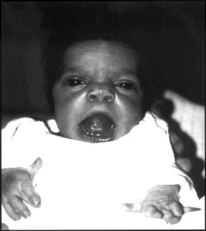
<span class="mw-page-title-main">Multiple epiphyseal dysplasia</span> Rare genetic disorder

Multiple epiphyseal dysplasia (MED), also known as Fairbank's disease, is a rare genetic disorder that affects the growing ends of bones. Long bones normally elongate by expansion of cartilage in the growth plate near their ends. As it expands outward from the growth plate, the cartilage mineralizes and hardens to become bone (ossification). In MED, this process is defective.

<span class="mw-page-title-main">Pseudoachondroplasia</span> Inherited disorder of bone growth

Pseudoachondroplasia is an inherited disorder of bone growth. It is a genetic autosomal dominant disorder. It is generally not discovered until 2–3 years of age, since growth is normal at first. Pseudoachondroplasia is usually first detected by a drop of linear growth in contrast to peers, a waddling gait or arising lower limb deformities.

<span class="mw-page-title-main">Matrix Gla protein</span>

Matrix Gla protein (MGP) is member of a family of vitamin K2 dependent, Gla-containing proteins. MGP has a high affinity binding to calcium ions, similar to other Gla-containing proteins. The protein acts as an inhibitor of vascular mineralization and plays a role in bone organization.

Williams–Campbell syndrome (WCS) is a disease of the airways where cartilage in the bronchi is defective. It is a form of congenital cystic bronchiectasis. This leads to collapse of the airways and bronchiectasis. It acts as one of the differential to allergic bronchopulmonary aspergillosis. WCS is a deficiency of the bronchial cartilage distally.

<span class="mw-page-title-main">Antley–Bixler syndrome</span> Medical condition

Antley–Bixler syndrome is a rare, severe autosomal recessive congenital disorder characterized by malformations and deformities affecting the majority of the skeleton and other areas of the body.

<span class="mw-page-title-main">Boomerang dysplasia</span> Medical condition

Boomerang dysplasia is a lethal form of osteochondrodysplasia known for a characteristic congenital feature in which bones of the arms and legs are malformed into the shape of a boomerang. Death usually occurs in early infancy due to complications arising from overwhelming systemic bone malformations.

<span class="mw-page-title-main">Gerodermia osteodysplastica</span> Medical condition

Gerodermia osteodysplastica (GO) is a rare autosomal recessive connective tissue disorder included in the spectrum of cutis laxa syndromes.

X-linked recessive chondrodysplasia punctata is a type of chondrodysplasia punctata that can involve the skin, hair, and cause short stature with skeletal abnormalities, cataracts, and deafness.

Congenital hypoplastic anemia is a congenital disorder that occasionally also includes leukopenia and thrombocytopenia and is characterized by deficiencies of red cell precursors.

<span class="mw-page-title-main">Gillespie syndrome</span> Medical condition

Gillespie syndrome, also called aniridia, cerebellar ataxia and mental deficiency, is a rare genetic disorder. The disorder is characterized by partial aniridia, ataxia, and, in most cases, intellectual disability. It is heterogeneous, inherited in either an autosomal dominant or autosomal recessive manner. Gillespie syndrome was first described by American ophthalmologist Fredrick Gillespie in 1965.

<span class="mw-page-title-main">Fryns syndrome</span> Medical condition

Fryns syndrome is an autosomal recessive multiple congenital anomaly syndrome that is usually lethal in the neonatal period. Fryns (1987) reviewed the syndrome.

<span class="mw-page-title-main">Amelogenesis imperfecta</span> Genetic disorder resulting in abnormal enamel

Amelogenesis imperfecta (AI) is a congenital disorder which presents with a rare abnormal formation of the enamel or external layer of the crown of teeth, unrelated to any systemic or generalized conditions. Enamel is composed mostly of mineral, that is formed and regulated by the proteins in it. Amelogenesis imperfecta is due to the malfunction of the proteins in the enamel as a result of abnormal enamel formation via amelogenesis.

Opsismodysplasia is a type of skeletal dysplasia first described by Zonana and associates in 1977, and designated under its current name by Maroteaux (1984). Derived from the Greek opsismos ("late"), the name "opsismodysplasia" describes a delay in bone maturation. In addition to this delay, the disorder is characterized by micromelia, particularly of the hands and feet, delay of ossification, platyspondyly, irregular metaphyses, an array of facial aberrations and respiratory distress related to chronic infection. Opsismodysplasia is congenital, being apparent at birth. It has a variable mortality, with some affected individuals living to adulthood. The disorder is rare, with an incidence of less than 1 per 1,000,000 worldwide. It is inherited in an autosomal recessive pattern, which means the defective (mutated) gene that causes the disorder is located on an autosome, and the disorder occurs when two copies of this defective gene are inherited. No specific gene has been found to be associated with the disorder. It is similar to spondylometaphyseal dysplasia, Sedaghatian type.

<span class="mw-page-title-main">Myhre syndrome</span> Medical condition

Myhre syndrome is a rare genetic disorder inherited in an autosomal dominant fashion. It is caused by mutation in SMAD4 gene.

<span class="mw-page-title-main">Spondyloenchondrodysplasia</span> Medical condition

Spondyloenchondrodysplasia is the medical term for a rare spectrum of symptoms that are inherited following an autosomal recessive inheritance pattern. Skeletal anomalies are the usual symptoms of the disorder, although its phenotypical nature is highly variable among patients with the condition, including symptoms such as muscle spasticity or thrombocytopenia purpura. It is a type of immunoosseous dysplasia.

References

  1. RESERVED, INSERM US14-- ALL RIGHTS. "Orphanet: Keutel syndrome". www.orpha.net. Retrieved 27 October 2019.{{cite web}}: CS1 maint: numeric names: authors list (link)
  2. 1 2 3 Munroe PB, Olgunturk RO, Fryns JP, et al. (1999). "Mutations in the gene encoding the human matrix Gla protein cause Keutel syndrome". Nat. Genet. 21 (1): 142–4. doi:10.1038/5102. PMID   9916809. S2CID   1244954.
  3. Potparic, Olivera; John Gibson (1995). A Dictionary of Congenital Malformations and Disorders. Informa Health Care. p. 98. ISBN   1-85070-577-1.
  4. 1 2 Teebi AS, Lambert DM, Kaye GM, Al-Fifi S, Tewfik TL, Azouz EM (1998). "Keutel syndrome: further characterization and review". Am J Med Genet. 78 (2): 182–7. doi:10.1002/(SICI)1096-8628(19980630)78:2<182::AID-AJMG18>3.0.CO;2-J. PMID   9674914.
  5. 1 2 Keutel J, Jorgensen G, Gabriel P (1972). "A new autosomal recessive syndrome: peripheral pulmonary stenoses, brachytelephalangism, neural hearing loss and abnormal cartilage calcifications-ossification". Birth Defects Orig Art Ser. VIII (5): 60–68.
  6. Ziereisen E, De Munter C, Perlmutter N (1993). "The Keutel syndrome: report of a case and review of the literature". Pediatric Radiology. 23 (4): 314–315. doi:10.1007/bf02010925. PMID   8414764. S2CID   31009506.
  7. 1 2 Parmar H, Blaser S, Unger S, Yoo SJ, Papsin B (2006). "Petrified ears in a patient with Keutel syndrome: temporal bone CT findings". Pediatric Radiology. 36 (3): 241–243. doi:10.1007/s00247-005-0036-7. PMID   16328322. S2CID   6833062.
  8. Cormode EJ, Dawson M, Lowry RB (1986). "Keutel syndrome: clinical report and literature review". American Journal of Medical Genetics. 24 (2): 289–294. doi:10.1002/ajmg.1320240209. PMID   3717211.
  9. Miller S (2003). "Brachytelephalangy with sparing of the fifth distal phalanx: a feature highly suggestive of Keutel syndrome". Pediatric Radiology. 33 (3): 186–189. doi:10.1007/s00247-002-0846-9. PMID   12612818. S2CID   9772501.
  10. Fryns JP, van Flateren A, Mattelaer P, et al. (1984). "Calcification of cartilages, brachytelephalangy, and peripheral pulmonary stenosis. Confirmation of the Keutel syndrome". European Journal of Pediatrics. 142 (3): 201–203. doi:10.1007/bf00442449. PMID   6468443. S2CID   19336365.
  11. Khosroshahi HE, Uluoglu O, Olgunturk R, Basaklar C (1989). "Keutel syndrome: a report of four cases". European Journal of Pediatrics. 149 (3): 188–191. doi:10.1007/bf01958278. PMID   2515061. S2CID   35206652.
  12. 1 2 Meier M, Weng LP, Alexandrakis E, Ruschoff J, Goeckenjan G (2001). "Tracheobronchial stenosis in Keutel syndrome". European Respiratory Journal. 17 (3): 566–569. doi: 10.1183/09031936.01.17305660 . PMID   11405537.
  13. Schurgers LJ, Aebert H, Vermeer C, Bultmann B, Janzen J (2004). "Oral anticoagulant treatment: friend or foe in cardiovascular disease?". Blood. 104 (10): 3231–3232. doi: 10.1182/blood-2004-04-1277 . PMID   15265793.
  14. 1 2 Hur DJ, Raymond GV, Kahler SG, Riegert-Johnson DL, Cohen BA, Boyadjiev SA (2005). "A novel MGP mutation in a consanguineous family: Review of the clinical and molecular characteristics of Keutel syndrome". American Journal of Medical Genetics. 135A (1): 36–40. doi:10.1002/ajmg.a.30680. PMID   15810001. S2CID   10466007.
  15. Luo G, Ducy P, McKee MD, Pinero GJ, Loyer E, Behringer RR, Karsenty G (1997). "Spontaneous calcification of arteries and cartilage in mice lacking matrix GLA protein". Nature. 386 (6620): 78–81. Bibcode:1997Natur.386...78L. doi:10.1038/386078a0. PMID   9052783. S2CID   4335985.
  16. Yagami K, Suh JY, Enomoto-Iwamoto M, Koyama E, Abrams WR, Shapiro IM, Pacifici M, Iwamoto M (1999). "Matrix GLA protein is a developmental regulator of chondroctye mineralization and, when constitutively expressed, blocks endochondral and intramembranous ossification in the limb". Journal of Cell Biology. 147 (5): 1097–1108. doi:10.1083/jcb.147.5.1097. PMC   2169349 . PMID   10579728.
  17. Crenenburg EC, Vermeer C, Koos R, Boumans ML, Hackeng TM, Bouwman FG, Kwaijtaal M, Brandenburg VM, Ketteler M, Schurgers LJ (2008). "The circulating inactive form of matrix gla protein (ucMGP) as a biomarker for cardiovascular calcification". Journal of Vascular Research. 45 (5): 427–436. doi:10.1159/000124863. PMID   18401181. S2CID   8737190.
  18. Berkner KL, Runge KW (2004). "The physiology of vitamin K nutriture and vitamin K-dependent protein function in atherosclerosis". Journal of Thrombosis and Haemostasis. 2 (12): 2118–2132. doi: 10.1111/j.1538-7836.2004.00968.x . PMID   15613016. S2CID   25194009.