Lead apron

Last updated
A rubber coated lead apron protects organs from exposure to x-rays. Lead Apron worn by Dr Danny Sands.jpg
A rubber coated lead apron protects organs from exposure to x-rays.
Thyroid gland protection. Mikael Haggstrom in 2019.jpg
Thyroid gland protection.

A lead apron or leaded apron is a type of protective clothing that acts as a radiation shield. It is constructed of a thin rubber exterior and an interior of lead in the shape of a hospital apron. The purpose of the lead apron is to reduce exposure of a hospital patient to x-rays to vital organs that are potentially exposed to ionizing radiation during medical imaging that uses x-rays (radiography, fluoroscopy, computed tomography).

Personal protective equipment

Personal protective equipment (PPE) is protective clothing, helmets, goggles, or other garments or equipment designed to protect the wearer's body from injury or infection. The hazards addressed by protective equipment include physical, electrical, heat, chemicals, biohazards, and airborne particulate matter. Protective equipment may be worn for job-related occupational safety and health purposes, as well as for sports and other recreational activities. "Protective clothing" is applied to traditional categories of clothing, and "protective gear" applies to items such as pads, guards, shields, or masks, and others.

Lead Chemical element with atomic number 82

Lead is a chemical element with the symbol Pb and atomic number 82. It is a heavy metal that is denser than most common materials. Lead is soft and malleable, and also has a relatively low melting point. When freshly cut, lead is silvery with a hint of blue; it tarnishes to a dull gray color when exposed to air. Lead has the highest atomic number of any stable element and three of its isotopes are endpoints of major nuclear decay chains of heavier elements.

X-ray form of electromagnetic radiation

X-rays make up X-radiation, a form of electromagnetic radiation. Most X-rays have a wavelength ranging from 0.01 to 10 nanometers, corresponding to frequencies in the range 30 petahertz to 30 exahertz (3×10¹⁶ Hz to 3×10¹⁹ Hz) and energies in the range 100 eV to 100 keV. X-ray wavelengths are shorter than those of UV rays and typically longer than those of gamma rays. In many languages, X-radiation is referred to with terms meaning Röntgen radiation, after the German scientist Wilhelm Röntgen who discovered these on November 8, 1895, who usually is credited as its discoverer, and who named it X-radiation to signify an unknown type of radiation. Spelling of X-ray(s) in the English language includes the variants x-ray(s), xray(s), and X ray(s).

Contents

Specific anatomic regions

Protection of the reproductive organs with a lead rubber apron is considered important because DNA changes to sperm or egg cells of the patient may pass on genetic defects to the offspring of the patient, causing serious and unnecessary hardship for child and parents.

DNA Molecule that encodes the genetic instructions used in the development and functioning of all known organisms and many viruses

Deoxyribonucleic acid is a molecule composed of two chains that coil around each other to form a double helix carrying genetic instructions for the development, functioning, growth and reproduction of all known organisms and many viruses. DNA and ribonucleic acid (RNA) are nucleic acids; alongside proteins, lipids and complex carbohydrates (polysaccharides), nucleic acids are one of the four major types of macromolecules that are essential for all known forms of life.

Sperm male reproductive cells

Sperm is the male reproductive cell. In the types of sexual reproduction known as anisogamy and its subtype oogamy, there is a marked difference in the size of the gametes with the smaller one being termed the "male" or sperm cell. A uniflagellar sperm cell that is motile is referred to as a spermatozoon, whereas a non-motile sperm cell is referred to as a spermatium. Sperm cells cannot divide and have a limited life span, but after fusion with egg cells during fertilization, a new organism begins developing, starting as a totipotent zygote. The human sperm cell is haploid, so that its 23 chromosomes can join the 23 chromosomes of the female egg to form a diploid cell. In mammals, sperm develops in the testicles, is stored in the epididymis, and released from the penis.

Egg organic vessel in which an embryo first begins to develop

The egg is the organic vessel containing the zygote in which an embryo develops until it can survive on its own; at which point the animal hatches. An egg results from fertilization of an egg cell. Most arthropods, vertebrates, and mollusks lay eggs, although some, such as scorpions do not.

The thyroid gland is especially vulnerable to x-ray exposure. Care should be taken to place a lead apron over the thyroid gland before taking dental radiographs. [1] [2] Aprons used for dental imaging should include thyroid collars.

Thyroid endocrine gland in the neck; secretes hormones that influence metabolism

The thyroid gland, or simply the thyroid, is an endocrine gland in the neck, consisting of two lobes connected by an isthmus. It is found at the front of the neck, below the Adam's apple. The thyroid gland secretes three hormones, namely the two thyroid hormones (thyroxine/T4 and triiodothyronine/T3), and calcitonin. The thyroid hormones primarily influence the metabolic rate and protein synthesis, but they also have many other effects, including effects on development. Calcitonin plays a role in calcium homeostasis.

Dentistry branch of medicine

Dentistry, also known as Dental and Oral Medicine, is a branch of medicine that consists of the study, diagnosis, prevention, and treatment of diseases, disorders, and conditions of the oral cavity, commonly in the dentition but also the oral mucosa, and of adjacent and related structures and tissues, particularly in the maxillofacial area. Although primarily associated with teeth among the general public, the field of dentistry or dental medicine is not limited to teeth but includes other aspects of the craniofacial complex including the temporomandibular joint and other supporting, muscular, lymphatic, nervous, vascular, and anatomical structures.

It is important to choose the correct thickness of lead-equivalent (Pbeq) aprons. This will depend on how long and how often you are working in an environment that requires protective lead rubber aprons. It is also contingent on what examinations are being performed. The minimum requirement is to wear 0.25 mm Pbeq if you are not behind lead shielding. If you are working in theatre using fluoroscopy (e.g. orthopaedics, cardiology or interventional) 0.35 or 0.5 mm lead may be appropriate because of the higher KV employed, and on how close to the primary beam you are working.3

Fluoroscopy

Fluoroscopy is an imaging technique that uses X-rays to obtain real-time moving images of the interior of an object. In its primary application of medical imaging, a fluoroscope allows a physician to see the internal structure and function of a patient, so that the pumping action of the heart or the motion of swallowing, for example, can be watched. This is useful for both diagnosis and therapy and occurs in general radiology, interventional radiology, and image-guided surgery. In its simplest form, a fluoroscope consists of an X-ray source and a fluorescent screen, between which a patient is placed. However, since the 1950s most fluoroscopes have included X-ray image intensifiers and cameras as well, to improve the image's visibility and make it available on a remote display screen. For many decades fluoroscopy tended to produce live pictures that were not recorded, but since the 1960s, as technology improved, recording and playback became the norm.

Interventional radiology medical specialty which provides minimally invasive image-guided diagnosis and treatment of disease

Interventional radiology (IR) is a medical specialty which provides minimally invasive image-guided diagnosis and treatment of disease. Although the range of procedures performed by interventional radiologists is broad, the unifying concept behind these procedures is the application of image guidance and minimally invasive techniques in order to minimize risk to the patient.

See also

Instruments used specially in radiology are as follows:

Lead shielding

Lead shielding refers to the use of lead as a form of radiation protection to shield people or objects from radiation so as to reduce the effective dose. Lead can effectively attenuate certain kinds of radiation because of its high density and high atomic number; principally, it is effective at stopping gamma rays and x-rays.

Related Research Articles

Radiography imaging technique

Radiography is an imaging technique using X-rays, gamma rays, or similar radiation to view the internal form of an object. To create the image, a beam of X-rays or other form of electromagnetic radiation is produced by an X-ray generator and is projected toward the object. A certain amount of the X-rays or other radiation is absorbed by the object, dependent on the object's density and structural composition. The X-rays that pass through the object are captured behind the object by a detector. The generation of flat two dimensional images by this technique is called projectional radiography. In computed tomography an X-ray source and its associated detectors rotate around the subject which itself moves through the conical X-ray beam produced. Any given point within the subject is crossed from many directions by many different beams at different times. Information regarding attenuation of these beams is collated and subjected to computation to generate two dimensional images in three planes which can be further processed to produce a three dimensional image.

Radiology specialty that uses medical imaging to diagnose and treat diseases seen within the body

Radiology is the medical specialty that uses medical imaging to diagnose and treat diseases within the human body.

Medical physics application of physics concepts, theories and methods to medicine or healthcare

Medical physics is, in general, the application of physics concepts, theories, and methods to medicine or healthcare. Medical physics departments may be found in hospitals or universities.

Medical imaging technique and process of creating visual representations of the interior of a body

Medical imaging is the technique and process of creating visual representations of the interior of a body for clinical analysis and medical intervention, as well as visual representation of the function of some organs or tissues (physiology). Medical imaging seeks to reveal internal structures hidden by the skin and bones, as well as to diagnose and treat disease. Medical imaging also establishes a database of normal anatomy and physiology to make it possible to identify abnormalities. Although imaging of removed organs and tissues can be performed for medical reasons, such procedures are usually considered part of pathology instead of medical imaging.

Radiation protection, also known as radiological protection, is defined by the International Atomic Energy Agency (IAEA) as "The protection of people from harmful effects of exposure to ionizing radiation, and the means for achieving this". The IAEA also states "The accepted understanding of the term radiation protection is restricted to protection of people. Suggestions to extend the definition to include the protection of non-human species or the protection of the environment are controversial". Exposure can be from a radiation source external to the human body or due to the bodily intake of a radioactive material.

Coronary catheterization minimally invasive procedure to access the coronary circulation and blood filled chambers of the heart using a catheter

A coronary catheterization is a minimally invasive procedure to access the coronary circulation and blood filled chambers of the heart using a catheter. It is performed for both diagnostic and interventional (treatment) purposes.

Radiocontrast agents are substances used to enhance the visibility of internal structures in X-ray-based imaging techniques such as computed tomography, projectional radiography, and fluoroscopy. Radiocontrast agents are typically iodine, barium-sulphate or gadolinium based compounds. They absorb external X-rays, resulting in decreased exposure on the X-ray detector. This is different from radiopharmaceuticals used in nuclear medicine which emit radiation.

An x-ray image intensifier (XRII) is an image intensifier that converts x-rays into visible light at higher intensity than the more traditional fluorescent screens can. Such intensifiers are used in x-ray imaging systems to allow low-intensity x-rays to be converted to a conveniently bright visible light output. The device contains a low absorbency/scatter input window, typically aluminum, input fluorescent screen, photocathode, electron optics, output fluorescent screen and output window. These parts are all mounted in a high vacuum environment within glass or more recently, metal/ceramic. By its intensifying effect, It allows the viewer to more easily see the structure of the object being imaged than fluorescent screens alone, whose images are dim. The XRII requires lower absorbed doses due to more efficient conversion of x-ray quanta to visible light. This device was originally introduced in 1948.

Radiation burn damage to the skin or other biological tissue caused by exposure to radiation

A radiation burn is damage to the skin or other biological tissue as an effect of radiation. The radiation types of greatest concern are thermal radiation, radio frequency energy, ultraviolet light and ionizing radiation.

Radiographer specialist in the imaging of human anatomy for diagnosis and treatment of pathology

Radiographers, also known as radiologic technologists, diagnostic radiographers and medical radiation technologists are healthcare professionals who specialise in the imaging of human anatomy for the diagnosis and treatment of pathology. Radiographers are infrequently, and almost always erroneously, known as x-ray technicians. In countries that use the title radiologic technologist they are often informally referred to as techs in the clinical environment; this phrase has emerged in popular culture such as television programmes. The term radiographer can also refer to a therapeutic radiographer, also known as a radiation therapist.

Industrial radiography

Industrial radiography is a method of non-destructive testing where many types of manufactured components can be examined to verify the internal structure and integrity of the specimen. Industrial Radiography can be performed utilizing either X-rays or gamma rays. Both are forms of electromagnetic radiation. The difference between various forms of electromagnetic energy is related to the wavelength. X and gamma rays have the shortest wavelength and this property leads to the ability to penetrate, travel through, and exit various materials such as carbon steel and other metals.

Dental radiography

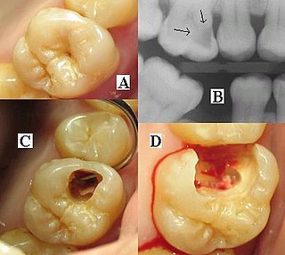
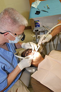
Dental radiographs are commonly called X-rays. Dentists use radiographs for many reasons: to find hidden dental structures, malignant or benign masses, bone loss, and cavities.

CT pulmonary angiogram angiogram for legs

CT pulmonary angiogram (CTPA) is a medical diagnostic test that employs computed tomography (CT) angiography to obtain an image of the pulmonary arteries. Its main use is to diagnose pulmonary embolism (PE). It is a preferred choice of imaging in the diagnosis of PE due to its minimally invasive nature for the patient, whose only requirement for the scan is an intravenous line.

Patients are exposed to ionizing radiations when they undergo diagnostic examinations using x-rays or radiopharmaceuticals, therapy of cancer or benign lesions using radiations emitted by radioisotopes or those by radiation generators; and in interventional procedures using fluoroscopy. There has been a tremendous increase in the use of ionizing radiation in medicine during recent decades. Health professionals and patients are concerned about the harmful effects of radiation. International Atomic Energy Agency (IAEA) has established a programme on radiological protection of patients in recognition of the increasing importance of this topic. The emphasis in the past had been on radiation protection of staff and this emphasis has helped to reduce radiation doses to staff to levels much below the dose limits prescribed by the International Commission on Radiological Protection (ICRP) and accepted by most countries. The recent emphasis on radiation protection of patients is helping in developing strategies to reduce radiation doses to patient without compromising on diagnostic or therapeutic purpose.

Panoramic radiograph

A panoramic radiograph is a panoramic scanning dental X-ray of the upper and lower jaw. It shows a two-dimensional view of a half-circle from ear to ear. Panoramic radiography is a form of focal plane tomography; thus, images of multiple planes are taken to make up the composite panoramic image, where the maxilla and mandible are in the focal trough and the structures that are superficial and deep to the trough are blurred.

Paediatric radiology is a subspecialty of radiology involving the imaging of fetuses, infants, children, adolescents, and young adults. Many paediatric radiologists practice at children's hospitals.

Cone beam computed tomography

Cone beam computed tomography is a medical imaging technique consisting of X-ray computed tomography where the X-rays are divergent, forming a cone.

Hybrid operating room

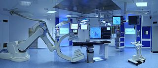
A hybrid operating room is a surgical theatre that is equipped with advanced medical imaging devices such as fixed C-Arms, CT scanners or MRI scanners. These imaging devices enable minimally-invasive surgery. Minimally-invasive surgery is intended to be less traumatic for the patient and minimize incisions on the patient and perform surgery procedure through one or several small cuts.

References

  1. A Patient's Guide to Medical Imaging By Ronald Eisenberg, JD, MD, FACR, Alexander Margulis, MD
  2. Current oral and maxillofacial imaging,Thomas F. Razmus, Gail F. Williamson, Page 107

3. Livingstone RS, Varghese A, Keshava SN. A Study on the Use of Radiation-Protective Apron among Interventionists in Radiology. J Clin Imaging Sci 2018;8:34 http://www.clinicalimagingscience.org/text.asp?2018/8/1/34/239707