
Magnetic resonance imaging (MRI) is a medical imaging technique used in radiology to form pictures of the anatomy and the physiological processes of the body. MRI scanners use strong magnetic fields, magnetic field gradients, and radio waves to generate images of the organs in the body. MRI does not involve X-rays or the use of ionizing radiation, which distinguishes it from CT and PET scans. MRI is a medical application of nuclear magnetic resonance (NMR). NMR can also be used for imaging in other NMR applications, such as NMR spectroscopy.

Spectroscopy is the study of the interaction between matter and electromagnetic radiation. Historically, spectroscopy originated through the study of visible light dispersed according to its wavelength, by a prism. Later the concept was expanded greatly to include any interaction with radiative energy as a function of its wavelength or frequency, predominantly in the electromagnetic spectrum, although matter waves and acoustic waves can also be considered forms of radiative energy; recently, with tremendous difficulty, even gravitational waves have been associated with a spectral signature in the context of the Laser Interferometer Gravitational-Wave Observatory (LIGO) and laser interferometry. Spectroscopic data are often represented by an emission spectrum, a plot of the response of interest, as a function of wavelength or frequency.
Magnetic resonance is a process by which a physical excitation (resonance) is set up via magnetism.
Microwave spectroscopy is the spectroscopy method that employs microwaves, i.e. electromagnetic radiation at GHz frequencies, for the study of matter.

Electron paramagnetic resonance (EPR) or electron spin resonance (ESR) spectroscopy is a method for studying materials with unpaired electrons. The basic concepts of EPR are analogous to those of nuclear magnetic resonance (NMR), but it is electron spins that are excited instead of the spins of atomic nuclei. EPR spectroscopy is particularly useful for studying metal complexes or organic radicals. EPR was first observed in Kazan State University by Soviet physicist Yevgeny Zavoisky in 1944, and was developed independently at the same time by Brebis Bleaney at the University of Oxford.

The National High Magnetic Field Laboratory (MagLab) is a facility at Florida State University, the University of Florida, and Los Alamos National Laboratory in New Mexico, that performs magnetic field research in physics, biology, bioengineering, chemistry, geochemistry, biochemistry. It is the only such facility in the US, and is among nine worldwide. The lab is supported by the National Science Foundation and the state of Florida, and works in collaboration with private industry.
Ferromagnetic resonance, or FMR, is coupling between an electromagnetic wave and the magnetization of a medium through which it passes. This coupling induces a significant loss of power of the wave. The power is absorbed by the precessing magnetization of the material and lost as heat. For this coupling to occur, the frequency of the incident wave must be equal to the precession frequency of the magnetization and the polarization of the wave must match the orientation of the magnetization.
Magnetic resonance force microscopy (MRFM) is an imaging technique that acquires magnetic resonance images (MRI) at nanometer scales, and possibly at atomic scales in the future. MRFM is potentially able to observe protein structures which cannot be seen using X-ray crystallography and protein nuclear magnetic resonance spectroscopy. Detection of the magnetic spin of a single electron has been demonstrated using this technique. The sensitivity of a current MRFM microscope is 10 billion times greater than a medical MRI used in hospitals.

Magnetic resonance microscopy is magnetic resonance imaging (MRI) at a microscopic level down to the scale of microns. The first definition of MRM was MRI having voxel resolutions of better than 100 μm.

Characterization, when used in materials science, refers to the broad and general process by which a material's structure and properties are probed and measured. It is a fundamental process in the field of materials science, without which no scientific understanding of engineering materials could be ascertained. The scope of the term often differs; some definitions limit the term's use to techniques which study the microscopic structure and properties of materials, while others use the term to refer to any materials analysis process including macroscopic techniques such as mechanical testing, thermal analysis and density calculation. The scale of the structures observed in materials characterization ranges from angstroms, such as in the imaging of individual atoms and chemical bonds, up to centimeters, such as in the imaging of coarse grain structures in metals.
In magnetic resonance, a spin echo is the refocusing of spin magnetisation by a pulse of resonant electromagnetic radiation. Modern nuclear magnetic resonance (NMR) and magnetic resonance imaging (MRI) make use of this effect.
Spin-polarized scanning tunneling microscopy (SP-STM) is a specialized application of scanning tunneling microscopy (STM) that can provide detailed information of magnetic phenomena on the single-atom scale additional to the atomic topography gained with STM. SP-STM opened a novel approach to static and dynamic magnetic processes as precise investigations of domain walls in ferromagnetic and antiferromagnetic systems, as well as thermal and current-induced switching of nanomagnetic particles.
Functional imaging, is a medical imaging technique of detecting or measuring changes in metabolism, blood flow, regional chemical composition, and absorption.
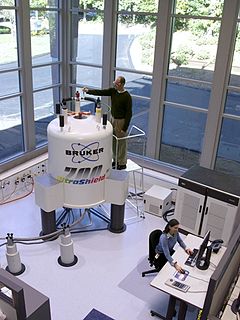
Nuclear magnetic resonance (NMR) is a method of physical observation in which nuclei in a strong constant magnetic field are perturbed by a weak oscillating magnetic field and respond by producing an electromagnetic signal with a frequency characteristic of the magnetic field at the nucleus. This process occurs near resonance, when the oscillation frequency matches the intrinsic frequency of the nuclei, which depends on the strength of the static magnetic field, the chemical environment, and the magnetic properties of the isotope involved; in practical applications with static magnetic fields up to ca. 20 tesla, the frequency is similar to VHF and UHF television broadcasts (60–1000 MHz). NMR results from specific magnetic properties of certain atomic nuclei. Nuclear magnetic resonance spectroscopy is widely used to determine the structure of organic molecules in solution and study molecular physics, crystals as well as non-crystalline materials. NMR is also routinely used in advanced medical imaging techniques, such as in magnetic resonance imaging (MRI).
Electron nuclear double resonance (ENDOR) is a magnetic resonance technique for elucidating the molecular and electronic structure of paramagnetic species. The technique was first introduced to resolve interactions in electron paramagnetic resonance (EPR) spectra. It is currently practiced in a variety of modalities, mainly in the areas of biophysics and heterogeneous catalysis.

Instrumental analysis is a field of analytical chemistry that investigates analytes using scientific instruments.
The following outline is provided as an overview of and topical guide to biophysics:
James S. Hyde is an American biophysicist. He holds the James S. Hyde chair in Biophysics at the Medical College of Wisconsin (MCW) where he specializes in magnetic resonance instrumentation and methodology development in two distinct areas: electron paramagnetic resonance (EPR) spectroscopy and magnetic resonance imaging (MRI). He is senior author of the widely cited 1995 paper by B.B. Biswal et al. reporting the discovery of resting state functional connectivity (fcMRI) in the human brain. He also serves as Director of the National Biomedical EPR Center, a Research Resource supported by the National Institutes of Health. He is author or more than 400 peer-reviewed papers and review articles and holds 35 U.S. Patents. He has been recognized by Festschrifts in both EPR and fcMRI.

Spectroelectrochemistry (SEC) is a set of multi-response analytical techniques in which complementary chemical information is obtained in a single experiment. Spectroelectrochemistry provides a whole vision of the phenomena that take place in the electrode process. The first spectroelectrochemical experiment was carried out by Kuwana in 1964.