Medial knee injuries

Last updated
Medial knee injuries
Knee diagram.svg
Knee diagram (normal)

Medial knee injuries (those to the inside of the knee) are the most common type of knee injury. [1] The medial ligament complex of the knee consists of: [2]

Contents

This complex is the major stabilizer of the medial knee. Injuries to the medial side of the knee are most commonly isolated to these ligaments. [1] [3] A thorough understanding of the anatomy and function of the medial knee structures, along with a detailed history and physical exam, are imperative to diagnosing and treating these injuries.

Symptoms

Patients often complain of pain and swelling over the medial aspect of the knee joint. They may also report instability with side-to-side movement and during athletic performance that involves cutting or pivoting. [4] [5]

Complications

Jacobson previously described the common problems to medial knee surgery. [6] It was stressed that adequate diagnosis is imperative and all possible injuries should be evaluated and addressed intraoperatively. Damage to the saphenous nerve and its infrapatellar branch is possible during medial knee surgery, potentially causing numbness or pain over the medial knee and leg. [7] As with all surgeries, there is a risk of bleeding, wound problems, deep vein thrombosis, and infection that can complicate the outcome and rehabilitation process. The long term complication of arthrofibrosis and heterotopic ossification (Pellegrini-Stieda syndrome) are problems that are best addressed with early range of motion and following defined rehabilitation protocols. [4] [5] [6] [8] Failure of graft due to intrinsic mechanical forces should be prevented with preoperative alignment assessment (osteotomy treatment) and proper rehabilitation. [4]

Causes

Medial knee injury is usually caused by a valgus knee force, a tibial external rotation force, or a combination thereof. This mechanism is often seen in sports that involve aggressive knee flexion like ice hockey, skiing, and football. [3] [4] [5]

Anatomy and function

Knee diagram Knee diagram.svg
Knee diagram

Structures on the medial side of the knee include the tibia, femur, vastus medialis obliquus muscle, semitendinosus tendon, gracilis tendon, sartorius tendon, adductor magnus tendon, medial head of the gastrocnemius muscle, semimembranosus tendon, medial meniscus, medial patellofemoral ligament (MPFL), sMCL, dMCL, and POL. It has been found that the most important structures for stabilization in this area of the knee are ligaments: sMCL, dMCL, and POL. [2] [3]

Bones

The bones of the knee are the femur, patella, tibia, and fibula. The fibula is on the lateral side of the knee and the patella has little effect on the medial side of the knee. The bony congruity of the medial knee consists of the opposing surfaces of the medial femoral condyle and the medial tibial plateau. On the medial femoral condyle there are three bony landmarks that are important: the medial epicondyle, adductor tubercle, and gastrocnemius tubercle. The medial epicondyle is the most distal and anterior prominence. The adductor tubercle is just proximal and posterior to the medial epicondyle. The gastrocnemius tubercle is just distal and posterior to the adductor tubercle. [2]

Ligaments and biomechanical function

Left knee joint from behind, showing interior ligaments. Gray348.png
Left knee joint from behind, showing interior ligaments.
Tibia muscle attachments (seen from front) Gray258.png
Tibia muscle attachments (seen from front)

The sMCL connects the femur to the tibia. It originates just proximal and posterior to the medial epicondyle (not directly on the epicondyle) and splits into two distinct sections. [9] [4] One tibial section attaches to soft tissue, 1 cm distal to the joint line. The other tibial section attaches directly to the tibia, anterior to the posteromedial tibial crest, 6 cm distal to the joint line. [2] [9] This distal attachment is the stronger of the two and makes up the floor of the pes anserine bursa. The proximal tibial attachment of the sMCL is the primary stabilizer to valgus force on the knee, whereas the distal tibial attachment is the primary stabilizer of external rotation at 30° of knee flexion. [3] [9]

The dMCL is a thickening of the medial aspect of the capsule surrounding the knee. It originates on the femur 1 cm distal to the sMCL origin and inserts 3–4 mm distal to the joint line. It runs parallel to and underneath the sMCL. [2] [9] The dMCL connects directly to the medial meniscus and therefore can be divided into meniscofemoral and meniscotibial ligament components.

The meniscofemoral ligament is longer than the meniscotibial ligament, which is shorter and thicker in nature. [2] The meniscofemoral ligament is a primary internal rotation stabilizer and a secondary external rotation stabilizer, activated when the sMCL fails. [3] [9] The meniscotibial ligament acts to secondarily stabilize internal rotation.

The POL (called by older texts: oblique portion of the sMCL) is a fascial expansion with three main components: superficial, central (tibial), and capsular. The central arm is the strongest and thickest. [2] [10] It arises from the semimembranosus tendon and connects anterior and distal to the gastrocnemius tubercle via the posterior joint capsule. The POL, therefore, is not a stand-alone structure, but a thickening of the posteromedial joint capsule. It stabilizes internal rotation of the knee through all degrees of flexion but bears the most load when internally rotated in full extension. It also acts as a secondary external rotation stabilizer. [3] [4] [11]

The MPFL arises from the fibers of the vastus medialis obliquus muscle and attaches distally to the superior medial aspect of the patella. [2] This ligament acts to keep the patella within the trochlear groove during flexion and extension. [2] It is rarely injured from a medial knee injury unless there is a concurrent lateral patellar subluxation or dislocation.

Tendons and muscles

The adductor magnus tendon attaches to the distal medial femoral condyle just posterior and proximal to the adductor tubercle. [2] It has a fascial expansion on the distal-medial aspect that attaches to the medial gastrocnemius tendon, capsular arm of the POL, and posteromedial joint capsule. The thick distal lateral aspect attaches to the medial supracondylar ridge. The adductor magnus tendon is an excellent, consistent landmark because it is rarely injured. The vastus medialis obliquus muscle courses over the anteromedial thigh, attaching along the adductor magnus anterior border and to the quadratus femoris tendon. The medial gastrocnemius tendon arises proximal and posterior to the gastrocnemius tubercle of the medial femoral condyle. [2] This is another important landmark because it is rarely injured and attaches close to the capsular arm of the POL, thus helping the surgeon locate the femoral attachment of the POL. [4]

Diagnosis

The majority of medial knee injuries are isolated ligamentous injuries. Most patients will relate a history of a traumatic blow to the lateral aspect of the knee (causing a valgus force) or a non-contact valgus force. Acute injuries are much easier to diagnose clinically, while chronic injuries may be less apparent due to difficulty in differentiating from a lateral knee injury, possibly requiring valgus stress radiographs. [4] [5]

Physical exam

The physical exam should always begin with a visual inspection of the joint for any outward signs of trauma. Palpation should follow paying close attention to effusion and subjective tenderness during the exam. The practitioner should also evaluate the contralateral (non-injured) knee to note any differences in gross appearance and landmarks. Palpation should focus specifically on the meniscofemoral and meniscotibial aspects of the sMCL. It has been reported that injury to one versus other has implications for healing, so localization of the site of injury is beneficial. Testing of the knee joint should be done using the following techniques and the findings compared to the contralateral, normal knee: [4] [5]

Classification

Grading of medial knee injuries is dependent on the amount of medial joint space gapping found upon valgus stress testing with the knee in 20° of flexion. Grade I injuries have no instability clinically and are associated with tenderness only, representing a mild sprain. Grade II injuries have broad tenderness over the medial knee and have some gapping with a firm end-point during valgus testing; this represents a partial tear of the ligaments. Grade III injuries have a complete ligamentous tear. There will be no end-point to valgus stress testing. [5] [6] [14] The historic quantified definition of grades I, II, and III represented 0–5 mm, 5–10 mm, and >10 mm of medial compartment gapping, respectively. [15] LaPrade et al. reported, however, that a simulated grade III sMCL injury showed only 3.2 mm of increased medial compartment gapping compared to the intact state. [15] Additionally, with the knee in full extension, if valgus stress testing reveals more than 1–2 mm of medial compartment gapping present, a concomitant anterior cruciate ligament (ACL) or posterior cruciate ligament (PCL) injury is suspected. [4] [5]

Radiographs

Anterior-posterior (AP) radiographs are useful for reliably assessing normal anatomical landmarks. Bilateral valgus stress AP images can show a difference in medial joint space gapping. It has been reported that an isolated grade III sMCL tear will show an increase in medial compartment gapping of 1.7 mm at 0° of knee flexion and 3.2 mm at 20° of knee flexion, compared to the contralateral knee. Additionally, a complete medial ligamentous disruption (sMCL, dMCL, and POL) will show increased gapping by 6.5 mm at 0° and 9.8 mm at 20° during valgus stress testing. [15] Pellegrini-Stieda syndrome can also be seen on AP radiographs. This finding is due to calcification of the sMCL (heterotopic ossification) caused by the chronic tear of the ligament. [5] [16]

MRI

Magnetic resonance imaging (MRI) can be helpful in assessing for a ligamentous injury to the medial side of the knee. [6] Milewski et al. has found that grade I to III classification can be seen on MRI. [17] With a high-quality image (1.5 tesla or 3 tesla magnet) and no previous knowledge of the patient's history, musculoskeletal radiologists were able to accurately diagnose medial knee injury 87% of the time. [18] MRI can also show associated bone bruises on the lateral side of the knee, which one study shows, happen in almost half of medial knee injuries. [19]

Knee MRIs should be avoided for knee pain without mechanical symptoms or effusion, and upon non-successful results from a functional rehabilitation program. [20]

Treatment

Treatment of medial knee injuries varies depending on location and classification of the injuries. [6] [21] The consensus of many studies is that isolated grade I, II, and III injuries are usually well suited to non-operative treatment protocols. Acute grade III injuries with concomitant multiligament injuries or knee dislocation involving medial side injury should undergo surgical treatment. Chronic grade III injuries should also undergo surgical treatment if the patient is experiencing rotational instability or side-to-side instability. [4] [5]

Nonoperative treatment

Conservative treatment of isolated medial knee injuries (grades I-III) begins with controlling swelling and protecting the knee. Swelling is managed well with rest, ice, elevation, and compression wraps. [22] Protection can be performed using a hinged brace that stabilizes against varus and valgus stress but allows full flexion and extension. The brace should be worn for the first four to six weeks of rehabilitation, especially during physical exercise to prevent trauma to the healing ligament. Stationary bike exercises are the recommended exercise for active range of motion and should be increased as tolerated by the patient. Side-to-side movements of the knee should be avoided. The patient is allowed to bear weight as tolerated and should perform quadriceps strengthening exercises along with range of motion exercises. The typical return-to-play time frame for most athletes with a grade III medial knee injury undergoing a rehabilitation program is 5 to 7 weeks. [4] [23] [5] [14]

Operative treatment

It has been reported that severe acute and chronic grade III medial knee injuries often involve the sMCL in combination with the POL. [10] [24] Direct surgical repair or reconstruction, therefore, should be performed for both of these ligaments because they both play an important role in static medial knee stability. [25] The biomechanically validated approach is to reconstruct both the POL and both divisions of the sMCL. [26] [27]

Severe acute tears

Surgery involving direct repair (with or without augmentation from a hamstring autograft), among other previously used techniques, have not been biomechanically tested. An anatomical reconstruction of the sMCL and POL has been biomechanically validated. [26]

Chronic instability

Underlying causes of chronic medial knee instability must be identified before surgical reconstruction is performed. More specifically, patients with genu valgum (knock-kneed) alignment must be evaluated and treated with an osteotomy(s) to establish balanced forces on knee ligaments, preventing premature failure of concurrent cruciate ligament reconstruction. These patients should be rehabilitated after the osteotomy heals before it can be verified that they do not still have functional limitations. Once proper alignment is achieved, reconstruction can be performed. [4]

Anatomic medial knee reconstruction

This technique, described in detail by LaPrade et al., uses two grafts in four separate tunnels. An incision is made over the medial knee 4 cm medial to the patella, and extended distally 7 to 8 cm past the joint line, directly over the pes anserinus tendons. [27]

Within the distal borders of the incision, the semitendinosus and gracilis tendons are found beneath the sartorius muscle fascia. The distal tibial attachment of the sMCL can be found under these identified tendons, making up the floor of the pes anserine bursa, 6 cm distal to the joint line. Once identified, the remaining soft tissue is removed from the attachment site. [27] An eyelet pin is then drilled through attachment site transversely through the tibia, making sure the starting point is located at the posterior aspect of the site to ensure better biomechanical outcomes. [27] Over the eyelet pin, a 7-mm reamer (6 mm considered in smaller patients) is reamed to a depth of 25 mm. Once prepared, attention is directed to preparing the reconstruction tunnel for the tibial attachment of the POL. Above the anterior arm attachment of the semimembranosus muscle tendon, the tibial attachment of the central arm of the POL is identified. This attachment is exposed by making a small incision parallel to the fibers along the posterior edge of the anterior arm of the semimembranosus tendon. Once exposed, an eyelet pin is drilled through the tibia toward Gerdy's tubercle (anterolateral tibia). After verifying the correct anatomic eyelet pin placement, a 7-mm reamer is used over the pin to drill a tunnel depth of 25 mm. [27]

Moving to the femoral attachments of the ligaments, the first step is identifying the adductor magnus muscle tendon, and its corresponding attachment site, near the adductor tubercle. Just distal and slightly anterior to this tubercle is the bony prominence of the medial epicondyle. The attachment site of the sMCL can be identified slightly proximal and posterior to the epicondyle. An eyelet pin can now be passed transversely through the femur at this site. The tunnel at this location, however, should be drilled after identifying the POL attachment site. [27]

The next step of identifying the POL femoral attachment is done by locating the gastrocnemius tubercle (2.6 mm distal and 3.1 mm anterior to the medial gastrocnemius tendon attachment on the femur). If the posteromedial capsule is not intact, the POL attachment site is located 7.7 mm distal and 2.9 mm anterior to the gastrocnemius tubercle. With the capsule intact, however, an incision is made along the posterior aspect of the sMCL, parallel to its fibers. The central arm of the POL can then be found at its femoral attachment site. Once identified, an eyelet pin is passed transversely through the femur. The distances between the femoral attachment site of the POL and the sMCL (on average, 11mm) should now be measured to verify that the anatomic attachment sites have been correctly identified. [2] Once this is done, the femoral tunnels for the sMCL and POL can be reamed to a depth of 25 mm using a 7-mm reamer. [27]

The next aspect of the surgery is preparation and placement of the reconstruction grafts. The preparation can be done while the other steps are being completed by another surgeon or physician's assistant. The semitendinosus tendon can be harvested using a hamstring stripper for use as the reconstruction autograft. [26] The autograft is sectioned into a 16-cm length for the sMCL reconstruction and 12-cm length for the POL reconstruction. These lengths are also used if the surgery is done with cadaver allograft. The sMCL and POL grafts are pulled into their respective femoral tunnels and each secured with a cannulated bioabsorbable screw. The grafts are passed distally along their native courses to the tibial attachments. The sMCL is passed under sartorius fascia (and any remaining sMCL fibers). Both grafts are passed (but not yet secured) into their respective tibial tunnels using the existing eyelet pins. If simultaneous cruciate ligament surgery is underway, the cruciate reconstructions are secured before securing the medial ligaments. [27]

Securing the POL graft is done in full knee extension. The graft is pulled tight and fixed using a bioabsorbable screw. The knee is then flexed to 20°. Making sure the tibia remains in neutral rotation, a varus force is used to ensure there is no medial compartment gapping of the knee. The sMCL graft is then tightened and fixed with a bioabsorbable screw. [27]

The final step of reconstruction ligament fixation is the proximal tibial attachment of the sMCL. This soft-tissue attachment can be reproduced with a suture anchor [28] placed 12.2 mm distal to the medial joint line (average location), directly medial to the anterior arm of the semimembranosus tibial attachment. [27] Once this aspect of the sMCL is secured to the suture anchor, the knee is put through range of motion testing by the physician to determine the "safe zone" of knee motion which is used during the first post-operative day rehabilitation (below). [27]

Rehabilitation

Future research

Future research with regard to medial knee injuries should evaluate clinical outcomes between different reconstruction techniques. [8] Determining the advantages and disadvantages of these techniques would also be beneficial for optimizing treatment.

Related Research Articles

<span class="mw-page-title-main">Human leg</span> Lower extremity or limb of the human body (foot, lower leg, thigh and hip)

The leg is the entire lower limb of the human body, including the foot, thigh or sometimes even the hip or buttock region. The major bones of the leg are the femur, tibia, and adjacent fibula. The thigh is between the hip and knee, while the calf (rear) and shin (front) are between the knee and foot.

<span class="mw-page-title-main">Knee</span> Leg joint in primates

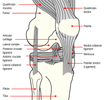
In humans and other primates, the knee joins the thigh with the leg and consists of two joints: one between the femur and tibia, and one between the femur and patella. It is the largest joint in the human body. The knee is a modified hinge joint, which permits flexion and extension as well as slight internal and external rotation. The knee is vulnerable to injury and to the development of osteoarthritis.

<span class="mw-page-title-main">Hamstring</span> Any of the three tendons in the thigh

In human anatomy, a hamstring is any one of the three posterior thigh muscles between the hip and the knee.

<span class="mw-page-title-main">Tibia</span> Leg bone in vertebrates

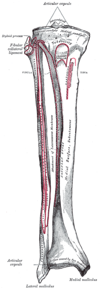
The tibia, also known as the shinbone or shankbone, is the larger, stronger, and anterior (frontal) of the two bones in the leg below the knee in vertebrates ; it connects the knee with the ankle. The tibia is found on the medial side of the leg next to the fibula and closer to the median plane. The tibia is connected to the fibula by the interosseous membrane of leg, forming a type of fibrous joint called a syndesmosis with very little movement. The tibia is named for the flute tibia. It is the second largest bone in the human body, after the femur. The leg bones are the strongest long bones as they support the rest of the body.

<span class="mw-page-title-main">Fibula</span> Leg bone in vertebrates

The fibula or calf bone is a leg bone on the lateral side of the tibia, to which it is connected above and below. It is the smaller of the two bones and, in proportion to its length, the most slender of all the long bones. Its upper extremity is small, placed toward the back of the head of the tibia, below the knee joint and excluded from the formation of this joint. Its lower extremity inclines a little forward, so as to be on a plane anterior to that of the upper end; it projects below the tibia and forms the lateral part of the ankle joint.

<span class="mw-page-title-main">Ankle</span> Region where the foot and the leg meet

The ankle, the talocrural region or the jumping bone (informal) is the area where the foot and the leg meet. The ankle includes three joints: the ankle joint proper or talocrural joint, the subtalar joint, and the inferior tibiofibular joint. The movements produced at this joint are dorsiflexion and plantarflexion of the foot. In common usage, the term ankle refers exclusively to the ankle region. In medical terminology, "ankle" can refer broadly to the region or specifically to the talocrural joint.

<span class="mw-page-title-main">Posterior cruciate ligament</span> One of four major ligaments of the knee

The posterior cruciate ligament (PCL) is a ligament in each knee of humans and various other animals. It works as a counterpart to the anterior cruciate ligament (ACL). It connects the posterior intercondylar area of the tibia to the medial condyle of the femur. This configuration allows the PCL to resist forces pushing the tibia posteriorly relative to the femur.

<span class="mw-page-title-main">Anterior cruciate ligament</span> Type of cruciate ligament in the human knee

The anterior cruciate ligament (ACL) is one of a pair of cruciate ligaments in the human knee. The two ligaments are also called "cruciform" ligaments, as they are arranged in a crossed formation. In the quadruped stifle joint, based on its anatomical position, it is also referred to as the cranial cruciate ligament. The term cruciate translates to cross. This name is fitting because the ACL crosses the posterior cruciate ligament to form an "X". It is composed of strong, fibrous material and assists in controlling excessive motion. This is done by limiting mobility of the joint. The anterior cruciate ligament is one of the four main ligaments of the knee, providing 85% of the restraining force to anterior tibial displacement at 30 and 90° of knee flexion. The ACL is the most injured ligament of the four located in the knee.

<span class="mw-page-title-main">Popliteal artery</span> Continuation of the femoral artery that supplies the lower leg

The popliteal artery is a deeply placed continuation of the femoral artery opening in the distal portion of the adductor magnus muscle. It courses through the popliteal fossa and ends at the lower border of the popliteus muscle, where it branches into the anterior and posterior tibial arteries.

<span class="mw-page-title-main">Segond fracture</span> Avulsion fracture of the lateral tibial condyle of the knee

The Segond fracture is a type of avulsion fracture from the lateral tibial plateau of the knee, immediately below the articular surface of the tibia.

<span class="mw-page-title-main">Medial collateral ligament</span> Ligament on the inner side of the knee joint

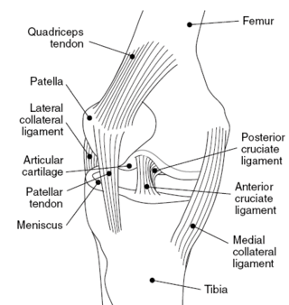
The medial collateral ligament (MCL), also called the superficial medial collateral ligament (sMCL) or tibial collateral ligament (TCL), is one of the major ligaments of the knee. It is on the medial (inner) side of the knee joint and occurs in humans and other primates. Its primary function is to resist valgus forces on the knee.

The biceps femoris is a muscle of the thigh located to the posterior, or back. As its name implies, it consists of two heads; the long head is considered part of the hamstring muscle group, while the short head is sometimes excluded from this characterization, as it only causes knee flexion and is activated by a separate nerve.

<span class="mw-page-title-main">Plantaris muscle</span> One of the superficial muscles of the superficial posterior compartment of the leg

The plantaris is one of the superficial muscles of the superficial posterior compartment of the leg, one of the fascial compartments of the leg.

<span class="mw-page-title-main">Medial meniscus</span> Cartilage formation in the human knee

The medial meniscus is a fibrocartilage semicircular band that spans the knee joint medially, located between the medial condyle of the femur and the medial condyle of the tibia. It is also referred to as the internal semilunar fibrocartilage. The medial meniscus has more of a crescent shape while the lateral meniscus is more circular. The anterior aspects of both menisci are connected by the transverse ligament. It is a common site of injury, especially if the knee is twisted.

The knee examination, in medicine and physiotherapy, is performed as part of a physical examination, or when a patient presents with knee pain or a history that suggests a pathology of the knee joint.

<span class="mw-page-title-main">Lower extremity of femur</span>

The lower extremity of femur is the lower end of the femur in human and other animals, closer to the knee. It is larger than the upper extremity of femur, is somewhat cuboid in form, but its transverse diameter is greater than its antero-posterior; it consists of two oblong eminences known as the lateral condyle and medial condyle.

<span class="mw-page-title-main">Unhappy triad</span> Medical condition of the knee

The unhappy triad, also known as a blown knee among other names, is an injury to the anterior cruciate ligament, medial collateral ligament, and meniscus. Analysis during the 1990s indicated that this 'classic' O'Donoghue triad is actually an unusual clinical entity among athletes with knee injuries. Some authors mistakenly believe that in this type of injury, "combined anterior cruciate and medial collateral ligament disruptions that were incurred during athletic endeavors" always present with concomitant medial meniscus injury. However, the 1990 analysis showed that lateral meniscus tears are more common than medial meniscus tears in conjunction with sprains of the ACL.

<span class="mw-page-title-main">Malleolus</span> Ankle bone protrusion

A malleolus is the bony prominence on each side of the human ankle.

Posterolateral corner injuries of the knee are injuries to a complex area formed by the interaction of multiple structures. Injuries to the posterolateral corner can be debilitating to the person and require recognition and treatment to avoid long term consequences. Injuries to the PLC often occur in combination with other ligamentous injuries to the knee; most commonly the anterior cruciate ligament (ACL) and posterior cruciate ligament (PCL). As with any injury, an understanding of the anatomy and functional interactions of the posterolateral corner is important to diagnosing and treating the injury.

<span class="mw-page-title-main">Tibial plateau fracture</span> Medical condition

A tibial plateau fracture is a break of the upper part of the tibia (shinbone) that involves the knee joint. This could involve the medial, lateral, central, or bicondylar. Symptoms include pain, swelling, and a decreased ability to move the knee. People are generally unable to walk. Complication may include injury to the artery or nerve, arthritis, and compartment syndrome.

References

  1. 1 2 Pedowitz, R. A.; O'Connor, J. J.; Akeson, W. H. (2003). Daniel's Knee Injuries: Ligament and Cartilage Structure, Function, Injury, and Repair (2nd ed.). Philadelphia, PA: Lippincott Williams & Wilkins. ISBN   978-0-7817-1817-2.
  2. 1 2 3 4 5 6 7 8 9 10 11 12 LaPrade, R. F.; Engebretsen, A. H.; Ly, T. V.; Johansen, S.; Wentorf, F. A.; Engebretsen, L. (2007). "The anatomy of the medial part of the knee". J Bone Joint Surg Am . 89 (9): 2000–2010. doi:10.2106/JBJS.F.01176. PMID   17768198. S2CID   46253119.
  3. 1 2 3 4 5 6 Marchant, M. H.; Tibor, L. M.; Sekiya, J. K.; Hardaker, W. T. Jr.; Garrett, W. E. Jr.; Taylor, D. C. (2010). "Management of medial-sided knee injuries, part 1: Medial collateral ligament". Am J Sports Med. 39 (5): 1102–1113. doi:10.1177/0363546510385999. PMID   21148144. S2CID   19863321.
  4. 1 2 3 4 5 6 7 8 9 10 11 12 13 14 15 16 17 18 19 LaPrade, R. F.; Wijdicks, C. A. (2012). "The management of injuries to the medial side of the knee". J Orthop Sports Phys Ther. 42 (3): 221–233. doi:10.2519/jospt.2012.3624. PMID   22382986.
  5. 1 2 3 4 5 6 7 8 9 10 11 12 13 14 15 Widjicks CA, Griffin CJ, Johansen S, Engebretsen L, LaPrade RF (2010). "Injuries to the medial collateral ligament and associated medial structures of the knee". J Bone Joint Surg Am. 92 (5): 1266–1280. doi:10.2106/jbjs.i.01229. PMID   20439679.
  6. 1 2 3 4 5 Jacobson KE (1999). "Technical pitfalls of collateral ligament surgery". Clinical Sports Med. 18 (4): 847–882. doi:10.1016/s0278-5919(05)70188-5. PMID   10553239.
  7. Widjicks CA, Westerhaus BD, Brand EJ, Johansen S, Engebretsen L, LaPrade RF (2010). "Sartorial branch of the saphenous nerve in relation to a medial knee ligament repair or reconstruction". Knee Surg Sports Traumatol Arthrosc. 18 (8): 1105–1109. doi:10.1007/s00167-009-0934-6. PMID   19859696. S2CID   17909895.
  8. 1 2 3 Lind M, Jakobsen BW, Lund B, Hansen MS, Abdallah O, Christiansen SE (2009). "Anatomical reconstruction of the medial collateral ligament and posteromedial corner of the knee in patients with chronic medial collateral ligament instability". Am J Sports Med. 37 (6): 1116–1122. doi:10.1177/0363546509332498. PMID   19336612. S2CID   13657821.{{cite journal}}: CS1 maint: multiple names: authors list (link)
  9. 1 2 3 4 5 Griffith, C. J.; LaPrade, R. F.; Johansen, S.; Armitage, B.; Wijdicks, C.; Engebretsen, L. (2009). "Medial knee injury: Part 1, static function of the individual components of the main medial knee structures". Am J Sports Med . 37 (9): 1762–1770. doi:10.1177/0363546509333852. PMID   19609008. S2CID   34594420.
  10. 1 2 Hughston, J. C.; Eilers, A. F. (1973). "The role of the posterior oblique ligament in repairs of acute medial (collateral) ligament tears of the knee" (PDF). J Bone Joint Surg Am. 55 (5): 923–940. doi:10.2106/00004623-197355050-00002. PMID   4760100.
  11. Tibor LM, Marchant MH, Sekiya JK, Hardaker WT Jr, Garrett WE Jr, Taylor DC (2010). "Management of medial-sided knee injuries, part 2: Posteromedial corner". Am J Sports Med. 39 (6): 1332–1340. doi:10.1177/0363546510387765. PMID   21173192. S2CID   29818907.
  12. Hughston, J. C.; Norwood, L. A. (1980). "The posterolateral drawer test and external rotation recurvatum test for posterolateral rotational instability of the knee". Clin Orthop. 147 (147): 82–87. doi:10.1097/00003086-198003000-00014. PMID   7371321.
  13. LaPrade RF, Terry GC (1997). "Injuries to the posterolateral aspect of the knee: Association of injuries with clinical instability". Am J Sports Med. 25 (4): 433–438. doi:10.1177/036354659702500403. PMID   9240974. S2CID   40756823.
  14. 1 2 Petermann J, von Garrel T, Gotzen L (1993). "Non-operative treatment of acute medial collateral ligament lesions of the knee joint". Knee Surg Sports Traumatol Arthrosc. 1 (2): 93–96. doi:10.1007/bf01565459. PMID   8536015. S2CID   8781323.
  15. 1 2 3 4 LaPrade RF, Bernhardson AS, Griffith CJ, Macalena JA, Wijdicks CA (2009). "Correlation of valgus stress radiographs with medial knee ligament injuries: An in vitro biomechanical study". Am J Sports Med. 38 (2): 330–338. doi:10.1177/0363546509349347. PMID   19966093. S2CID   28747464.
  16. Altschuler EL, Bryce TN (2006). "Images in clinical medicine. Pellegrini-Stieda syndrome". N Engl J Med. 354 (1): e1. doi:10.1056/nejmicm040406. PMID   16394294.
  17. Milewski MD, Sanders TG, Miller MD (2011) MRI-arthroscopy correlation: the knee. J Bone Joint Surg 93-A:1735-1745
  18. Yao L, Dungan D, Seeger LL (1994). "MR imaging of tibial collateral ligament injury: Comparison with clinical examination". Skeletal Radiol (Submitted manuscript). 23 (7): 52–524. doi:10.1007/bf00223082. PMID   7824979. S2CID   33430768.
  19. Miller MD, Osborne JR, Gordon WT, Hinkin DT, Brinker MR (1998). "The natural history of bone bruises: A prospective study of magnetic resonance imaging-detected trabecular microfractures in patients with isolated medial collateral ligament injuries". Am J Sports Med. 26 (1): 15–19. doi:10.1177/03635465980260011001. PMID   9474396. S2CID   5657067.
  20. --> American Medical Society for Sports Medicine (24 April 2014), "Five Things Physicians and Patients Should Question", Choosing Wisely: an initiative of the ABIM Foundation , American Medical Society for Sports Medicine, retrieved 29 July 2014
  21. Frank CB, Loitz BJ, Shrive NG (1995). "Injury location affects ligament healing: a morphologic and mechanical study of the healing rabbit medial collateral ligament". Acta Orthop Scand. 66 (5): 455–462. doi:10.3109/17453679508995587. PMID   7484130.
  22. 1 2 Schroder D, Passler HH (1994). "Combination of cold and compression after knee surgery: a prospective randomized study". Knee Surg Sports Traumatol Arthrosc. 2 (3): 158–165. doi:10.1007/bf01467918. PMID   7584198. S2CID   22890342.
  23. Ballmer PM, Jakob RP (1988). "The non-operative treatment of isolated complete tears of the medial collateral ligament of the knee: a prospective study". Arch Orthop Trauma Surg. 107 (5): 273–276. doi:10.1007/bf00451505. PMID   3178440. S2CID   6637768.
  24. Hugston JC (1994). "The importance of the posterior oblique ligament in repairs of acute tears of the medial ligaments in knees with and without an associated rupture of the anterior cruciate ligament: Results of long-term follow-up". J Bone Joint Surg Am. 76 (9): 1328–1344. doi:10.2106/00004623-199409000-00008. PMID   8077263.
  25. Griffith CJ, Wijdicks CA, LaPrade RF, Armitage BM, Johansen S, Engebretsen L (2009). "Force measurements on the posterior oblique ligament and superficial medial collateral ligament proximal and distal divisions to applied loads". Am J Sports Med. 37 (1): 140–148. doi:10.1177/0363546508322890. PMID   18725650. S2CID   18684131.
  26. 1 2 3 Coobs BR, Wijdicks CA, Armitage BM, Spiridonov SI, Westerhaus BD, Johansen S, Engebretsen L, LaPrade RF (2010). "An in vitro analysis of an anatomical medial knee reconstruction". Am J Sports Med. 38 (2): 339–347. doi:10.1177/0363546509347996. PMID   19966100. S2CID   26972568.
  27. 1 2 3 4 5 6 7 8 9 10 11 LaPrade RF, Wijdicks CA (2012). "Surgical technique: Development of an anatomic medial knee reconstruction". Clin Orthop Relat Res. 470 (3): 806–814. doi:10.1007/s11999-011-2061-1. PMC   3270176 . PMID   21909850.
  28. Widjicks CA, Brand EJ, Nuckley DJ, Johansen S, LaPrade RF, Engebretsen L (2010). "Biomechanical evaluation of a medial knee reconstruction with comparison of bioabsorbable interference screw constructs and optimization with a cortical button". Knee Surg Sports Traumatol Arthrosc. 18 (11): 1532–1541. doi:10.1007/s00167-010-1127-z. PMID   20563561. S2CID   2438119.