Related Research Articles

The brachial plexus is a network of nerves formed by the anterior rami of the lower four cervical nerves and first thoracic nerve. This plexus extends from the spinal cord, through the cervicoaxillary canal in the neck, over the first rib, and into the armpit, it supplies afferent and efferent nerve fibers to the chest, shoulder, arm, forearm, and hand.

A spinal nerve is a mixed nerve, which carries motor, sensory, and autonomic signals between the spinal cord and the body. In the human body there are 31 pairs of spinal nerves, one on each side of the vertebral column. These are grouped into the corresponding cervical, thoracic, lumbar, sacral and coccygeal regions of the spine. There are eight pairs of cervical nerves, twelve pairs of thoracic nerves, five pairs of lumbar nerves, five pairs of sacral nerves, and one pair of coccygeal nerves. The spinal nerves are part of the peripheral nervous system.

In human anatomy, the spinal canal, vertebral canal or spinal cavity is an elongated body cavity enclosed within the dorsal bony arches of the vertebral column, which contains the spinal cord, spinal roots and dorsal root ganglia. It is a process of the dorsal body cavity formed by alignment of the vertebral foramina. Under the vertebral arches, the spinal canal is also covered anteriorly by the posterior longitudinal ligament and posteriorly by the ligamentum flavum. The potential space between these ligaments and the dura mater covering the spinal cord is known as the epidural space. Spinal nerves exit the spinal canal via the intervertebral foramina under the corresponding vertebral pedicles.

Spondylosis is the degeneration of the vertebral column from any cause. In the more narrow sense it refers to spinal osteoarthritis, the age-related degeneration of the spinal column, which is the most common cause of spondylosis. The degenerative process in osteoarthritis chiefly affects the vertebral bodies, the neural foramina and the facet joints. If severe, it may cause pressure on the spinal cord or nerve roots with subsequent sensory or motor disturbances, such as pain, paresthesia, imbalance, and muscle weakness in the limbs.

Degenerative disc disease (DDD) is a medical condition typically brought on by the aging process in which there are anatomic changes and possibly a loss of function of one or more intervertebral discs of the spine. DDD can take place with or without symptoms, but is typically identified once symptoms arise. The root cause is thought to be loss of soluble proteins within the fluid contained in the disc with resultant reduction of the oncotic pressure, which in turn causes loss of fluid volume. Normal downward forces cause the affected disc to lose height, and the distance between vertebrae is reduced. The anulus fibrosus, the tough outer layers of a disc, also weakens. This loss of height causes laxity of the longitudinal ligaments, which may allow anterior, posterior, or lateral shifting of the vertebral bodies, causing facet joint malalignment and arthritis; scoliosis; cervical hyperlordosis; thoracic hyperkyphosis; lumbar hyperlordosis; narrowing of the space available for the spinal tract within the vertebra ; or narrowing of the space through which a spinal nerve exits with resultant inflammation and impingement of a spinal nerve, causing a radiculopathy.

The superior orbital fissure is a foramen or cleft of the skull between the lesser and greater wings of the sphenoid bone. It gives passage to multiple structures, including the oculomotor nerve, trochlear nerve, ophthalmic nerve, abducens nerve, ophthalmic veins, and sympathetic fibres from the cavernous plexus.

The iliopsoas muscle refers to the joined psoas major and the iliacus muscles. The two muscles are separate in the abdomen, but usually merge in the thigh. They are usually given the common name iliopsoas. The iliopsoas muscle joins to the femur at the lesser trochanter. It acts as the strongest flexor of the hip.

A nerve plexus is a plexus of intersecting nerves. A nerve plexus is composed of afferent and efferent fibers that arise from the merging of the anterior rami of spinal nerves and blood vessels. There are five spinal nerve plexuses, except in the thoracic region, as well as other forms of autonomic plexuses, many of which are a part of the enteric nervous system. The nerves that arise from the plexuses have both sensory and motor functions. These functions include muscle contraction, the maintenance of body coordination and control, and the reaction to sensations such as heat, cold, pain, and pressure. There are several plexuses in the body, including:

The human back, also called the dorsum, is the large posterior area of the human body, rising from the top of the buttocks to the back of the neck. It is the surface of the body opposite from the chest and the abdomen. The vertebral column runs the length of the back and creates a central area of recession. The breadth of the back is created by the shoulders at the top and the pelvis at the bottom.
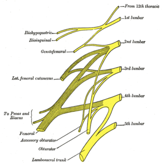
The lumbar nerves are the five pairs of spinal nerves emerging from the lumbar vertebrae. They are divided into posterior and anterior divisions.

The middle cranial fossa is formed by the sphenoid bones, and the temporal bones. It lodges the temporal lobes, and the pituitary gland. It is deeper than the anterior cranial fossa, is narrow medially and widens laterally to the sides of the skull. It is separated from the posterior cranial fossa by the clivus and the petrous crest.

Ramus communicans is the Latin term used for a nerve which connects two other nerves, and can be translated as "communicating branch".

The intervertebral foramen is an opening between two pedicles of adjacent vertebra in the articulated spine. Each intervertebral foramen gives passage to a spinal nerve and spinal blood vessels, and lodges a posterior (dorsal) root ganglion. Cervical, thoracic, and lumbar vertebrae all have intervertebral foramina.

A spinal disc herniation is an injury to the intervertebral disc between two spinal vertebrae, usually caused by excessive strain or trauma to the spine. It may result in back pain, pain or sensation in different parts of the body, and physical disability. The most conclusive diagnostic tool for disc herniation is MRI, and treatment may range from painkillers to surgery. Protection from disc herniation is best provided by core strength and an awareness of body mechanics including good posture.
The dorsal ramus of spinal nerve, posterior ramus of spinal nerve, or posterior primary division is the posterior division of a spinal nerve. The dorsal rami provide motor innervation to the deep muscles of the back, and sensory innervation to the skin of the posterior portion of the head, neck and back.
The ventral ramus is the anterior division of a spinal nerve. The ventral rami supply the antero-lateral parts of the trunk and the limbs. They are mainly larger than the dorsal rami.
The facet joints are a set of synovial, plane joints between the articular processes of two adjacent vertebrae. There are two facet joints in each spinal motion segment and each facet joint is innervated by the recurrent meningeal nerves.
Posterior ramus syndrome, also referred to as thoracolumbar junction syndrome, Maigne syndrome and dorsal ramus syndrome is caused by the unexplained activation of the primary division of a posterior ramus of a spinal nerve. This nerve irritation causes referred pain in a well described tri-branched pattern. The diagnosis is made clinically with the variable presence of four criteria.

The cervical spinal nerve 1 (C1) is a spinal nerve of the cervical segment. C1 carries predominantly motor fibres, but also a small meningeal branch that supplies sensation to parts of the dura around the foramen magnum.
References
- Drake RL, Vogl W, Mitchell AWM. Gray's Anatomy for Students. New York: Elsevier; 2005:69-70.
- Ropper AH, Samuels MA. Adams and Victor's Principles of Neurology. Ninth Edition. New York: McGraw Hill; 2009.
| | This neuroanatomy article is a stub. You can help Wikipedia by expanding it. |