Microsporum nanum

Last updated

Microsporum nanum
Scientific classification OOjs UI icon edit-ltr.svg
Domain: Eukaryota
Kingdom: Fungi
Division: Ascomycota
Class: Eurotiomycetes
Order: Onygenales
Family: Arthrodermataceae
Genus: Microsporum
Species:
M. nanum
Binomial name
Microsporum nanum
C.A.Fuentes (1956)
Synonyms
  • Microsporum gypseum var. nanumC.A.Fuentes, R.Aboulafia, R.J.Vidal (1954)
  • Nannizzia obtusa C.O.Dawson, Gentles (1961)
  • Arthroderma obtusum(C.O.Dawson & Gentles) Weitzman, McGinnis, A.A.Padhye & Ajello (1986)

Microsporum nanum is a pathogenic fungus in the family Arthrodermataceae. It is a type of dermatophyte that causes infection in dead keratinized tissues such as skin, hair, and nails. [1] [2] Microsporum nanum is found worldwide and is both zoophilic and geophilic. Animals such as pigs and sheep are the natural hosts for the fungus; however, infection of humans is also possible. Majority of the human cases reported are associated with pig farming. The fungus can invade the skin of the host; if it is scratched off by the infected animal, the fungus is still capable of reproducing in soil. [3]

Contents

When grown on Sabouraud's Dextrose agar at 25 °C, M. nanum produces a thin, powdery, and soft fibrous colony that appears white at the center becoming light yellowish-brown towards the colony margin. The reverse side appears brownish-orange in young colony and reddish-brown in older colony. [4] [5] The mitochondrial genome of M. nanum consists of 15 protein-coding genes, 2 rRNAs, 25 tRNAs, one intron and one intronic ORF. Approximately 84% of the mitochondrial genome are the structural genes. [6] Microsporum nanum infections include tinea capitis, tinea corporis, tinea cruris, and tinea faciei. [3] Griseofulvin, clotrimazole, miconazole, [7] enilconazole [8] and many herbal treatments, such as extracts from Azadirachta indica , [9] essential oil from Curcuma longa [10] and Eucalyptus pauciflora [11] have been reported to be effective in inhibiting the fungus.

Taxonomy and naming

Microsporum nanum was first thought to be a variant of M. gypseum by Fuentes, Aboulafia, and Vidal who named it M. gypseum var. nanum in 1954. [12] Significant morphological differences between the macroconidia of M. gypseum var. nanum and M. gypseum var. gypseum prompted Fuentes to elevate the former to the species-level as M. nanum two years later. [13] The sexual reproductive (teleomorph) stage of M. nanum was first described by Dawson and Gentles as Nannizzia obtusa in 1961 who isolated it from skin lesions in pigs in Kenya. [14] The name was changed to Arthroderma obtusum by Weitzman, McGinnis, Padhye and Ajello in 1986. [15]

Description

Microsporum nanum is a mesophile and grows moderately rapid on Sabouraud's Dextrose agar. Its macroconidia are ovoid in shape and, unlike those of most other species of Microsporum, normally consist of no more than three cells. Rare to moderate numbers of rough-walled microconidia can also be found in the colony. [3] Microsporum nanum grows optimally at 25 °C but is capable of growth at temperatures up to 37 °C. Physiological tests have shown that M. nanum does not require vitamin supplementation for its growth. Like many other dermatophytes, M. nanum is tolerant of the antifungal agent cycloheximide. In addition, M. nanum also exhibits soil association characters such as urease activity and the formation of perforating organ on hair shafts. [16]

Mitochondrial genome

Mitochondrial DNA sequences have been used for phylogenetic studies in the dermatophytes. A typical fungal mitochondrial DNA usually contains 14 conserved protein-coding genes, 22 to 26 tRNA gene, and 2 rRNA genes. The difference of mitochondrial DNA between fungal species is due to the variations in intergenic region, intron, and gene order. The complete sequences of Trichophyton rubrum , T. mentagrophytes , T. ajelloi , M. canis , M. nanum, and Epidermophyton floccosum show that these 6 species in dermatophytes are closely related because their mitochondrial genomes are highly conserved. Genome size of mitochondrial DNA in M. nanum is 24,105 basepairs. The DNA is AT-rich and the GC content is only 24.47%. M. nanum mtDNA has 15 protein-coding genes, including ATP synthase subunits atp6, atp8, and atp9, cytochrome oxidase subunits cox1, cox2, and cox3, apocytochrome b cob, a ribosomal protein rps5, and NADH dehydrogenase subunits nad1, nad2, nad3, nad4, nad5, and nad6. The gene order of the 15 conserved protein coding genes in M. nanum mtDNA is identical to that of T. rubrum, T. mentagrophytes, T. ajelloi, and M. canis. The Microsporum nanum mtDNA also contains 2 rRNAs and 25 tRNAs. In addition, this mitochondrial genome is highly compact because 83.76% are structural genes. Moreover, the genome contains one intron and one intronic ORF. [6]

Distribution

The distribution of M. nanum is worldwide, including North and South Americas, Canada, Europe, the Middle East, Australia, and Asia. [3] [17] [18] [19] [20] [21] [22] [23] Microsporum nanum and several other dermatophytes were isolated from eleven soil samples of hospitals and public places at Gulbarga, India. Keratinous wastes such as human dander and feathers of birds were suspected to provide a growth medium for these keratinophilic fungi. Microscporum nanum was observed much more frequently in hospital soil than in public places; the percentage was 45.4% and 33.3% of the total number of the observed keratinophilic fungi respectively. [24]

Pathogenicity

Microsporum nanum is geophilic and zoophilic. There are records of isolation from both pig farm soil and the infected pigs. Like M. canis , M. audouinii , and M. ferrugineum , M. nanum causes ectothrix infection. [25] Its growth is usually stops at the keratinized layer of the skin. However, there is some literature suggesting that Microsporum nanum may form endothrix infection under certain circumstances. [13] Unlike other species of Microsporum, Wood's Light Examinationof the skin yields inconsistent findings and fluorescence may or may not be observed. [3]

At the keratinized layer, M. nanum secretes many metabolic products, which trigger inflammation and result in chronic inflammatory infection in swine. [8] The skin lesions are characterized by large brownish spots. [25] Moreover, the infections are also found in cattle, [26] dogs, [27] mice, guinea pigs, and rabbits. [28] Although uncommon, it is known to cause infection in humans following contact with infected animals or contaminated soil. [8] [16] Examples of some common human diseases caused by Microsporum nanum including tinea capitis, tinea corporis, tinea cruris, and tinea faciei. [3] Symptoms might include erythematous papular rash with scaling or inflamed patches on skin. [7] Since M. nanum is only associated with mild, readily-treatable human disease, it is classified at Biosafety Level 2 in many jurisdictions. [25]

Treatment

Griseofulvin, clotrimazole, and miconazole are antimycotic agents that are used to treat M. nanum and T. rubrum . Of three human cases of M. nanum infection reported in 1986, all resolved with treatments by griseofulvin, clotrimazole or miconazole cream. [7] Enilconazole is a fungicide that can be used as veterinary medicine. 0.2% of enilconazole in tap water was reported to be effective in clearing M. nanum in infected sows. [8]

Plant extracts

Azadirachta indica

Azadirachta indica , also known as neem, is used as an antifungal treatment against many dermatophytes such as M. nanum. It is known for its immunomodulatory, anti-inflammatory, antihyperglycemic, antiulcer, antimalarial, antibacterial, antioxidant, antimutagenic and anticarcinogenic activities. [9] Both neem seed and neem leaves are capable of killing M. nanum. The minimum inhibitory concentration (MIC) of seed extract was lower than that of leaf extract when treating M. nanum and other dermatophytes, which were 31 and 15 μg/mL respectively. The application of the neem extract significantly inhibits the growth of the treated dermatophytes when compared with the control (untreated fungi). [29] [30]

Curcuma longa L.

The Curcuma longa L. is a perennial herb, which belongs to the ginger family, Zingiberaceae. [31] The essential oil from the leaves of the herb is effective in treating many pathogenic fungi, such as Epidermophyton floccosum , M. gypseum , M. nanum, T. mentagrophytes, T. rubrum , and T. violaceum . [10] [32] The essential oil is mainly composed of 26.4% terpinolene, 8% α-phellandrene, 7.4% terpinen-4-ol, and 3.2% sabinyl acetate. The oil is fungistatic and fungicidal to Microsporum nanum at concentrations of 2.0 and 2.5 μL/mL respectively. The treatment completely inhibits the mycelia growth of the ringworm caused by Microsporum nanum, Epidermophyton floccosum , Trichophyton rubrum , and Trichophyton violaceum . No adverse effect is reported yet. In addition, the juice from the rhizome of Curcuma longa also be used to treat skin infections, indolent ulcers, inflamed joints, and in purulent ophthalmia. [10]

Eucalyptus pauciflora

Eucalyptus pauciflora , commonly known as snow gum, is a flowering plant that grows in eastern Australia. The essential oil from Eucalyptus pauciflora has a strong antifungal activity. [33] It is fungicidal to M. nanum, E. floccosum , M. audouinii , M. canis , M. gypseum , T. mentagrophytes , T. rubrum , T. tonsurans , and T. violaceum at a concentration of 1.0 μL/mL. No adverse effects on mammalian skin have been reported. [11]

Related Research Articles

<span class="mw-page-title-main">Tinea cruris</span> Fungal infection

Tinea cruris, also known as jock itch, is a common type of contagious, superficial fungal infection of the groin and buttocks region, which occurs predominantly but not exclusively in men and in hot-humid climates.

<span class="mw-page-title-main">Athlete's foot</span> Skin infection caused by fungus

Athlete's foot, known medically as tinea pedis, is a common skin infection of the feet caused by a fungus. Signs and symptoms often include itching, scaling, cracking and redness. In rare cases the skin may blister. Athlete's foot fungus may infect any part of the foot, but most often grows between the toes. The next most common area is the bottom of the foot. The same fungus may also affect the nails or the hands. It is a member of the group of diseases known as tinea.

Dermatophyte is a common label for a group of fungus of Arthrodermataceae that commonly causes skin disease in animals and humans. Traditionally, these anamorphic mold genera are: Microsporum, Epidermophyton and Trichophyton. There are about 40 species in these three genera. Species capable of reproducing sexually belong in the teleomorphic genus Arthroderma, of the Ascomycota. As of 2019 a total of nine genera are identified and new phylogenetic taxonomy has been proposed.

<span class="mw-page-title-main">Tinea capitis</span> Cutaneous fungal infection of the scalp

Tinea capitis is a cutaneous fungal infection (dermatophytosis) of the scalp. The disease is primarily caused by dermatophytes in the genera Trichophyton and Microsporum that invade the hair shaft. The clinical presentation is typically single or multiple patches of hair loss, sometimes with a 'black dot' pattern, that may be accompanied by inflammation, scaling, pustules, and itching. Uncommon in adults, tinea capitis is predominantly seen in pre-pubertal children, more often boys than girls.

<span class="mw-page-title-main">Dermatophytosis</span> Fungal infection of the skin

Dermatophytosis, also known as tinea and ringworm, is a fungal infection of the skin, that may affect skin, hair, and nails. Typically it results in a red, itchy, scaly, circular rash. Hair loss may occur in the area affected. Symptoms begin four to fourteen days after exposure. The types of dermatophytosis are typically named for area of the body that they affect. Multiple areas can be affected at a given time.

<span class="mw-page-title-main">Onychomycosis</span> Medical condition

Onychomycosis, also known as tinea unguium, is a fungal infection of the nail. Symptoms may include white or yellow nail discoloration, thickening of the nail, and separation of the nail from the nail bed. Fingernails may be affected, but it is more common for toenails. Complications may include cellulitis of the lower leg. A number of different types of fungus can cause onychomycosis, including dermatophytes and Fusarium. Risk factors include athlete's foot, other nail diseases, exposure to someone with the condition, peripheral vascular disease, and poor immune function. The diagnosis is generally suspected based on the appearance and confirmed by laboratory testing.

<span class="mw-page-title-main">Kerion</span> Medical condition

Kerion or kerion celsi is an acute inflammatory process which is the result of the host's response to a fungal ringworm infection of the hair follicles of the scalp that can be accompanied by secondary bacterial infection(s). It usually appears as raised, spongy lesions, and typically occurs in children. This honeycomb is a painful inflammatory reaction with deep suppurative lesions on the scalp. Follicles may be seen discharging pus. There may be sinus formation and rarely mycetoma-like grains are produced. It is usually caused by dermatophytes such as Trichophyton verrucosum, T. mentagrophytes, and Microsporum canis. Treatment with oral griseofulvin common.

<span class="mw-page-title-main">Tinea manuum</span> Medical condition

Tinea manuum is a fungal infection of the hand, mostly a type of dermatophytosis, often part of two feet-one hand syndrome. There is diffuse scaling on the palms or back of usually one hand and the palmer creases appear more prominent. When both hands are affected, the rash looks different on each hand, with palmer creases appearing whitish if the infection has been present for a long time. It can be itchy and look slightly raised. Nails may also be affected.

<span class="mw-page-title-main">Butenafine</span> Chemical compound

Butenafine, sold under the brand names Lotrimin Ultra, Mentax, and Butop (India), is a synthetic benzylamine antifungal. It is structurally related to synthetic allylamine antifungals such as terbinafine.

<i>Trichophyton rubrum</i> Species of fungus

Trichophyton rubrum is a dermatophytic fungus in the phylum Ascomycota. It is an exclusively clonal, anthropophilic saprotroph that colonizes the upper layers of dead skin, and is the most common cause of athlete's foot, fungal infection of nail, jock itch, and ringworm worldwide. Trichophyton rubrum was first described by Malmsten in 1845 and is currently considered to be a complex of species that comprises multiple, geographically patterned morphotypes, several of which have been formally described as distinct taxa, including T. raubitschekii, T. gourvilii, T. megninii and T. soudanense.

<i>Trichophyton</i> Genus of Fungi

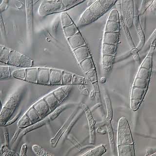
Trichophyton is a genus of fungi, which includes the parasitic varieties that cause tinea, including athlete's foot, ringworm, jock itch, and similar infections of the nail, beard, skin and scalp. Trichophyton fungi are molds characterized by the development of both smooth-walled macro- and microconidia. Macroconidia are mostly borne laterally directly on the hyphae or on short pedicels, and are thin- or thick-walled, clavate to fusiform, and range from 4 to 8 by 8 to 50 μm in size. Macroconidia are few or absent in many species. Microconidia are spherical, pyriform to clavate or of irregular shape, and range from 2 to 3 by 2 to 4 μm in size.

<span class="mw-page-title-main">Fungal folliculitis</span> Inflammation of hair follicles due to fungal infection

Majocchi's granuloma is a skin condition characterized by deep, pustular plaques, and is a form of tinea corporis. It is a localized form of fungal folliculitis. Lesions often have a pink and scaly central component with pustules or folliculocentric papules at the periphery. The name comes from Domenico Majocchi, who discovered the disorder in 1883. Majocchi was a professor of dermatology at the University of Parma and later the University of Bologna. The most common dermatophyte is called Trichophyton rubrum.

<i>Microsporum gypseum</i> Species of fungus

Microsporum gypseum is a soil-associated dermatophyte that occasionally is known to colonise and infect the upper dead layers of the skin of mammals. The name refers to an asexual "form-taxon" that has been associated with four related biological species of fungi: the pathogenic taxa Arthroderma incurvatum, A. gypsea, A. fulva and the non-pathogenic saprotroph A. corniculata. More recent studies have restricted M. gypseum to two teleomorphic species A. gypseum and A. incurvatum. The conidial states of A. fulva and A. corniculata have been assigned to M. fulvum and M. boullardii. Because the anamorphic states of these fungi are so similar, they can be identified reliably only by mating. Two mating strains have been discovered, "+" and "–". The classification of this species has been based on the characteristically rough-walled, blunt, club-shaped, multicelled macroconidia. Synonyms include Achorion gypseum, Microsporum flavescens, M. scorteum, and M. xanthodes. There has been past nomenclatural confusion in the usage of the generic names Microsporum and Microsporon.

<i>Trichophyton mentagrophytes</i> Species of fungus

Trichophyton mentagrophytes is a species in the fungal genus Trichophyton. It is one of three common fungi which cause ringworm in companion animals. It is also the second-most commonly isolated fungus causing tinea infections in humans, and the most common or one of the most common fungi that cause zoonotic skin disease. Trichophyton mentagrophytes is being frequently isolated from dogs, cats, rabbits, guinea pigs and other rodents, though at least some genetic variants possess the potential of human-to-human transmission, e.g. Type VII and Type VIII. Particular genetic variants of the fungus have distinct geographic ranges.

<i>Microsporum gallinae</i> Species of fungus

Microsporum gallinae is a fungus of the genus Microsporum that causes dermatophytosis, commonly known as ringworm. Chickens represent the host population of Microsporum gallinae but its opportunistic nature allows it to enter other populations of fowl, mice, squirrels, cats, dogs and monkeys. Human cases of M. gallinae are rare, and usually mild, non-life-threatening superficial infections.

<span class="mw-page-title-main">Luliconazole</span> Chemical compound

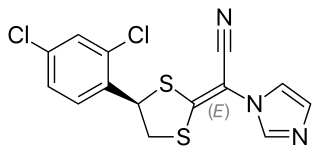
Luliconazole, trade names Luzu among others, is an imidazole antifungal medication. As a 1% topical cream, It is indicated for the treatment of athlete's foot, jock itch, and ringworm caused by dermatophytes such as Trichophyton rubrum, Microsporum gypseum, and Epidermophyton floccosum.

<span class="mw-page-title-main">Favus</span> Medical condition

Favus or tinea favosa is the severe form of tinea capitis, a skin infectious disease caused by the dermatophyte fungus Trichophyton schoenleinii. Typically the species affects the scalp, but occasionally occurs as onychomycosis, tinea barbae, or tinea corporis.

<i>Epidermophyton floccosum</i> Species of fungus

Epidermophyton floccosum is a filamentous fungus that causes skin and nail infections in humans. This anthropophilic dermatophyte can lead to diseases such as tinea pedis, tinea cruris, tinea corporis and onychomycosis. Diagnostic approaches of the fungal infection include physical examination, culture testing, and molecular detection. Topical antifungal treatment, such as the use of terbinafine, itraconazole, voriconazole, and ketoconazole, is often effective.

Microsporum fulvum is a wildly-distributed dermatophyte species in the Fungi Kingdom. It is known to be a close relative to other dermatophytes such as Trichophyton andEpidermophyton. The fungus is common within soil environments and grows well on keratinized material, such as hair, nails and dead skin. It is recognized as an opportunistic fungal pathogen capable of causing cutaneous mycoses in humans and animals. Originally, the fungus was thought to be Microsporum gypseum until enhanced genetic examination separated the two as distinct species in 1963.

References

  1. Ajello, Libero; Edith Varsavsky; Oliver J. Ginther; George Bubash (Nov–Dec 1964). "The natural history of Microsporum nanum". Mycologia. 56 (6): 873–884. doi:10.2307/3756652. JSTOR   3756652.
  2. Weitzman, I; Summerbell, RC (April 1995). "The Dermatophytes". Clinical Microbiology Reviews. 8 (2): 240–259. doi:10.1128/CMR.8.2.240. PMC   172857 . PMID   7621400.
  3. 1 2 3 4 5 6 Land, Geoffrey A (1997). Chapter 7 The Genus Microsporum. Korea: Star Publishing Company.
  4. Fuentes. "Microsporum nanum Species". DoctorFungus. Retrieved 12 October 2013.
  5. "Microsporum nanum". Mycology Online. Retrieved 1 November 2013.
  6. 1 2 Wu, Y; Yang J; Yang F; Liu T; Leng W; Chu Y; Jin Q (May 2009). "Recent dermatophyte divergence revealed by comparative and phylogenetic analysis of mitochondrial genomes". BMC Genomics. 10: 238. doi: 10.1186/1471-2164-10-238 . PMC   2693141 . PMID   19457268.
  7. 1 2 3 Roller, JA; Westblom TU (November 1986). "Microsporum nanum infection in hog farmers". J Am Acad Dermatol. 15 (5 Pt 1): 935–9. doi:10.1016/s0190-9622(86)70252-1. PMID   3782533.
  8. 1 2 3 4 Garcia-Sanchez, A; Bazan, J; de Mendoza, JH; Martinez, R; Sanchez, S; de Mendoza, MH (March 2011). "Outbreak of ringworm in a traditional Iberian pig farm in Spain". Mycoses. 54 (2): 179–181. doi:10.1111/j.1439-0507.2009.01776.x.
  9. 1 2 Mahmoud, DA; Hassanein NM; Youssef KA; Abou Zeid MA (July 2011). "Antifungal activity of different neem leaf extracts and the nimonol against some important human pathogens". Braz J Microbiol. 42 (3): 1007–1016. doi:10.1590/S1517-83822011000300021. PMC   3768785 . PMID   24031718.
  10. 1 2 3 Pandey, KP; Mishra RK; Kamran A; Mishra P; Bajaj AK; Dikshit A (Apr 2010). "Studies on antidermatophytic activity of waste leaves of Curcuma longa L." Physiol Mol Biol Plants. 16 (2): 177–185. doi:10.1007/s12298-010-0019-5. PMC   3550606 . PMID   23572967.
  11. 1 2 Shahi, SK; Shukla AC; Bajaj AK; Banerjee U; Rimek D; Midgely G; Dikshit A (Jan–Feb 2000). "Broad spectrum herbal therapy against superficial fungal infections". Skin Pharmacol Appl Skin Physiol. 13 (1): 60–64. doi:10.1159/000029909. PMID   10657767.
  12. Fuentes, CA; Aboulafia, R; Vidal, RJ (1954). "A dwarf form of Microsporum gypseum". Journal of Investigative Dermatology. 23 (1): 51–61. doi: 10.1038/jid.1954.82 .
  13. 1 2 FUENTES, CA (1956). "A NEW SPECIES OF MlCROSPORUM". Mycologia. 48 (4): 613–614. doi:10.2307/3755345. JSTOR   3755345.
  14. Dawson, CO; Gentles JC. (Jan 1961). "The perfect states of Keratinomyces ajelloi Vanbreuseghem, Trichophyton terrestre Durie & Frey and Microsporum nanum Fuentes". Sabouraudia. 1: 49–57. doi:10.1080/00362176285190111. PMID   13720313.
  15. "Microsporum nanum". MycoBank. Retrieved 17 October 2013.
  16. 1 2 St-Germain, Guy; Summerbell, Richard (2010). Identifying Fungi: A Clinical Laboratory Handbook. Korea: Star Publishing Company, Inc. ISBN   978-0898633115.
  17. Brock, JM (1961). "Microsporum nanum – a cause of tinea capitis – a case report". Archives of Dermatology. 84 (3): 504–505. doi:10.1001/archderm.1961.01580150150027.
  18. Londero, A; Benevenga J (1972). "Human infection by Microsporum nanum in Brazil". Rev Inst Med Trop Sao Paulo. 14 (6): 388–391. PMID   4651713.
  19. Alteras, I (1970). "First case of tinea infection by Microsporum nanum in Romania". Mykosen. 13 (9): 447–450. doi:10.1111/j.1439-0507.1970.tb01322.x. PMID   5529052.
  20. Garg, AK; Mulay DN (Feb 1972). "Isolation of Microsporum nanum from man in India". Hindustan Antibiot Bull. 14 (3): 137–139. PMID   4636285.
  21. Baxter, M (Jul 1969). "Ringworm caused by Microsporum nanum in New Zealand". N Z Med J. 70 (446): 24–26. PMID   5258849.
  22. O'Keeffe, MF (Aug 1973). "A report of three human infections due to Microsporum nanum". Australasian Journal of Dermatology. 14 (2): 73–74. doi:10.1111/j.1440-0960.1973.tb00703.x. PMID   4759452.
  23. Long, JR; Brandenburg AC; Oliver PG (Jul 1972). "Case report. Microsporum nanum: a cause of porcine ringworm in Ontario". Can Vet J. 13 (7): 164–166. PMC   1695825 . PMID   5041149.
  24. Vidyasagar, GM; Hosmani N; Shivkumar D (January 2005). "Keratinophilic fungi isolated from hospital dust and soils of public places at Gulbarga, India". Mycopathologia. 159 (1): 13–21. doi:10.1007/s11046-004-9483-1. PMID   15750728.
  25. 1 2 3 De Hoog, G. S. (2000). Atlas of Clinical Fungi. CBS, Utrecht. p. 758. ISBN   978-9070351434.
  26. Smith, J; Rush-Munro F; McCarthy M (1969). "Animals as a reservoir of human ringworm in New Zealand". Australasian Journal of Dermatology. 10 (3): 169–182. doi:10.1111/j.1440-0960.1969.tb01187.x.
  27. Muhammed, S; Mbogwa S (1974). "The isolation of M. nahum from a dog with skin lesions". Vet Rec. 95 (25–26): 573. doi:10.1136/vr.95.25-26.573.
  28. Refai, M; Ali A; Abdallah I (1970). "Incidence of an experimental infection with Keratinomyces ajelloi and Microsporum nanum in laboratory animals". Bulletin of Pharmaceutical Research Institute. 85 (86): 7–9.
  29. Natarajan, V; Pushkala S; Karuppiah VP; Prasad PV (July 2002). "Anti dermatophytic activity of Azardirachta indica (neem) by in vitro study". Indian J Pathol Microbiol. 45 (3): 311–3. PMID   12785173.
  30. Natarajan, V; Venugopal PV; Menon T (2003). "Effect of Azadirachta indica (neem) on the growth pattern of dermatophytes". Indian Journal of Medical Microbiology. 21 (2): 98–101. PMID   17642990.
  31. Smitinand, T (2001). Thai Plant Names. Bangkok, Thailand: Forest Herbarium, Royal Forest Department.
  32. Apisariyakul, A; Vanittanakom N; Buddhasukh D (1995). "Antifungal activity of turmeric oil extracted from Curcuma longa (Zingiberaceae)". Journal of Ethnopharmacology. 49 (3): 163–169. doi:10.1016/0378-8741(95)01320-2. PMID   8824742.
  33. Reuter, Juliane; Merfort I; Schempp CM (2010). "Botanicals in Dermatology: An Evidence-based Review". American Journal of Clinical Dermatology. 11 (4): 247–267. doi:10.2165/11533220-000000000-00000. PMID   20509719.