NT5M

Last updated
NT5M
Available structures
PDB Ortholog search: PDBe RCSB
Identifiers
Aliases NT5M , dNT-2, dNT2, mdN, 5',3'-nucleotidase, mitochondrial
External IDs OMIM: 605292 MGI: 1917127 HomoloGene: 10622 GeneCards: NT5M
Orthologs
SpeciesHumanMouse
Entrez
Ensembl
UniProt
RefSeq (mRNA)

NM_020201

NM_134029

RefSeq (protein)

NP_064586

NP_598790

Location (UCSC) Chr 17: 17.3 – 17.35 Mb Chr 11: 59.73 – 59.77 Mb
PubMed search [3] [4]
Wikidata
View/Edit Human View/Edit Mouse

5',3'-nucleotidase, mitochondrial, also known as 5'(3')-deoxyribonucleotidase, mitochondrial (mdN) or deoxy-5'-nucleotidase 2 (dNT-2), is an enzyme that in humans is encoded by the NT5M gene. This gene encodes a 5' nucleotidase that localizes to the mitochondrial matrix. This enzyme dephosphorylates the 5'- and 2'(3')-phosphates of uracil and thymine deoxyribonucleotides. The gene is located within the Smith–Magenis syndrome region on chromosome 17. [5] [6]

Structure

The cDNA of mdN encodes a 25.9-kDa polypeptide, and the crystal structure of this enzymes reveals a 196-long amino acid sequence in the mature protein. [7] [8] The first 32 amino acids, which contain the mitochondrial targeting sequence, are removed during the processing of the premature protein for its import into the mitochondrial matrix. The enzyme is likely a dimer protein formed by the interaction of alpha and beta loops between the cores of the two monomers. Each monomer is composed of a large and small domain connected by two loops. The large domain forms an alpha/beta Rossmann fold as well as 2 helix loops in the fold, whereas the small domain forms a truncated four-helix bundle which is inserted between a beta strand and alpha-helix in the large domain. The active site is found in a cleft between the two domains and binds a magnesium ion that is coordinated by three exogenous ligands, a phosphate ion, and two water molecules in an octahedral shape. [8]

mdN is one of seven 5' nucleotidases identified in humans, all of which differ in tissue specificity, subcellular location, primary structure and substrate specificity. [8] [9] Of the seven, the cytosolic counterpart of mdN, cdN, is the most closely related to mdN. Their genes, NT5M and NT5C, share the same exon/intron organization, and their amino acid sequences are 52% identical. [7] [8] In addition, mdN structurally resembles members of the HAD family despite no significant sequence similarity. [8]

Function

This enzyme functions in dephosphorylating nucleoside triphosphates, especially the 5′- and 2′(3′)-phosphates of uracil and thymine deoxyribonucleotides (dUMPs and dTMPs). [7] [8] [10] Due to this function, mdN regulates the size of pyrimidine deoxyribonucleotide pools within mitochondria, in conjunction with the mitochondrial thymidine kinase, as part of the thymidine (dTTP)/dTMP substrate cycle. Since excess dTTP leads to aberrant mitochondrial DNA replication, the regulatory role of mdN serves to maintain dTTP levels to ensure proper mitochondrial DNA replication. [9] [10]

Similar to other mitochondrial enzymes, mdN mRNA is found in heart, brain, and muscle, and to a lesser degree in kidney and pancreas, while it is absent from placenta, liver, and lung. [7] Though the enzyme is ubiquitous, mdN activity has only been detected in brain and heart tissue. [7] [8]

Clinical significance

Since the NT5M gene is located in the Smith–Magenis syndrome region of chromosome 17, mutations in this gene could contribute to the disease. Moreover, its location may indicate that the disease involves a mitochondrial component. [7] The protein mdN is essential to counteract dTTP accumulation, as excess dTTP has been linked to mitochondrial genetic disease. [10] In addition, this enzyme's dephosphorylation function could be applied to anticancer and antiviral treatments which use nucleoside analogs. [8] [9] These treatments rely on the kinase activation of the analogs, which then are incorporated into the DNA of the tumor cell or virus to act as DNA chain terminators. [9] mdN can be used to maintain the concentrations of nucleoside analogs at low levels to avoid mitochondrial toxicity. Thus, only analogs whose 5' phosphates are rapidly and specifically degraded by mdN should be used. [8] [9]

Interactions

mdN binds and dephosphorylates uracil and thymine deoxyribonucleotides. [7] [8]

See also

Related Research Articles

<span class="mw-page-title-main">Nucleic acid</span> Class of large biomolecules essential to all known life

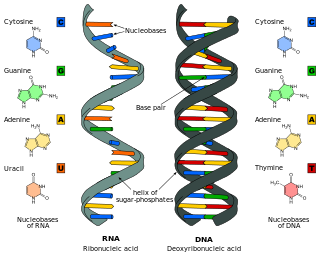
Nucleic acids are biopolymers, macromolecules, essential to all known forms of life. They are composed of nucleotides, which are the monomers made of three components: a 5-carbon sugar, a phosphate group and a nitrogenous base. The two main classes of nucleic acids are deoxyribonucleic acid (DNA) and ribonucleic acid (RNA). If the sugar is ribose, the polymer is RNA; if the sugar is the ribose derivative deoxyribose, the polymer is DNA.

<span class="mw-page-title-main">Nucleotide</span> Biological molecules that form the building blocks of nucleic acids

Nucleotides are organic molecules consisting of a nucleoside and a phosphate. They serve as monomeric units of the nucleic acid polymers – deoxyribonucleic acid (DNA) and ribonucleic acid (RNA), both of which are essential biomolecules within all life-forms on Earth. Nucleotides are obtained in the diet and are also synthesized from common nutrients by the liver.

<span class="mw-page-title-main">Thymine</span> Chemical compound of DNA

Thymine is one of the four nucleobases in the nucleic acid of DNA that are represented by the letters G–C–A–T. The others are adenine, guanine, and cytosine. Thymine is also known as 5-methyluracil, a pyrimidine nucleobase. In RNA, thymine is replaced by the nucleobase uracil. Thymine was first isolated in 1893 by Albrecht Kossel and Albert Neumann from calf thymus glands, hence its name.

<span class="mw-page-title-main">Thymidine</span> Chemical compound

Thymidine, also known as deoxythymidine, deoxyribosylthymine, or thymine deoxyriboside, is a pyrimidine deoxynucleoside. Deoxythymidine is the DNA nucleoside T, which pairs with deoxyadenosine (A) in double-stranded DNA. In cell biology it is used to synchronize the cells in G1/early S phase. The prefix deoxy- is often left out since there are no precursors of thymine nucleotides involved in RNA synthesis.

<span class="mw-page-title-main">Ribonucleotide</span> Nucleotide containing ribose as its pentose component

In biochemistry, a ribonucleotide is a nucleotide containing ribose as its pentose component. It is considered a molecular precursor of nucleic acids. Nucleotides are the basic building blocks of DNA and RNA. Ribonucleotides themselves are basic monomeric building blocks for RNA. Deoxyribonucleotides, formed by reducing ribonucleotides with the enzyme ribonucleotide reductase (RNR), are essential building blocks for DNA. There are several differences between DNA deoxyribonucleotides and RNA ribonucleotides. Successive nucleotides are linked together via phosphodiester bonds.

<span class="mw-page-title-main">Phosphatase</span> Enzyme which catalyzes the removal of a phosphate group from a molecule

In biochemistry, a phosphatase is an enzyme that uses water to cleave a phosphoric acid monoester into a phosphate ion and an alcohol. Because a phosphatase enzyme catalyzes the hydrolysis of its substrate, it is a subcategory of hydrolases. Phosphatase enzymes are essential to many biological functions, because phosphorylation and dephosphorylation serve diverse roles in cellular regulation and signaling. Whereas phosphatases remove phosphate groups from molecules, kinases catalyze the transfer of phosphate groups to molecules from ATP. Together, kinases and phosphatases direct a form of post-translational modification that is essential to the cell's regulatory network.

A salvage pathway is a pathway in which a biological product is produced from intermediates in the degradative pathway of its own or a similar substance. The term often refers to nucleotide salvage in particular, in which nucleotides are synthesized from intermediates in their degradative pathway.

A nucleoside triphosphate is a nucleotide containing a nitrogenous base bound to a 5-carbon sugar, with three phosphate groups bound to the sugar. They are the molecular precursors of both DNA and RNA, which are chains of nucleotides made through the processes of DNA replication and transcription. Nucleoside triphosphates also serve as a source of energy for cellular reactions and are involved in signalling pathways.

<span class="mw-page-title-main">Purine nucleoside phosphorylase</span> Enzyme

Purine nucleoside phosphorylase, PNP, PNPase or inosine phosphorylase is an enzyme that in humans is encoded by the NP gene. It catalyzes the chemical reaction

A nucleotidase is a hydrolytic enzyme that catalyzes the hydrolysis of a nucleotide into a nucleoside and a phosphate.

<span class="mw-page-title-main">Nucleic acid metabolism</span> Process

Nucleic acid metabolism is the process by which nucleic acids are synthesized and degraded. Nucleic acids are the polymers of nucleotides. Nucleotide synthesis is an anabolic mechanism generally involving the chemical reaction of phosphate, pentose sugar, and a nitrogenous base. Destruction of nucleic acid is a catabolic reaction. Additionally, parts of the nucleotides or nucleobases can be salvaged to recreate new nucleotides. Both synthesis and degradation reactions require enzymes to facilitate the event. Defects or deficiencies in these enzymes can lead to a variety of diseases.

<span class="mw-page-title-main">Deoxycytidine kinase</span>

Deoxycytidine kinase (dCK) is an enzyme which is encoded by the DCK gene in humans. dCK predominantly phosphorylates deoxycytidine (dC) and converts dC into deoxycytidine monophosphate. dCK catalyzes one of the initial steps in the nucleoside salvage pathway and has the potential to phosphorylate other preformed nucleosides, specifically deoxyadenosine (dA) and deoxyguanosine (dG), and convert them into their monophosphate forms. There has been recent biomedical research interest in investigating dCK's potential as a therapeutic target for different types of cancer.

dUTP diphosphatase Enzyme

In Enzymology, a dUTP diphosphatase (EC 3.6.1.23) is an enzyme that catalyzes the chemical reaction

<span class="mw-page-title-main">Thymidine phosphorylase</span>

Thymidine phosphorylase is an enzyme that is encoded by the TYMP gene and catalyzes the reaction:

<span class="mw-page-title-main">Uracil-DNA glycosylase</span> Enzyme that repairs DNA damage

Uracil-DNA glycosylase is also known as UNG or UDG. Its most important function is to prevent mutagenesis by eliminating uracil from DNA molecules by cleaving the N-glycosidic bond and initiating the base-excision repair (BER) pathway.

<span class="mw-page-title-main">DGUOK</span> Protein-coding gene in the species Homo sapiens

Deoxyguanosine kinase, mitochondrial is an enzyme that in humans is encoded by the DGUOK gene.

<span class="mw-page-title-main">NT5C3</span>

Cytosolic 5'-nucleotidase 3 (NTC53), also known as cytosolic 5'-nucleotidase 3A, pyrimidine 5’-nucleotidase, and p56, is an enzyme that in humans is encoded by the NT5C3, or NT5C3A, gene on chromosome 7.

<span class="mw-page-title-main">NT5C</span> Protein-coding gene in the species Homo sapiens

5', 3'-nucleotidase, cytosolic, also known as 5'(3')-deoxyribonucleotidase, cytosolic type (cdN) or deoxy-5'-nucleotidase 1 (dNT-1), is an enzyme that in humans is encoded by the NT5C gene on chromosome 17.

<span class="mw-page-title-main">SUCLG1</span>

Succinyl-CoA ligase [GDP-forming] subunit alpha, mitochondrial is an enzyme that in humans is encoded by the SUCLG1 gene.

<span class="mw-page-title-main">NT5C1A</span> Protein-coding gene in the species Homo sapiens

5'-nucleotidase, cytosolic IA is a protein that in humans is encoded by the NT5C1A gene.

References

  1. 1 2 3 GRCh38: Ensembl release 89: ENSG00000205309 - Ensembl, May 2017
  2. 1 2 3 GRCm38: Ensembl release 89: ENSMUSG00000032615 - Ensembl, May 2017
  3. "Human PubMed Reference:". National Center for Biotechnology Information, U.S. National Library of Medicine.
  4. "Mouse PubMed Reference:". National Center for Biotechnology Information, U.S. National Library of Medicine.
  5. "UniProtKB: Q9NPB1 (NT5M_HUMAN)".
  6. "Entrez Gene: NT5M 5',3'-nucleotidase, mitochondrial".
  7. 1 2 3 4 5 6 7 Rampazzo C, Gallinaro L, Milanesi E, Frigimelica E, Reichard P, Bianchi V (Jul 2000). "A deoxyribonucleotidase in mitochondria: involvement in regulation of dNTP pools and possible link to genetic disease". Proceedings of the National Academy of Sciences of the United States of America. 97 (15): 8239–44. Bibcode:2000PNAS...97.8239R. doi: 10.1073/pnas.97.15.8239 . PMC   26931 . PMID   10899995.
  8. 1 2 3 4 5 6 7 8 9 10 Rinaldo-Matthis A, Rampazzo C, Reichard P, Bianchi V, Nordlund P (Oct 2002). "Crystal structure of a human mitochondrial deoxyribonucleotidase". Nature Structural Biology. 9 (10): 779–87. doi:10.1038/nsb846. PMID   12352955. S2CID   29533643.
  9. 1 2 3 4 5 Walldén K, Ruzzenente B, Rinaldo-Matthis A, Bianchi V, Nordlund P (Jul 2005). "Structural basis for substrate specificity of the human mitochondrial deoxyribonucleotidase". Structure. 13 (7): 1081–8. doi: 10.1016/j.str.2005.04.023 . PMID   16004879.
  10. 1 2 3 Gallinaro L, Crovatto K, Rampazzo C, Pontarin G, Ferraro P, Milanesi E, Reichard P, Bianchi V (Sep 2002). "Human mitochondrial 5'-deoxyribonucleotidase. Overproduction in cultured cells and functional aspects". The Journal of Biological Chemistry. 277 (38): 35080–7. doi: 10.1074/jbc.M203755200 . PMID   12124385.