Related Research Articles

Tylopilus felleus, commonly known as the bitter bolete or the bitter tylopilus, is a fungus of the bolete family. Its distribution includes east Asia, Europe, and eastern North America, extending south into Mexico and Central America. A mycorrhizal species, it grows in deciduous and coniferous woodland, often fruiting under beech and oak. Its fruit bodies have convex to flat caps that are some shade of brown, buff, or tan, and typically measure up to 15 cm (6 in) in diameter. The pore surface is initially white before turning pinkish with age. Like most boletes it lacks a ring, and it may be distinguished from Boletus edulis and other similar species by its unusual pink pores and the prominent dark brown netlike pattern on its stalk.
Mycobacterium arupense is a slowly growing mycobacterium first isolated from soil and human sputum samples in Spain. Etymology: arupense, pertaining to the ARUP Institute for Clinical and Experimental Pathology, where the type strain was characterized.
Mycobacterium kubicae is a Gram-positive, nonmotile and acid-fast bacterial species. Cells are typically rod-shaped, with some coccoid forms. Colonies of M. kubicae on solid media are generally smooth and domed, with a yellow scotochromogenic pigment. On Löwenstein-Jensen media they appear film-like. This species is not known to be pathogenic to humans. The species is named after American mycobacteriologist George Kubica.

Photobacterium profundum is a deep sea Gammaproteobacterium, belonging to the family Vibrionaceae and genus Photobacterium. Like other members of this genus, P. profundum is a marine organism and has two circular chromosomes. P. profundum is a gram-negative rod with the ability for growth at temperatures from 0 °C to 25 °C and pressures from 0.1 MPa to 70 MPa depending on the strain. It has a requirement for salt, is able to metabolise a wide range of simple and complex carbohydrates and has two flagella systems. Cells are rod shape, 2-4μm long and 0.8-1.0μm wide, with a single unsheathed flagella. This bacterium was originally isolated in 1986 from the Sulu Sea and there are currently 4 cultured wild-type strains of P. profundum,.
Herminiimonas glaciei is a species of ultramicrobacterium in the family Oxalobacteraceae. These small gram-negative cells have a variable number of long flagella at the ends and sides of their rod-shaped bodies. With dimensions of 0.5–0.9 by 0.3–0.4 µm, H. glaciei is roughly 10 to 50 times smaller than Escherichia coli. Discovered in 2009, the species was isolated from 120,000 years old glacial ice, 3,042 metres (1.9 mi) deep, from Greenland. It was revived after a long-term incubation—seven months of oxygen-free growth at 2 °C, followed by growth on agar plates at 5 °C for almost five months. DNA sequence analysis suggests that with a sequence similarity of 99.6%, H. glaciei is most closely related to H. saxobsidens, a species originally isolated from lichen-colonized rock. Loveland-Curtze, head of the team of scientists from Pennsylvania State University who found the species, speculates that it may offer insight into the existence of organisms in extraterrestrial habitats.
Staphylococcus succinus is a Gram-positive coccoid bacterium belonging to the genus Staphylococcus.
Geobacillus thermoglucosidasius is a thermophilic gram-positive bacterium, and a member of the Firmicutes phylum. It was first isolated from soil in Japan in 1983.
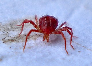
Acaricomes phytoseiuli is a bacterium which is thought to be a pathogen of the mite Phytoseiulus persimilis. A. phytoseiuli causes a set of symptoms in the mite, known as nonresponding syndrome or NR syndrome. Dramatic changes in longevity, fecundity, and behavior are characteristic with this disease. The bacteria accumulate in the lumen of the mite's digestive tract and cause extreme degeneration of its epithelium. Infection with A. phytoseiuli greatly reduces the mite's attraction to herbivore-induced plant volatiles, and the mite is more prone to leave patches with ample prey. The disease is transmitted horizontally by means of feces and debris. The strain that was isolated was “CSC”. Differences between strain CSC compared to its closest phylogenetic neighbors are as follows: CSC uses glucose-1-phosphate and L-glutamic acid, and its colonies are more yellow in appearance as compared to its phylogenetic neighbors which are more cream/white in color.
Armatimonas rosea is a Gram-negative bacterium and also the first species to be characterized within the phylum Armatimonadetes. The Armatimonadetes were previously known as candidate phylum OP10. OP10 was composed solely of environmental 16S rRNA gene clone sequences prior to A. rosea's discovery.
Chthonomonas calidirosea is a Gram-negative bacterium and also the first representative of the new class Chthonomonadetes within the phylum Armatimonadetes. The Armatimonadetes were previously known as candidate phylum OP10. OP10 was composed solely of environmental 16S rRNA gene clone sequences prior to C. calidirosea's relative, Armatimonas rosea's discovery. It is now known that bacterial communities from geothermal environments, are generally constituted by, at least 5–10% of bacteria belonging to Armatimonadetes.
Fimbriimonas ginsengisoli is a Gram-negative bacterium and also the first representative of the new class Fimbriimonadia within the phylum Armatimonadetes. The Armatimonadetes were previously known as candidate phylum OP10. OP10 was composed solely of environmental 16S rRNA gene clone sequences prior to F. ginsengisoli's relative, Armatimonas rosea's discovery.
Bordetella trematum is a species of Gram-negative bacteria identified in 1996 by comparison of 10 strains of B. trematum against other well characterized Bordetella and Alcaligenes species. The term trema refers to something pierced or penetrated, or to a gap. "Trematum" pertains to open things, and refers to the presence of bacteria in wounds and other exposed parts of the body. Strain LMG 13506T is the reference strain for this species.
Rhodoferax is a genus of Betaproteobacteria belonging to the purple nonsulfur bacteriarophic. Originally, Rhodoferax species were included in the genus Rhodocyclus as the Rhodocyclus gelatinous-like group. The genus Rhodoferax was first proposed in 1991 to accommodate the taxonomic and phylogenetic discrepancies arising from its inclusion in the genus Rhodocyclus. Rhodoferax currently comprises four described species: R. fermentans, R. antarcticus, R. ferrireducens, and R. saidenbachensis. R. ferrireducens, lacks the typical phototrophic character common to two other Rhodoferax species. This difference has led researchers to propose the creation of a new genus, Albidoferax, to accommodate this divergent species. The genus name was later corrected to Albidiferax. Based on geno- and phenotypical characteristics, A. ferrireducens was reclassified in the genus Rhodoferax in 2014. R. saidenbachensis, a second non-phototrophic species of the genus Rhodoferax was described by Kaden et al. in 2014.
Desulfurobacterium atlanticum is a thermophilic, anaerobic and chemolithoautotrophic bacterium from the family Aquificaceae. In 2006 it was isolated from marine hydrothermal systems and proposed to become a new bacterial species.
Corynebacterium uropygiale is a bacterium described in 2016 following thorough investigations using a polyphasic approach including MALDI-TOF mass spectrometry, phylogeny of 16S rRNA and rpoB genes and DNA fingerprinting. To date, it has been regarded as endemic to preen gland secretions of healthy turkeys . It is member of the genus Corynebacterium and belongs to the phylum Actinobacteria. Although, a large number of bacteria including Corynebacteria have been reported as part of the normal microbiome of birds, C. uropygiale is the only member of the genus that has been recovered in preen gland secretions of birds. It is one of three bacterial species which have been found to colonize preen gland secretions of birds.
Salisediminibacterium halotolerans is a gram-positive, alkalitolerant, and halophilic bacterium from the family Bacillaceae and genus of Salisediminibacterium, which was one of three bacterial strains, and the only novel species, isolated from sediments from the Xiarinaoer soda lake in Mongolia in 2012.
Propionispira raffinosivorans is a motile, obligate anaerobic, gram-negative bacteria. It was originally isolated from spoiled beer and believed to have some causative effect in beer spoilage. Since then, it has been taxonomically reclassified and proven to play a role in anaerobic beer spoilage, because of its production of acids, such as acetic and propionic acid, during fermentation
Endozoicomonas gorgoniicola is a Gram-negative and facultative anaerobic bacterium from the genus of Endozoicomonas. Individual cells are motile and rod-shaped. Bacteria in this genus are symbionts of coral. E. gorgoniicola live specifically with soft coral and were originally isolated from a species of Plexaura, an octocoral, off the coast of Bimini in the Bahamas. The presence of this bacterium in a coral microbiome is associated with coral health.
Thiosocius is a genus of bacteria that lives in symbiosis with the giant shipworm Kuphus polythalamius. It contains a single species, Thiosocius teredinicola, which was isolated from the gills of the shipworm. The specific name derives from the Latin terms teredo (shipworm) and incola (dweller).
Arthrobacter bussei is a pink-coloured, aerobic, coccus-shaped, Gram-stain-positive, oxidase-positive and catalase-positive bacterium isolated from cheese made of cow´s milk. A. bussei is non-motile and does not form spores. Rod–coccus life cycle is not observed. Cells are 1.1–1.5 µm in diameter. On trypticase soy agar it forms pink-coloured, raised and round colonies, which are 1.0 mm in diameter after 5 days at 30 °C The genome of the strain A. bussei KR32T has been fully sequenced.
References
- 1 2 3 4 5 6 Kuisiene N, Raugalas J, Spröer C, Kroppenstedt RM, Stuknyte M, Chitavichius D (2008). "Paenibacillus tylopili sp.nov., a chitinolytic bacterium isolated from the mycorhizosphere of Tylopilus felleus". Folia Microbiologica. 53 (5): 433–37. doi:10.1007/s12223-008-0066-2. PMID 19085079. S2CID 7997315.

- ↑ de Castro AL, Vollú RE, Peixoto RS, Grigorevski-Lima AL, Coelho RR, Bon EP, Rosado AS, Seldin L (2011). "Cellulolytic potential of a novel strain of Paenibacillus sp. isolated from the armored catfish Parotocinclus maculicauda gut". Brazilian Journal of Microbiology. 42 (4): 1608–15. doi:10.1590/S1517-83822011000400048. PMC 3768713 . PMID 24031795.
