Agar plate

Last updated
Agar plate
Uses Microbiological culture
Art
Related items Petri dish
Growth medium
Contamination on an agar plate Contamination on agar plate.jpg
Contamination on an agar plate

An agar plate is a Petri dish that contains a growth medium solidified with agar, used to culture microorganisms. Sometimes selective compounds are added to influence growth, such as antibiotics. [1]

Contents

96 pinner used to perform spot assays with yeast, fungal or bacterial cells 96pinner.jpg
96 pinner used to perform spot assays with yeast, fungal or bacterial cells

Individual microorganisms placed on the plate will grow into individual colonies, each a clone genetically identical to the individual ancestor organism (except for the low, unavoidable rate of mutation). Thus, the plate can be used either to estimate the concentration of organisms in a liquid culture or a suitable dilution of that culture using a colony counter, or to generate genetically pure cultures from a mixed culture of genetically different organisms.

Several methods are available to plate out cells. One technique is known as "streaking". In this technique, a drop of the culture on the end of a thin, sterile loop of wire, sometimes known as an inoculator, is streaked across the surface of the agar leaving organisms behind, a higher number at the beginning of the streak and a lower number at the end. At some point during a successful "streak", the number of organisms deposited will be such that distinct individual colonies will grow in that area which may be removed for further culturing, using another sterile loop.

Another way of plating organisms, next to streaking, on agar plates is the spot analysis. This type of analysis is often used to check the viability of cells and performed with pinners (often also called froggers). A third used technique is the use of sterile glass beads to plate out cells. In this technique cells are grown in a liquid culture of which a small volume is pipetted on the agar plate and then spread out with the beads. Replica plating is another technique in order to plate out cells on agar plates. These four techniques are the most common, but others are also possible. It is crucial to work in a sterile manner in order to prevent contamination on the agar plates. [1] Plating is thus often done in a laminar flow cabinet or on the working bench next to a bunsen burner. [2]

History

In 1881, Fanny Hesse, who was working as a technician for her husband Walther Hesse in the laboratory of Robert Koch, suggested agar as an effective setting agent, since it had been commonplace in jam making for some time. [3]

Types

An agar plate being viewed in an electronic colony counter Quintote colony counter.jpg
An agar plate being viewed in an electronic colony counter
An agar culture of E. coli colonies Ecoli colonies.png
An agar culture of E. coli colonies
Example of a workup algorithm of possible bacterial infection in cases with no specifically requested targets (non-bacteria, mycobacteria etc.), with most common situations and agents seen in a New England community hospital setting. Different agar plates are used for different specimen sources as seen in upper left quadrant. Diagnostic algorithm of possible bacterial infection.png
Example of a workup algorithm of possible bacterial infection in cases with no specifically requested targets (non-bacteria, mycobacteria etc.), with most common situations and agents seen in a New England community hospital setting. Different agar plates are used for different specimen sources as seen in upper left quadrant.

Like other growth media, the formulations of agar used in plates may be classified as either "defined" or "undefined"; a defined medium is synthesized from individual chemicals required by the organism so the exact molecular composition is known, whereas an undefined medium is made from natural products such as yeast extract, where the precise composition is unknown. [4]

Agar plates may be formulated as either permissive, with the intent of allowing the growth of whatever organisms are present, or restrictive or selective, with the intent of only allowing growth a particular subset of those organisms. [5] This may take the form of a nutritional requirement, for instance providing a particular compound such as lactose as the only source of carbon and thereby selecting only organisms which can metabolize that compound, or by including a particular antibiotic or other substance to select only organisms which are resistant to that substance. This correlates to some degree with defined and undefined media; undefined media, made from natural products and containing an unknown combination of very many organic molecules, is typically more permissive in terms of supplying the needs of a wider variety of organisms, while defined media can be precisely tailored to select organisms with specific properties.

Agar plates may also be indicator plates, in which the organisms are not selected on the basis of growth, but are instead distinguished by a color change in some colonies, typically caused by the action of an enzyme on some compound added to the medium. [6]

The plates are incubated for 12 hours up to several days depending on the test that is performed.

Commonly used types of agar plates include:

Red blood cells on an agar plate are used to diagnose infection. On the left is a positive Staphylococcus infection, on the right a positive Streptococcus culture. Agarplate redbloodcells edit.jpg
Red blood cells on an agar plate are used to diagnose infection. On the left is a positive Staphylococcus infection, on the right a positive Streptococcus culture.

Blood agar

Hemolyses of Streptococcus spp. (left) a-hemolysis (S. mitis); (middle) b-hemolysis (S. pyogenes); (right) g-hemolysis (= nonhemolytic, S. salivarius) Streptococcal hemolysis.jpg
Hemolyses of Streptococcus spp. (left) α-hemolysis (S. mitis); (middle) β-hemolysis ( S. pyogenes ); (right) γ-hemolysis (= nonhemolytic, S. salivarius )

Blood agar plate

Blood agar plates (BAPs) contain mammalian blood (usually sheep or horse), typically at a concentration of 5–10%. BAPs are enriched, differential media used to isolate fastidious organisms and detect hemolytic activity. β-Hemolytic activity will show lysis and complete digestion of red blood cell contents surrounding a colony. Examples include Streptococcus haemolyticus. α-Hemolysis will only cause partial lysis of the red blood cells (the cell membrane is left intact) and will appear green or brown, due to the conversion of hemoglobin to methemoglobin. An example of this would be Streptococcus viridans. γ-Hemolysis (or nonhemolytic) is the term referring to a lack of hemolytic activity. [7] BAPs also contain meat extract or yeast extract, tryptone, sodium chloride, and agar. [8]

Chocolate agar

Chocolate agar is a type of blood agar plate in which the blood cells have been lysed by heating the cells to 80 °C. It is used for growing fastidious respiratory bacteria, such as Haemophilus influenzae . Chocolate agar is named for its color, and no chocolate is actually contained in the plate.

Horse blood agar

Horse blood agar is a type of blood-enriched microbiological culture media. As it is enriched, it allows the growth of certain fastidious bacteria, and allows indication of haemolytic activity in these bacterial cultures.

Thayer–Martin agar

Thayer–Martin agar is a chocolate agar designed to isolate Neisseria gonorrhoeae and Neisseria meningitidis .

Thiosulfate–citrate–bile salts–sucrose agar

Thiosulfate–citrate–bile salts–sucrose agar enhances growth of Vibrio spp., including Vibrio cholerae . [9]

General bacterial media

Four types of agar plate demonstrating differential growth depending on bacterial metabolism Agarplates.jpg
Four types of agar plate demonstrating differential growth depending on bacterial metabolism

Fungal media

Moss media

Yeast media

Mega Plate

the yeast Candida albicans growing both as yeast cells and filamentous cells on YPD agar Candida albicans growing as yeast cells and filamentous (hypha) cells.jpg
the yeast Candida albicans growing both as yeast cells and filamentous cells on YPD agar

See also

Different specific types of agar:

Related Research Articles

<span class="mw-page-title-main">Microbiological culture</span> Method of allowing microorganisms to multiply in a controlled medium

A microbiological culture, or microbial culture, is a method of multiplying microbial organisms by letting them reproduce in predetermined culture medium under controlled laboratory conditions. Microbial cultures are foundational and basic diagnostic methods used as research tools in molecular biology.

<span class="mw-page-title-main">Bacteriological water analysis</span>

Bacteriological water analysis is a method of analysing water to estimate the numbers of bacteria present and, if needed, to find out what sort of bacteria they are. It represents one aspect of water quality. It is a microbiological analytical procedure which uses samples of water and from these samples determines the concentration of bacteria. It is then possible to draw inferences about the suitability of the water for use from these concentrations. This process is used, for example, to routinely confirm that water is safe for human consumption or that bathing and recreational waters are safe to use.

<span class="mw-page-title-main">Blood culture</span> Test to detect bloodstream infections

A blood culture is a medical laboratory test used to detect bacteria or fungi in a person's blood. Under normal conditions, the blood does not contain microorganisms: their presence can indicate a bloodstream infection such as bacteremia or fungemia, which in severe cases may result in sepsis. By culturing the blood, microbes can be identified and tested for resistance to antimicrobial drugs, which allows clinicians to provide an effective treatment.

<span class="mw-page-title-main">Coliform bacteria</span> Group of bacterial species

Coliform bacteria are defined as either motile or non-motile Gram-negative non-spore forming bacilli that possess β-galactosidase to produce acids and gases under their optimal growth temperature of 35–37 °C. They can be aerobes or facultative aerobes, and are a commonly used indicator of low sanitary quality of foods, milk, and water. Coliforms can be found in the aquatic environment, in soil and on vegetation; they are universally present in large numbers in the feces of warm-blooded animals as they are known to inhabit the gastrointestinal system. While coliform bacteria are not normally causes of serious illness, they are easy to culture, and their presence is used to infer that other pathogenic organisms of fecal origin may be present in a sample, or that said sample is not safe to consume. Such pathogens include disease-causing bacteria, viruses, or protozoa and many multicellular parasites. Every drinking water source must be tested for the presence of these total coliform bacteria.

<span class="mw-page-title-main">Eosin methylene blue</span> Biological stain

Eosin methylene blue is a selective and differential media used for the identification of Gram-negative bacteria, specifically the Enterobacteriaceae. EMB inhibits the growth of most Gram-positive bacteria. EMB is often used to confirm the presence of coliforms in a sample. It contains two dyes, eosin and methylene blue in the ratio of 6:1. EMB is a differential microbiological media, which inhibits the growth of Gram-positive bacteria and differentiates bacteria that ferment lactose from those that do not. Organisms that ferment lactose appear dark/black or green often with "nucleated colonies"—colonies with dark centers. Organisms that do not ferment lactose will appear pink and often mucoid.

<span class="mw-page-title-main">Growth medium</span> Solid, liquid or gel used to grow microorganisms or cells

A growth medium or culture medium is a solid, liquid, or semi-solid designed to support the growth of a population of microorganisms or cells via the process of cell proliferation or small plants like the moss Physcomitrella patens. Different types of media are used for growing different types of cells.

<span class="mw-page-title-main">MacConkey agar</span> Differential media

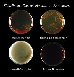
MacConkey agar is a selective and differential culture medium for bacteria. It is designed to selectively isolate Gram-negative and enteric bacteria and differentiate them based on lactose fermentation. Lactose fermenters turn red or pink on MacConkey agar, and nonfermenters do not change color. The media inhibits growth of Gram-positive organisms with crystal violet and bile salts, allowing for the selection and isolation of gram-negative bacteria. The media detects lactose fermentation by enteric bacteria with the pH indicator neutral red.

<i>Shigella dysenteriae</i> Bacterial species

Shigella dysenteriae is a species of the rod-shaped bacterial genus Shigella. Shigella species can cause shigellosis. Shigellae are Gram-negative, non-spore-forming, facultatively anaerobic, nonmotile bacteria. S. dysenteriae has the ability to invade and replicate in various species of epithelial cells and enterocytes.

Bacillary dysentery is a type of dysentery, and is a severe form of shigellosis. It is associated with species of bacteria from the family Enterobacteriaceae. The term is usually restricted to Shigella infections.

<span class="mw-page-title-main">XLD agar</span> Selective culture medium

Xylose Lysine Deoxycholate agar is a selective growth medium used in the isolation of Salmonella and Shigella species from clinical samples and from food. The agar was developed by Welton Taylor in 1965. It has a pH of approximately 7.4, leaving it with a bright pink or red appearance due to the indicator phenol red. Sugar fermentation lowers the pH and the phenol red indicator registers this by changing to yellow. Most gut bacteria, including Salmonella, can ferment the sugar xylose to produce acid; Shigella colonies cannot do this and therefore remain red. After exhausting the xylose supply Salmonella colonies will decarboxylate lysine, increasing the pH once again to alkaline and mimicking the red Shigella colonies. Salmonellae metabolise thiosulfate to produce hydrogen sulfide, which leads to the formation of colonies with black centers and allows them to be differentiated from the similarly coloured Shigella colonies.

<span class="mw-page-title-main">Streaking (microbiology)</span> Method for isolation of bacterial strains

In microbiology, streaking is a technique used to isolate a pure strain from a single species of microorganism, often bacteria. Samples can then be taken from the resulting colonies and a microbiological culture can be grown on a new plate so that the organism can be identified, studied, or tested.

<span class="mw-page-title-main">Mannitol salt agar</span> Culture medium used in microbiology

Mannitol salt agar or MSA is a commonly used selective and differential growth medium in microbiology. It encourages the growth of a group of certain bacteria while inhibiting the growth of others. It contains a high concentration of salt (NaCl) which is inhibitory to most bacteria - making MSA selective against most Gram-negative and selective for some Gram-positive bacteria that tolerate high salt concentrations. It is also a differential medium for mannitol-fermenting staphylococci, containing the sugar alcohol mannitol and the indicator phenol red, a pH indicator for detecting acid produced by mannitol-fermenting staphylococci. Staphylococcus aureus produces yellow colonies with yellow zones, whereas other coagulase-negative staphylococci produce small pink or red colonies with no colour change to the medium. If an organism can ferment mannitol, an acidic byproduct is formed that causes the phenol red in the agar to turn yellow. It is used for the selective isolation of presumptive pathogenic (pp) Staphylococcus species.

<span class="mw-page-title-main">Thayer–Martin agar</span>

Thayer–Martin agar is a Mueller–Hinton agar with 5% chocolate sheep blood and antibiotics. It is used for culturing and primarily isolating pathogenic Neisseria bacteria, including Neisseria gonorrhoeae and Neisseria meningitidis, as the medium inhibits the growth of most other microorganisms. When growing Neisseria meningitidis, one usually starts with a normally sterile body fluid, so a plain chocolate agar is used. Thayer–Martin agar was initially developed in 1964, with an improved formulation published in 1966.

<span class="mw-page-title-main">Pathogenic bacteria</span> Disease-causing bacteria

Pathogenic bacteria are bacteria that can cause disease. This article focuses on the bacteria that are pathogenic to humans. Most species of bacteria are harmless and are often beneficial but others can cause infectious diseases. The number of these pathogenic species in humans is estimated to be fewer than a hundred. By contrast, several thousand species are part of the gut flora present in the digestive tract.

<span class="mw-page-title-main">Hektoen enteric agar</span> Selective and differential agar

Hektoen enteric agar is a selective and differential agar primarily used to recover Salmonella and Shigella from patient specimens. HEA contains indicators of lactose fermentation and hydrogen sulfide production; as well as inhibitors to prevent the growth of Gram-positive bacteria. It is named after the Hektoen Institute in Chicago, where researchers developed the agar.

<span class="mw-page-title-main">Mueller–Hinton agar</span> Culture medium used in microbiology

Mueller Hinton agar is a type of growth medium used in microbiology to culture bacterial isolates and test their susceptibility to antibiotics. This medium was first developed in 1941 by John Howard Mueller and Jane Hinton, who were microbiologists working at Harvard University. However, Mueller Hinton agar is made up of a couple of components, including beef extract, acid hydrolysate of casein, and starch, as well as agar to solidify the mixture. The composition of Mueller Hinton agar can vary depending on the manufacturer and the intended use, but the medium is generally nutrient-rich and free of inhibitors that could interfere with bacterial growth.                                

<span class="mw-page-title-main">New York City agar</span>

The NYC medium or GC medium agar is used for isolating Gonococci.

<span class="mw-page-title-main">Granada medium</span>

Granada medium is a selective and differential culture medium designed to selectively isolate Streptococcus agalactiae and differentiate it from other microorganisms. Granada Medium was developed by Manuel Rosa-Fraile et al. at the Service of Microbiology in the Hospital Virgen de las Nieves in Granada (Spain).

In microbiology, the term isolation refers to the separation of a strain from a natural, mixed population of living microbes, as present in the environment, for example in water or soil, or from living beings with skin flora, oral flora or gut flora, in order to identify the microbe(s) of interest. Historically, the laboratory techniques of isolation first developed in the field of bacteriology and parasitology, before those in virology during the 20th century.

Diagnostic microbiology is the study of microbial identification. Since the discovery of the germ theory of disease, scientists have been finding ways to harvest specific organisms. Using methods such as differential media or genome sequencing, physicians and scientists can observe novel functions in organisms for more effective and accurate diagnosis of organisms. Methods used in diagnostic microbiology are often used to take advantage of a particular difference in organisms and attain information about what species it can be identified as, which is often through a reference of previous studies. New studies provide information that others can reference so that scientists can attain a basic understanding of the organism they are examining.

References

  1. 1 2 Madigan M, Martinko J, eds. (2005). Brock Biology of Microorganisms (11th ed.). Prentice Hall. ISBN   0-13-144329-1.
  2. Sanders, Erin R. (11 May 2012). "Aseptic Laboratory Techniques: Plating Methods". Journal of Visualized Experiments (63): e3064. doi:10.3791/3064. PMC   4846335 . PMID   22617405. Archived from the original on 14 November 2017. Retrieved 3 May 2018.
  3. "History of the agar plate". Laboratory News . Archived from the original on 11 February 2010. Retrieved 2010-02-22.
  4. Baron S; et al., eds. (1996). Baron's Medical Microbiology (4th ed.). University of Texas Medical Branch. ISBN   0-9631172-1-1. (via NCBI Bookshelf).
  5. Ryan KJ; Ray CG, eds. (2004). Sherris Medical Microbiology (4th ed.). McGraw Hill. ISBN   0-8385-8529-9.
  6. "Indicator Plates" . Retrieved 12 July 2018.
  7. "Blood Agar Plates and Hemolysis Protocols". Archived from the original on 2012-02-02. Retrieved 2014-10-28.
  8. "Blood Agar- Composition, Preparation, Uses and Pictures", Microbiology Info.com
  9. 1 2 Fisher, Bruce; Harvey, Richard P.; Champe, Pamela C. (2007). Lippincott's Illustrated Reviews: Microbiology (Lippincott's Illustrated Reviews Series). Hagerstwon, MD: Lippincott Williams & Wilkins. ISBN   978-0-7817-8215-9.
  10. Miller, J. H. (1972). Experiments in molecular genetics. Cold Spring Harbor Laboratory, Cold Spring Harbor, New York.
  11. Jung, Benjamin; Hoilat, Gilles J. (2022), "MacConkey Medium", StatPearls, Treasure Island (FL): StatPearls Publishing, PMID   32491326 , retrieved 2022-12-12
  12. Reski, Ralf; Abel, Wolfgang O. (1985). "Induction of budding on chloronemata and caulonemata of the moss, Physcomitrella patens, using isopentenyladenine". Planta. 165 (3): 354–358. doi:10.1007/bf00392232. PMID   24241140. S2CID   11363119.
  13. "A cinematic approach to drug resistance". Harvard Gazette. 2016-09-08. Retrieved 2021-04-08.