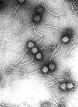
Viral culture is a laboratory technique [1] in which samples of a virus are placed to different cell lines which the virus being tested for its ability to infect. If the cells show changes, known as cytopathic effects, then the culture is positive. [2]
Traditional viral culture has been generally superseded by shell vial culture, in which the sample is centrifuged onto a single layer of cells and viral growth is measured by antigen detection methods. This greatly reduces the time to detection for slow growing viruses such as cytomegalovirus, for which the method was developed. [3] In addition, the centrifugation step in shell vial culture enhances the sensitivity of this method because after centrifugation, the viral particles of the sample are in close proximity to the cells.
Human and monkey cells are used in both traditional viral culture and shell vial culture.
Human virus types that can be identified by viral culture include adenovirus, cytomegalovirus, enteroviruses, herpes simplex virus, influenza virus, parainfluenza virus, rhinovirus, respiratory syncytial virus, varicella zoster virus, measles and mumps. [4] For these, the final identification method is generally by immunofluorescence, with exception of cytomegalovirus and rhinovirus, whose identification in a viral culture are determined by cytopathic effects. [4]
Preliminary research (i.e. not yet peer-reviewed at the time of writing, 29 September 2020) exploring the potential suitability of viral culture testing of SARS-CoV-2 has been conducted. [5]
Virology is the scientific study of biological viruses. It is a subfield of microbiology that focuses on their detection, structure, classification and evolution, their methods of infection and exploitation of host cells for reproduction, their interaction with host organism physiology and immunity, the diseases they cause, the techniques to isolate and culture them, and their use in research and therapy.

The rhinovirus is a positive-sense, single-stranded RNA virus belonging to the genus Enterovirus in the family Picornaviridae. Rhinovirus is the most common viral infectious agent in humans and is the predominant cause of the common cold.
Viral load, also known as viral burden, is a numerical expression of the quantity of virus in a given volume of fluid, including biological and environmental specimens. It is not to be confused with viral titre or viral titer, which depends on the assay. When an assay for measuring the infective virus particle is done, viral titre often refers to the concentration of infectious viral particles, which is different from the total viral particles. Viral load is measured using body fluids sputum and blood plasma. As an example of environmental specimens, the viral load of norovirus can be determined from run-off water on garden produce. Norovirus has not only prolonged viral shedding and has the ability to survive in the environment but a minuscule infectious dose is required to produce infection in humans: less than 100 viral particles.

Picornaviruses are a group of related nonenveloped RNA viruses which infect vertebrates including fish, mammals, and birds. They are viruses that represent a large family of small, positive-sense, single-stranded RNA viruses with a 30 nm icosahedral capsid. The viruses in this family can cause a range of diseases including the common cold, poliomyelitis, meningitis, hepatitis, and paralysis.
A helper dependent virus, also termed a gutless virus, is a synthetic viral vector dependent on the assistance of a helper virus in order to replicate, and can be used for purposes such as gene therapy. Naturally-occurring satellite viruses are also helper virus dependent, and can sometimes be modified to become viral vectors.

Bovine viral diarrhea (BVD), bovine viral diarrhoea or mucosal disease, previously referred to as bovine virus diarrhea (BVD), is an economically significant disease of cattle that is found in the majority of countries throughout the world. Worldwide reviews of the economically assessed production losses and intervention programs incurred by BVD infection have been published. The causative agent, bovine viral diarrhea virus (BVDV), is a member of the genus Pestivirus of the family Flaviviridae.
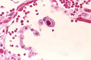
Human betaherpesvirus 5, also called human cytomegalovirus (HCMV), is species of virus in the genus Cytomegalovirus, which in turn is a member of the viral family known as Herpesviridae or herpesviruses. It is also commonly called CMV. Within Herpesviridae, HCMV belongs to the Betaherpesvirinae subfamily, which also includes cytomegaloviruses from other mammals. CMV is a double-stranded DNA virus.

Cytopathic effect or cytopathogenic effect refers to structural changes in host cells that are caused by viral invasion. The infecting virus causes lysis of the host cell or when the cell dies without lysis due to an inability to replicate. Both of these effects occur due to CPEs. If a virus causes these morphological changes in the host cell, it is said to be cytopathogenic. Common examples of CPE include rounding of the infected cell, fusion with adjacent cells to form syncytia, and the appearance of nuclear or cytoplasmic inclusion bodies.

Vincent R. Racaniello is a Higgins Professor in the Department of Microbiology and Immunology at Columbia University's College of Physicians and Surgeons. He is a co-author of a textbook on virology, Principles of Virology.
In the diagnostic laboratory, virus infections can be confirmed by a myriad of methods. Diagnostic virology has changed rapidly due to the advent of molecular techniques and increased clinical sensitivity of serological assays.
Human betaherpesvirus 7 (HHV-7) is one of nine known members of the Herpesviridae family that infects humans. HHV-7 is a member of Betaherpesvirinae, a subfamily of the Herpesviridae that also includes HHV-6 and Cytomegalovirus. HHV-7 often acts together with HHV-6, and the viruses together are sometimes referred to by their genus, Roseolovirus. HHV-7 was first isolated in 1990 from CD4+ T cells taken from peripheral blood lymphocytes.
Neurovirology is an interdisciplinary field which represents a melding of clinical neuroscience, virology, immunology, and molecular biology. The main focus of the field is to study viruses capable of infecting the nervous system. In addition to this, the field studies the use of viruses to trace neuroanatomical pathways, for gene therapy, and to eliminate detrimental populations of neural cells.
Virus quantification is counting or calculating the number of virus particles (virions) in a sample to determine the virus concentration. It is used in both research and development (R&D) in academic and commercial laboratories as well as in production situations where the quantity of virus at various steps is an important variable that must be monitored. For example, the production of virus-based vaccines, recombinant proteins using viral vectors, and viral antigens all require virus quantification to continually monitor and/or modify the process in order to optimize product quality and production yields and to respond to ever changing demands and applications. Other examples of specific instances where viruses need to be quantified include clone screening, multiplicity of infection (MOI) optimization, and adaptation of methods to cell culture.
The plaque reduction neutralization test is used to quantify the titer of neutralizing antibody for a virus.
Avian orthoreovirus, also known as avian reovirus, is an orthoreovirus from the Reoviridae family. Infection causes arthritis and tenosynovitis in poultry. It can also cause respiratory disease.

Indirect immunoperoxidase assay (IPA) is a laboratory technique used to detect and titrate viruses that do not cause measurable cytopathic effects and cannot be measured by classical plaque assays. These viruses include human coronavirus 229E and OC43.
Aichivirus A formerly Aichi virus (AiV) belongs to the genus Kobuvirus in the family Picornaviridae. Six species are apart of the genus Kobuvirus, Aichivirus A-F. Within Aichivirus A, there are six different types including human Aichi virus, canine kobuvirus, murine kobuvirus, Kathmandu sewage kobuvirus, roller kobuvirus, and feline kobuvirus. Three different genotypes are found in human Aichi virus, represented as genotype A, B, and C.
This glossary of virology is a list of definitions of terms and concepts used in virology, the study of viruses, particularly in the description of viruses and their actions. Related fields include microbiology, molecular biology, and genetics.

Nodamura virus (NoV) is a member of the family Nodaviridae, which was originally isolated from mosquitoes in Japan near the village of Nodamura in 1956. Other members of Nodaviridae are flock house virus (FHV) and black beetle virus (BBV). NoV has been found to multiply in several insect and tick species; however, these infected individuals seem to be asymptomatic. Nodamura virus is the only member of the genus Alphanodavirus that can infect insects, fish, and mammals.
In microbiology, the term isolation refers to the separation of a strain from a natural, mixed population of living microbes, as present in the environment, for example in water or soil, or from living beings with skin flora, oral flora or gut flora, in order to identify the microbe(s) of interest. Historically, the laboratory techniques of isolation first developed in the field of bacteriology and parasitology, before those in virology during the 20th century.
| Components | 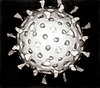 | |
|---|---|---|
| Viral life cycle | ||
| Genetics | ||
| By host | ||
| Other | ||
| | This medical diagnostic article is a stub. You can help Wikipedia by expanding it. |
| | This virus-related article is a stub. You can help Wikipedia by expanding it. |