Laboratory diagnosis of viral infections

Last updated
Laboratory diagnosis of viral infections
Purposetest for viral infection

In the diagnostic laboratory, virus infections can be confirmed by a myriad of methods. Diagnostic virology has changed rapidly due to the advent of molecular techniques and increased clinical sensitivity of serological assays. [1]

Contents

Sampling

A wide variety of samples can be used for virological testing. The type of sample sent to the laboratory often depends on the type of viral infection being diagnosed and the test required. Proper sampling technique is essential to avoid potential pre-analytical errors. For example, different types of samples must be collected in appropriate tubes to maintain the integrity of the sample and stored at appropriate temperatures (usually 4 °C) to preserve the virus and prevent bacterial or fungal growth. Sometimes multiple sites may also be sampled.[ citation needed ]

Types of samples include the following:

For example, a nasal mucus test may be done to diagnose rhinovirus. [2]

Virus isolation

Viruses are often isolated from the initial patient sample. This allows the virus sample to be grown into larger quantities and allows a larger number of tests to be run on them. This is particularly important for samples that contain new or rare viruses for which diagnostic tests are not yet developed.[ citation needed ]

Many viruses can be grown in cell culture in the lab. To do this, the virus sample is mixed with cells, a process called adsorption, after which the cells become infected and produce more copies of the virus. [3] Although different viruses often only grow in certain types of cells, there are cells that support the growth of a large variety of viruses and are a good starting point, for example, the African monkey kidney cell line (Vero cells), human lung fibroblasts (MRC-5), and human epidermoid carcinoma cells (HEp-2). One means of determining whether the cells are successfully replicating the virus is to check for a change in cell morphology or for the presence of cell death using a microscope.

Other viruses may require alternative methods for growth such as the inoculation of embryonated chicken eggs (e.g. avian influenza viruses [4] ) or the intracranial inoculation of virus using newborn mice (e.g. lyssaviruses [5] ).

Nucleic acid based methods

Molecular techniques are the most specific and sensitive diagnostic tests. [6] They are capable of detecting either the whole viral genome or parts of the viral genome. In the past nucleic acid tests have mainly been used as a secondary test to confirm positive serological results. [3] However, as they become cheaper and more automated, they are increasingly becoming the primary tool for diagnostics and can also be use for monitoring of treatment of viral infected individuals t. [3]

Polymerase chain reaction

Detection of viral RNA and DNA genomes can be performed using polymerase chain reaction. This technique makes many copies of the virus genome using virus-specific probes. Variations of PCR such as nested reverse transcriptase PCR and real time PCR can also be used to determine viral loads in patient serum. This is often used to monitor treatment success in HIV cases.[ citation needed ]

Sequencing

Sequencing is the only diagnostic method that will provide the full sequence of a virus genome. Hence, it provides the most information about very small differences between two viruses that would look the same using other diagnostic tests. Currently it is only used when this depth of information is required. For example, sequencing is useful when specific mutations in the patient are tested for in order to determine antiviral therapy and susceptibility to infection. However, as the tests are getting cheaper, faster and more automated, sequencing will likely become the primary diagnostic tool in the future.[ citation needed ]

Microscopy based methods

Immunofluorescence or immunoperoxidase

Immunofluorescence or immunoperoxidase assays are commonly used to detect whether a virus is present in a tissue sample. These tests are based on the principle that if the tissue is infected with a virus, an antibody specific to that virus will be able to bind to it. To do this, antibodies that are specific to different types of viruses are mixed with the tissue sample. After the tissue is exposed to a specific wavelength of light or a chemical that allows the antibody to be visualized.[ citation needed ]

These tests require specialized antibodies that are produced and purchased from commercial companies. These commercial antibodies are usually well characterized and are known to bind to only one specific type of virus. They are also conjugated to a special kind of tag that allows the antibody to be visualized in the lab, i.e.so that it will emit fluorescence or a color. Hence, immunofluorescence refers to the detection of a fluorescent antibody (immuno) and immunoperoxidase refers to the detection of a colored antibody (peroxidase produces a dark brown color).[ citation needed ]

Immunofluorescence assayImmunoperoxidase assay

Electron microscopy

Electron microscopy is a method that can take a picture of a whole virus and can reveal its shape and structure. It is not typically used as a routine diagnostic test as it requires a highly specialized type of sample preparation, microscope and technical expertise. However, electron microscopy is highly versatile due to its ability to analyze any type of sample and identify any type of virus. Therefore, it remains the gold standard for identifying viruses that do not show up on routine diagnostic tests or for which routine tests present conflicting results. [7]

Electron microscopy of the Sapovirus Sappovirus.jpg
Electron microscopy of the Sapovirus

Host antibody detection

A person who has recently been infected by a virus will produce antibodies in their bloodstream that specifically recognize that virus. This is called humoral immunity. Two types of antibodies are important. The first called IgM is highly effective at neutralizing viruses but is only produced by the cells of the immune system for a few weeks. The second, called, IgG is produced indefinitely. Therefore, the presence of IgM in the blood of the host is used to test for acute infection, whereas IgG indicates an infection sometime in the past. [8] Both types of antibodies are measured when tests for immunity are carried out. [9]

Antibody testing has become widely available. It can be done for individual viruses (e.g. using an ELISA assay) but automated panels that can screen for many viruses at once are becoming increasingly common.[ citation needed ]

Hemagglutination assay

Some viruses attach to molecules present on the surface of red blood cells, for example, influenza virus. [10] A consequence of this is that – at certain concentrations – a viral suspension may bind together (agglutinate) the red blood cells thus preventing them from settling out of suspension.[ citation needed ]

See also

Related Research Articles

<span class="mw-page-title-main">Virology</span> Study of viruses

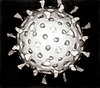
Virology is the scientific study of biological viruses. It is a subfield of microbiology that focuses on their detection, structure, classification and evolution, their methods of infection and exploitation of host cells for reproduction, their interaction with host organism physiology and immunity, the diseases they cause, the techniques to isolate and culture them, and their use in research and therapy.

<span class="mw-page-title-main">Diagnosis of HIV/AIDS</span> Immunological test

HIV tests are used to detect the presence of the human immunodeficiency virus (HIV), the virus that causes acquired immunodeficiency syndrome (AIDS), in serum, saliva, or urine. Such tests may detect antibodies, antigens, or RNA.

<span class="mw-page-title-main">Seroconversion</span> Development of specific antibodies in the blood serum as a result of infection or immunization

In immunology, seroconversion is the development of specific antibodies in the blood serum as a result of infection or immunization, including vaccination. During infection or immunization, antigens enter the blood, and the immune system begins to produce antibodies in response. Before seroconversion, the antigen itself may or may not be detectable, but the antibody is absent. During seroconversion, the antibody is present but not yet detectable. After seroconversion, the antibody is detectable by standard techniques and remains detectable unless the individual seroreverts, in a phenomenon called seroreversion, or loss of antibody detectability, which can occur due to weakening of the immune system or decreasing antibody concentrations over time. Seroconversion refers the production of specific antibodies against specific antigens, meaning that a single infection could cause multiple waves of seroconversion against different antigens. Similarly, a single antigen could cause multiple waves of seroconversion with different classes of antibodies. For example, most antigens prompt seroconversion for the IgM class of antibodies first, and subsequently the IgG class.

Serology is the scientific study of serum and other body fluids. In practice, the term usually refers to the diagnostic identification of antibodies in the serum. Such antibodies are typically formed in response to an infection, against other foreign proteins, or to one's own proteins. In either case, the procedure is simple.

<span class="mw-page-title-main">Hemagglutination assay</span> Measure of virus or bacteria concentration

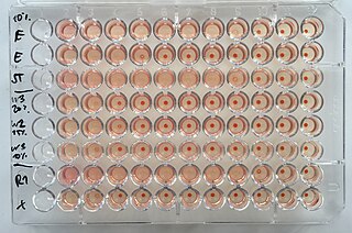
The hemagglutination assay or haemagglutination assay (HA) and the hemagglutination inhibition assay were developed in 1941–42 by American virologist George Hirst as methods for quantifying the relative concentration of viruses, bacteria, or antibodies.

Heterophile antibodies are antibodies induced by external antigens.

<i>Influenza C virus</i> Genus of viruses in the family Orthomyxoviridae

Influenza C virus is the only species in the genus Gammainfluenzavirus, in the virus family Orthomyxoviridae, which like other influenza viruses, causes influenza.

Porcine parvovirus (PPV), a virus in the species Ungulate protoparvovirus 1 of genus Protoparvovirus in the virus family Parvoviridae, causes reproductive failure of swine characterized by embryonic and fetal infection and death, usually in the absence of outward maternal clinical signs. The disease develops mainly when seronegative dams are exposed oronasally to the virus anytime during about the first half of gestation, and conceptuses are subsequently infected transplacentally before they become immunocompetent. There is no definitive evidence that infection of swine other than during gestation is of any clinical or economic significance. The virus is ubiquitous among swine throughout the world and is enzootic in most herds that have been tested. Diagnostic surveys have indicated that PPV is the major infectious cause of embryonic and fetal death. In addition to its direct causal role in reproductive failure, PPV can potentiate the effects of porcine circovirus type II (PCV2) infection in the clinical course of postweaning multisystemic wasting syndrome (PMWS).

<span class="mw-page-title-main">Psittacine beak and feather disease</span> Viral disease affecting parrots

Psittacine beak and feather disease (PBFD) is a viral disease affecting all Old World and New World parrots. The causative virus—beak and feather disease virus (BFDV)—belongs to the taxonomic genus Circovirus, family Circoviridae. It attacks the feather follicles and the beak and claw matrices of the bird, causing progressive feather, claw and beak malformation and necrosis. In later stages of the disease, feather shaft constriction occurs, hampering development until eventually all feather growth stops. It occurs in an acutely fatal form and a chronic form.

<span class="mw-page-title-main">Bovine viral diarrhea</span> Significant economic disease of cattle caused by two species of Pestivirus

Bovine viral diarrhea (BVD), bovine viral diarrhoea or mucosal disease, previously referred to as bovine virus diarrhea (BVD), is an economically significant disease of cattle that is found in the majority of countries throughout the world. Worldwide reviews of the economically assessed production losses and intervention programs incurred by BVD infection have been published. The causative agent, bovine viral diarrhea virus (BVDV), is a member of the genus Pestivirus of the family Flaviviridae.

<i>Human betaherpesvirus 5</i> Species of virus

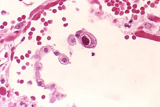
Human betaherpesvirus 5, also called human cytomegalovirus (HCMV), is species of virus in the genus Cytomegalovirus, which in turn is a member of the viral family known as Herpesviridae or herpesviruses. It is also commonly called CMV. Within Herpesviridae, HCMV belongs to the Betaherpesvirinae subfamily, which also includes cytomegaloviruses from other mammals. CMV is a double-stranded DNA virus.

Bovine leukemia virus (BLV) is a retrovirus which causes enzootic bovine leukosis in cattle. It is closely related to the human T‑lymphotropic virus type 1 (HTLV-I). BLV may integrate into the genomic DNA of B‑lymphocytes as a DNA intermediate, or exist as unintegrated circular or linear forms. Besides structural and enzymatic genes required for virion production, BLV expresses the Tax protein and microRNAs involved in cell proliferation and oncogenesis. In cattle, most infected animals are asymptomatic; leukemia is rare, but lymphoproliferation is more frequent (30%).

<span class="mw-page-title-main">Influenza</span> Infectious disease, often just "the flu"

Influenza, commonly known as "the flu", is an infectious disease caused by influenza viruses. Symptoms range from mild to severe and often include fever, runny nose, sore throat, muscle pain, headache, coughing, and fatigue. These symptoms begin from one to four days after exposure to the virus and last for about 2–8 days. Diarrhea and vomiting can occur, particularly in children. Influenza may progress to pneumonia, which can be caused by the virus or by a subsequent bacterial infection. Other complications of infection include acute respiratory distress syndrome, meningitis, encephalitis, and worsening of pre-existing health problems such as asthma and cardiovascular disease.

<span class="mw-page-title-main">Hemagglutinin</span>

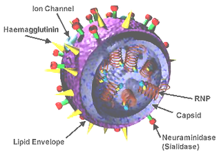
In molecular biology, hemagglutinins are receptor-binding membrane fusion glycoproteins produced by viruses in the Paramyxoviridae and Orthomyxoviridae families. Hemagglutinins are responsible for binding to receptors on red blood cells to initiate viral attachment and infection. The agglutination of red cells occurs when antibodies on one cell bind to those on others, causing amorphous aggregates of clumped cells.

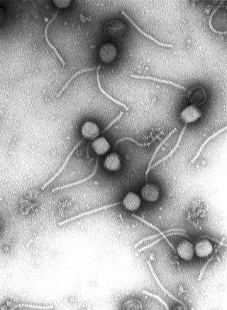
Virus quantification is counting or calculating the number of virus particles (virions) in a sample to determine the virus concentration. It is used in both research and development (R&D) in academic and commercial laboratories as well as in production situations where the quantity of virus at various steps is an important variable that must be monitored. For example, the production of virus-based vaccines, recombinant proteins using viral vectors, and viral antigens all require virus quantification to continually monitor and/or modify the process in order to optimize product quality and production yields and to respond to ever changing demands and applications. Other examples of specific instances where viruses need to be quantified include clone screening, multiplicity of infection (MOI) optimization, and adaptation of methods to cell culture.

The plaque reduction neutralization test is used to quantify the titer of neutralizing antibody for a virus.

Avian orthoreovirus, also known as avian reovirus, is an orthoreovirus from the Reoviridae family. Infection causes arthritis and tenosynovitis in poultry. It can also cause respiratory disease.

<span class="mw-page-title-main">Surround optical-fiber immunoassay</span>

Surround optical-fiber immunoassay (SOFIA) is an ultrasensitive, in vitro diagnostic platform incorporating a surround optical-fiber assembly that captures fluorescence emissions from an entire sample. The technology's defining characteristics are its extremely high limit of detection, sensitivity, and dynamic range. SOFIA's sensitivity is measured at the attogram level (10−18 g), making it about one billion times more sensitive than conventional diagnostic techniques. Based on its enhanced dynamic range, SOFIA is able to discriminate levels of analyte in a sample over 10 orders of magnitude, facilitating accurate titering.

<span class="mw-page-title-main">George Hirst (virologist)</span> American virologist and science administrator

George Keble Hirst, M.D. was an American virologist and science administrator who was among the first to study the molecular biology and genetics of animal viruses, especially influenza virus. He directed the Public Health Research Institute in New York City (1956–1981), and was also the founding editor-in-chief of Virology, the first English-language journal to focus on viruses. He is particularly known for inventing the hemagglutination assay, a simple method for quantifying viruses, and adapting it into the hemagglutination inhibition assay, which measures virus-specific antibodies in serum. He was the first to discover that viruses can contain enzymes, and the first to propose that virus genomes can consist of discontinuous segments. The New York Times described him as "a pioneer in molecular virology."

<i>Influenza D virus</i> Species of virus

Influenza D virus is a species in the virus genus Deltainfluenzavirus, in the family Orthomyxoviridae, that causes influenza.

References

  1. Leland, D. S.; Ginocchio, C. C. (2007). "Role of Cell Culture for Virus Detection in the Age of Technology". Clinical Microbiology Reviews. 20 (1): 49–78. doi:10.1128/CMR.00002-06. PMC   1797634 . PMID   17223623.
  2. Gwaltney JM, Hayden FG (February 1992). "Psychological stress and the common cold". N Engl J Med. 326 (9): 644–5, author reply 645–6. doi:10.1056/NEJM199202273260915. PMID   1310349.
  3. 1 2 3 "Diagnostic Methods in Virology, Virological Methods, Virus Culture, Virus Isolation". virology-online.com. Retrieved 2018-01-03.
  4. Brauer, Rena; Chen, Peter (2015). "Influenza Virus Propagation in Embryonated Chicken Eggs". Journal of Visualized Experiments (97). doi:10.3791/52421. PMC   4401370 . PMID   25867050.
  5. Kuzmin, Ivan V. (2015). "Virus Isolation in Animals". Current Laboratory Techniques in Rabies Diagnosis, Research and Prevention, Volume 2. pp. 13–23. doi:10.1016/B978-0-12-801919-1.00002-6. ISBN   9780128019191.
  6. Dhamad, AE; Abdal Rhida, MA (2020). "COVID-19: molecular and serological detection methods". PeerJ. 8: e10180. doi: 10.7717/peerj.10180 . PMC   7547594 . PMID   33083156.
  7. Hazelton, Paul R.; Gelderblom, Hans R. (2003). "Electron Microscopy for Rapid Diagnosis of Emerging Infectious Agents1". Emerging Infectious Diseases. 9 (3): 294–303. doi:10.3201/eid0903.020327. PMC   2958539 . PMID   12643823.
  8. Greer, Shaun; Alexander, Graeme J.M. (1995). "4 Viral serology and detection". Baillière's Clinical Gastroenterology. 9 (4): 689–721. doi:10.1016/0950-3528(95)90057-8. PMID   8903801.
  9. Laurence, Jeffrey C. (2005). "Hepatitis a and B immunizations of individuals infected with human immunodeficiency virus". The American Journal of Medicine. 118 (10): 75–83. doi:10.1016/j.amjmed.2005.07.024. PMID   16271546.
  10. "Influenza hemagglutination inhibition assay". www.virology.ws. Retrieved 19 October 2020.