
The lumbricals are intrinsic muscles of the hand that flex the metacarpophalangeal joints and extend the interphalangeal joints.
Flexor digitorum superficialis is an extrinsic flexor muscle of the fingers at the proximal interphalangeal joints.

Phalen's maneuver is a diagnostic test for carpal tunnel syndrome discovered by an American orthopedist named George S. Phalen.
The flexor pollicis longus is a muscle in the forearm and hand that flexes the thumb. It lies in the same plane as the flexor digitorum profundus.
In human anatomy, the extensor indicis [proprius] is a narrow, elongated skeletal muscle in the deep layer of the dorsal forearm, placed medial to, and parallel with, the extensor pollicis longus. Its tendon goes to the index finger, which it extends.

Within each osseo-aponeurotic canal, the tendons of the flexor digitorum superficialis and flexor digitorum profundus are connected to each other, and to the phalanges, by slender, tendinous bands, called vincula tendina. There are two sets of these:

The anterior interosseous nerve is a branch of the median nerve that supplies the deep muscles on the anterior of the forearm, except the ulnar (medial) half of the flexor digitorum profundus.
The posterior compartment of the forearm contains twelve muscles which are chiefly responsible for extension of the wrist and digits, and supination of the forearm. It is separated from the anterior compartment by the interosseous membrane between the radius and ulna.

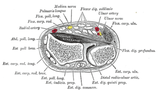
In the human body, the carpal tunnel or carpal canal is the passageway on the palmar side of the wrist that connects the forearm to the hand.
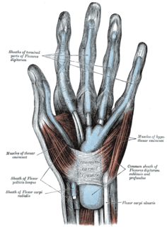
The common synovial sheath for the flexor tendons or the ulnar bursa is a synovial sheath in the carpal tunnel of the human hand.

An ulnar claw, also known as claw hand, or 'Spinster's Claw' is a deformity or an abnormal attitude of the hand that develops due to ulnar nerve damage causing paralysis of the lumbricals. A claw hand presents with a hyperextension at the metacarpo-phalangeal joints and flexion at the proximal and distal inter-phalangeal joints of the 4th and 5th fingers. The patients with this condition can make a full fist but when they extend their fingers, the hand posture is referred to as claw hand. The ring- and little finger can usually not fully extend at the proximal interphalangeal joint (PIP).
The hand of benediction, also known as benediction sign or preacher's hand, occurs as a result of prolonged compression or injury of the median nerve at the forearm or elbow.
Extensor digitorum brevis manus is an extra or accessory muscle on the backside (dorsum) of the hand. It was first described by Albinus in 1758. The muscles lies in the fourth extensor compartment of the wrist, and is relatively rare. It has a prevalence of 4% in the general population according to a meta-analysis. This muscle is commonly misdiagnosed as a ganglion cysta, synovial nodule or cyst.

Pronator teres syndrome is a compression neuropathy of the median nerve at the elbow. It is rare compared to compression at the wrist or isolated injury of the anterior interosseous branch of the median nerve.
Jersey finger is a finger-related tendon injury that is common in athletics and can result in permanent loss of flexion of the end of the finger if not surgically repaired. This injury often occurs in American football when a player grabs another player's jersey with the tips of one or more fingers while that player is pulling or running away.
Injuries to the arm, forearm or wrist area can lead to various nerve disorders. One such disorder is median nerve palsy. The median nerve controls the majority of the muscles in the forearm. It controls abduction of the thumb, flexion of hand at wrist, flexion of digital phalanx of the fingers, is the sensory nerve for the first three fingers, etc. Because of this major role of the median nerve, it is also called the eye of the hand. If the median nerve is damaged, the ability to abduct and oppose the thumb may be lost due to paralysis of the thenar muscles. Various other symptoms can occur which may be repaired through surgery and tendon transfers. Tendon transfers have been very successful in restoring motor function and improving functional outcomes in patients with median nerve palsy.

The extensor medii proprius is a rare anatomical variant in the extensor compartment of the forearm. The aberrant muscle is analogous to the extensor indicis with the insertion being the middle finger instead of the index finger.

Palmaris profundus is a rare anatomical variant in the anterior compartment of forearm. It was first described in 1908. It is usually found incidentally in cadaveric dissection or surgery. The prevalence of the palmaris longus was reported to be one in 1600 upper limbs.
Linburg–Comstock variation is an occasional tendinous connection between the flexor pollicis longus and the flexor digitorum profundus of the index, the middle finger or both. It is found in around 21% of the population. It is an anatomical variation in human, which may be viewed as a pathology if causes symptoms. It was recognised as early as the 1800s, but was first described by Linburg and Comstock in 1979.
