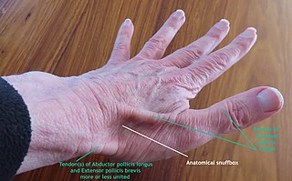
The anatomical snuff box or snuffbox or foveola radialis is a triangular deepening on the radial, dorsal aspect of the hand—at the level of the carpal bones, specifically, the scaphoid and trapezium bones forming the floor. The name originates from the use of this surface for placing and then sniffing powdered tobacco, or "snuff." It is sometimes referred to by its French name tabatière.

In human anatomy, the radial artery is the main artery of the lateral aspect of the forearm.
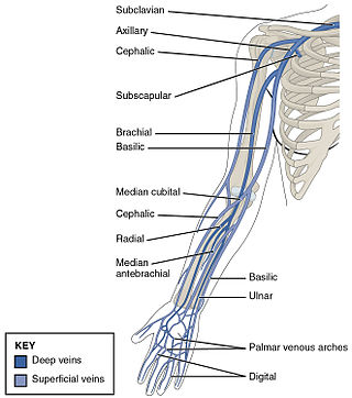
In human anatomy, the cephalic vein is a superficial vein in the arm. It originates from the radial end of the dorsal venous network of hand, and ascends along the radial (lateral) side of the arm before emptying into the axillary vein. At the elbow, it communicates with the basilic vein via the median cubital vein.
The ulnar artery is the main blood vessel, with oxygenated blood, of the medial aspects of the forearm. It arises from the brachial artery and terminates in the superficial palmar arch, which joins with the superficial branch of the radial artery. It is palpable on the anterior and medial aspect of the wrist.
The ulnar veins are venae comitantes of the ulnar artery. They drain the superficial venous palmar arch. They arise in the hand and terminate by uniting with the radial veins to form the brachial veins. They mostly drain the medial aspect of the forearm. They receive the venae comitantes of the anterior and posterior interosseous arteries near the elbow, as well as a large branch from the median cubital vein. The ulnar veins are larger than the radial veins.

The flexor pollicis brevis is a muscle in the hand that flexes the thumb. It is one of three thenar muscles. It has both a superficial part and a deep part.

The palmar aponeurosis invests the muscles of the palm, and consists of central, lateral, and medial portions.

The lateral cutaneous nerve of forearm is a sensory nerve representing the continuation of the musculocutaneous nerve beyond the lateral edge of the tendon of the biceps brachii muscle. The lateral cutaneous nerve provides sensory innervation to the skin of the lateral forearm. It pierces the deep fascia of forearm to enter the subcutaneous compartment before splitting into a volar branch and a dorsal branch.
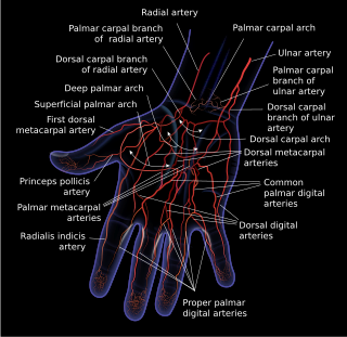
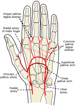
The superficial palmar arch is formed predominantly by the ulnar artery, with a contribution from the superficial palmar branch of the radial artery. However, in some individuals the contribution from the radial artery might be absent, and instead anastomoses with either the princeps pollicis artery, the radialis indicis artery, or the median artery, the former two of which are branches from the radial artery.
The deep palmar arch is an arterial network found in the palm. It is usually primarily formed from the terminal part of the radial artery. The ulnar artery also contributes through an anastomosis. This is in contrast to the superficial palmar arch, which is formed predominantly by the ulnar artery.

The superficial temporal vein is a vein of the side of the head which collects venous blood from the region of the temple. It arises from an anastomosing venous plexus on the side and vertex of the head. The superficial temporal vein terminates within the substance of the parotid gland by uniting with the maxillary vein to form the retromandibular vein.

The palmar metacarpal arteries are three or four arteries that arise from the convexity of the deep palmar arch.
The radialis indicis artery is a branch of the radial artery that provides blood to the index finger.
The dorsal carpal arch is an anatomical term for the combination (anastomosis) of dorsal carpal branch of the radial artery and the dorsal carpal branch of the ulnar artery near the back of the wrist.

The median antebrachial vein is a superficial vein of the (anterior) forearm. It arises from - and drains - the superficial palmar venous arch, ascending superficially along the anterior forearm before terminating by draining into either the basilic vein and/or median cubital vein; it may bifurcate distal to the elbow and proceed to drain into both aforementioned veins. A bifurcation of the median antebrachial vein produces the (medial) intermediate basilic vein and the (lateral) intermediate cephalic vein; the two veins produced by such a split may replace the median cubital vein.

Most of the dorsal metacarpal arteries arise from the dorsal carpal arch and run downward on the second, third, and fourth dorsal interossei of the hand and bifurcate into the dorsal digital arteries. Near their origin, they anastomose with the deep palmar arch by perforating arteries. They also anastomose with common palmar digital arteries, also via perforating arteries.

In the palm of the hand the median nerve is covered by the skin and the palmar aponeurosis, and rests on the tendons of the flexor muscles. Immediately after emerging from under the transverse carpal ligament the median nerve becomes enlarged and flattened and splits into a smaller, lateral, and a larger, medial portion.
The palmar carpal arch is a joining of an artery to an artery, a circulatory anastomosis, known as an arterio-arterial anastomosis. The two connected arteries are the palmar carpal branch of the radial artery and the palmar carpal branch of the ulnar artery.
A circulatory anastomosis is a connection between two blood vessels, such as between arteries, between veins or between an artery and a vein. Anastomoses between arteries and between veins result in a multitude of arteries and veins, respectively, serving the same volume of tissue. Such anastomoses occur normally in the body in the circulatory system, serving as back-up routes in a collateral circulation that allow blood to flow if one link is blocked or otherwise compromised, but may also occur pathologically.
The superficial palmar branch of the radial artery arises from the radial artery, just where this vessel is about to wind around the lateral side of the wrist.