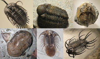
Trilobites are extinct marine arthropods that form the class Trilobita. Trilobites form one of the earliest known groups of arthropods. The first appearance of trilobites in the fossil record defines the base of the Atdabanian stage of the Early Cambrian period and they flourished throughout the lower Paleozoic before slipping into a long decline, when, during the Devonian, all trilobite orders except the Proetida died out. The last trilobites disappeared in the mass extinction at the end of the Permian about 251.9 million years ago. Trilobites were among the most successful of all early animals, existing in oceans for almost 270 million years, with over 22,000 species having been described.

The skull is a bone protective cavity for the brain. The skull is composed of three types of bone: cranial bones, facial bones, and ear ossicles. Two parts are more prominent: the cranium and the mandible. In humans, these two parts are the neurocranium (braincase) and the viscerocranium that includes the mandible as its largest bone. The skull forms the anterior-most portion of the skeleton and is a product of cephalisation—housing the brain, and several sensory structures such as the eyes, ears, nose, and mouth. In humans, these sensory structures are part of the facial skeleton.

A fontanelle is an anatomical feature of the infant human skull comprising soft membranous gaps (sutures) between the cranial bones that make up the calvaria of a fetus or an infant. Fontanelles allow for stretching and deformation of the neurocranium both during birth and later as the brain expands faster than the surrounding bone can grow. Premature complete ossification of the sutures is called craniosynostosis.

In botany, a capsule is a type of simple, dry, though rarely fleshy dehiscent fruit produced by many species of angiosperms.

The occipital bone is a cranial dermal bone and the main bone of the occiput. It is trapezoidal in shape and curved on itself like a shallow dish. The occipital bone overlies the occipital lobes of the cerebrum. At the base of the skull in the occipital bone, there is a large oval opening called the foramen magnum, which allows the passage of the spinal cord.

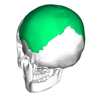
The parietal bones are two bones in the skull which, when joined at a fibrous joint known as a cranial suture, form the sides and roof of the neurocranium. In humans, each bone is roughly quadrilateral in form, and has two surfaces, four borders, and four angles. It is named from the Latin paries (-ietis), wall.

Goniatids, informally goniatites, are ammonoid cephalopods that form the order Goniatitida, derived from the more primitive Agoniatitida during the Middle Devonian some 390 million years ago. Goniatites (goniatitids) survived the Late Devonian extinction to flourish during the Carboniferous and Permian only to become extinct at the end of the Permian some 139 million years later.

Craniosynostosis is a condition in which one or more of the fibrous sutures in a young infant's skull prematurely fuses by turning into bone (ossification), thereby changing the growth pattern of the skull. Because the skull cannot expand perpendicular to the fused suture, it compensates by growing more in the direction parallel to the closed sutures. Sometimes the resulting growth pattern provides the necessary space for the growing brain, but results in an abnormal head shape and abnormal facial features. In cases in which the compensation does not effectively provide enough space for the growing brain, craniosynostosis results in increased intracranial pressure leading possibly to visual impairment, sleeping impairment, eating difficulties, or an impairment of mental development combined with a significant reduction in IQ.

The sagittal suture, also known as the interparietal suture and the sutura interparietalis, is a dense, fibrous connective tissue joint between the two parietal bones of the skull. The term is derived from the Latin word sagitta, meaning arrow.

The lambdoid suture, or lambdoidal suture, is a dense, fibrous connective tissue joint on the posterior aspect of the skull that connects the parietal bones with the occipital bone. It is continuous with the occipitomastoid suture.

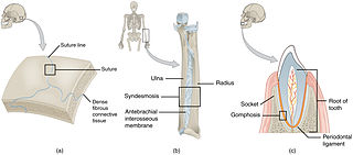
In anatomy, fibrous joints are joints connected by fibrous tissue, consisting mainly of collagen. These are fixed joints where bones are united by a layer of white fibrous tissue of varying thickness. In the skull, the joints between the bones are called sutures. Such immovable joints are also referred to as synarthroses.

In anatomy, a suture is a fairly rigid joint between two or more hard elements of an organism, with or without significant overlap of the elements.

In botany, a follicle is a dry unilocular fruit formed from one carpel, containing two or more seeds. It is usually defined as dehiscing by a suture in order to release seeds, for example in Consolida, peony and milkweed (Asclepias).
Dimeroceratoidea, formerly Dimerocerataceae, is one of six superfamilies in the goniatitid suborder Tornoceratina which lived during the Devonian. Five families are included, the Dimeroceratidae being the type family.
Prolobitidae is a family of middle and upper Devonian ammonoid cephalopods currently included in the goniatitid suborder Tornoceratina and superfamily Dimeroceratoidea, but previously included in the ancestral Anarcestida.
Cheiloceratidae is a family of ammonoid cephalopods included in the goniatitid suborder Tornoceratina in which the suture has 4 to 12 lobes, the ventral one undivided and those in the lateral areas originating as subdivisions of internal and external lateral saddles.
Tornoceratoidea, also known as Tornocerataceae, is a superfamily of goniatitid ammonoids included in the suborder Tornoceratina. Tornoceratoidea, or Tornocerataceae, is essentially the Cheilocerataceae of Miller, Furnish, and Schindewolf (1957) in the Treatise Part L, revised to accommodate new taxa and new perspectives on the phylogeny.
Tornoceratidae is a family of goniatitid ammonoids from the middle and upper Devonian. The family is included in the suborder Tornoceratina and the superfamily Tornoceratoidea.

A surgical suture, also known as a stitch or stitches, is a medical device used to hold body tissues together and approximate wound edges after an injury or surgery. Application generally involves using a needle with an attached length of thread. There are numerous types of suture which differ by needle shape and size as well as thread material and characteristics. Selection of surgical suture should be determined by the characteristics and location of the wound or the specific body tissues being approximated.

Prolecanitoidea is a taxonomic superfamily of ammonoids in the order Prolecanitida. Prolecanitoidea is one of two superfamilies in the order, along with the younger and more complex Medlicottioidea. The Prolecanitoidea were a low-diversity and morphologically conservative group. They lived from the Lower Carboniferous up to the Middle Permian. Their shells are generally smooth and discoidal, with a rounded lower edge, a moderate to large umbilicus, and goniatitic to ceratitic sutures. Suture complexity varies from 10 up to 22 total lobes ; new lobes are added from subdivision of saddles adjacent to the original main umbilical lobe.


