The heart is a muscular organ in most animals. This organ pumps blood through the blood vessels of the circulatory system. The pumped blood carries oxygen and nutrients to the body, while carrying metabolic waste such as carbon dioxide to the lungs. In humans, the heart is approximately the size of a closed fist and is located between the lungs, in the middle compartment of the chest, called the mediastinum.

A heart valve is a one-way valve that allows blood to flow in one direction through the chambers of the heart. Four valves are usually present in a mammalian heart and together they determine the pathway of blood flow through the heart. A heart valve opens or closes according to differential blood pressure on each side.

The aortic valve is a valve in the heart of humans and most other animals, located between the left ventricle and the aorta. It is one of the four valves of the heart and one of the two semilunar valves, the other being the pulmonary valve. The aortic valve normally has three cusps or leaflets, although in 1–2% of the population it is found to congenitally have two leaflets. The aortic valve is the last structure in the heart the blood travels through before stopping the flow through the systemic circulation.

The ventricular system is a set of four interconnected cavities known as cerebral ventricles in the brain. Within each ventricle is a region of choroid plexus which produces the circulating cerebrospinal fluid (CSF). The ventricular system is continuous with the central canal of the spinal cord from the fourth ventricle, allowing for the flow of CSF to circulate.

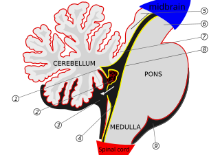
The fourth ventricle is one of the four connected fluid-filled cavities within the human brain. These cavities, known collectively as the ventricular system, consist of the left and right lateral ventricles, the third ventricle, and the fourth ventricle. The fourth ventricle extends from the cerebral aqueduct to the obex, and is filled with cerebrospinal fluid (CSF).

In the brain, the interventricular foramina are channels that connect the paired lateral ventricles with the third ventricle at the midline of the brain. As channels, they allow cerebrospinal fluid (CSF) produced in the lateral ventricles to reach the third ventricle and then the rest of the brain's ventricular system. The walls of the interventricular foramina also contain choroid plexus, a specialized CSF-producing structure, that is continuous with that of the lateral and third ventricles above and below it.

The posterior inferior cerebellar artery (PICA) is the largest branch of the vertebral artery. It is one of the three main arteries that supply blood to the cerebellum, a part of the brain. Blockage of the posterior inferior cerebellar artery can result in a type of stroke called lateral medullary syndrome.

The central canal is the cerebrospinal fluid-filled space that runs through the spinal cord. The central canal lies below and is connected to the ventricular system of the brain, from which it receives cerebrospinal fluid, and shares the same ependymal lining. The central canal helps to transport nutrients to the spinal cord as well as protect it by cushioning the impact of a force when the spine is affected.

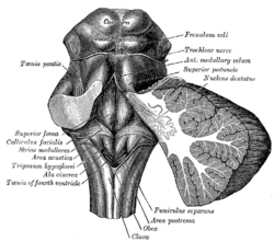
The facial colliculus is an elevated area located in the pontine tegmentum, within the floor of the fourth ventricle. It is formed by fibres from the facial motor nucleus looping over the abducens nucleus. The facial colliculus is an essential landmark of the rhomboid fossa.
The lateral aperture is a paired structure in human anatomy. It is an opening in each lateral extremity of the lateral recess of the fourth ventricle of the human brain, which also has a single median aperture. The two lateral apertures provide a conduit for cerebrospinal fluid to flow from the brain's ventricular system into the subarachnoid space; specifically into the pontocerebellar cistern at the cerebellopontine angle. The structure is also called the lateral aperture of the fourth ventricle or the foramen of Luschka after anatomist Hubert von Luschka.

The trabeculae carneae are rounded or irregular muscular columns which project from the inner surface of the right and left ventricle of the heart. These are different from the pectinate muscles, which are present in the atria of the heart. In development, trabeculae carneae are among the first of the cardiac structures to develop in the embryonic cardiac tube. Further, throughout development some trabeculae carneae condense to form the myocardium, papillary muscles, chordae tendineae, and septum.

The rhomboid fossa is a rhombus-shaped depression that is the anterior part of the fourth ventricle. Its anterior wall, formed by the back of the pons and the medulla oblongata, constitutes the floor of the fourth ventricle.

The superior medullary velum is a thin, transparent lamina of white matter, which stretches between the superior cerebellar peduncles; on the dorsal surface of its lower half the folia and lingula are prolonged.
The sulcus limitans is found in the fourth ventricle of the brain. It separates the cranial nerve motor nuclei (medial) from the sensory nuclei (lateral). It can also be located by searching laterally from the medial eminence. It is parallel to the median sulcus.
In the brain, the taenia of the fourth ventricle are two narrow bands of white matter, one on either side, which complete the lower part of the roof of the fourth ventricle.

The laryngeal ventricle, is a fusiform fossa, situated between the vestibular and vocal folds on either side, and extending nearly their entire length. There is also a sinus of Morgagni in the pharynx.
The inferior medullary velum is a thin layer of white substance, prolonged from the white center of the cerebellum, above and on either side of the nodule; it forms the infero-posterior part of the fourth ventricle.

The posterior median sulcus of medulla oblongata is a narrow groove; and exists only in the closed part of the medulla oblongata; it becomes gradually shallower from below upward, and finally ends about the middle of the medulla oblongata, where the central canal expands into the cavity of the fourth ventricle.
In the upper part of the medulla oblongata, the hypoglossal nucleus approaches the rhomboid fossa, where it lies close to the middle line, under an eminence named the hypoglossal trigone. It is a slight elevation in the floor of the inferior recess of the fourth ventricle, beneath which is the nucleus of origin of the twelfth cranial nerve.
The roof of fourth ventricle is the dorsal surface of the fourth ventricle.