
Vibrissae, more generally called whiskers, are a type of stiff, functional hair used by mammals to sense their environment. These hairs are finely specialised for this purpose, whereas other types of hair are coarser as tactile sensors. Although whiskers are specifically those found around the face, vibrissae are known to grow in clusters at various places around the body. Most mammals have them, including all non-human primates and especially nocturnal mammals.
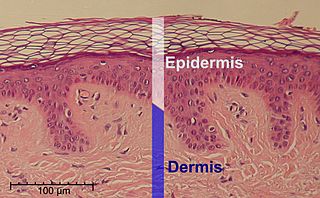
The epidermis is the outermost of the three layers that comprise the skin, the inner layers being the dermis and hypodermis. The epidermis layer provides a barrier to infection from environmental pathogens and regulates the amount of water released from the body into the atmosphere through transepidermal water loss.

The sensory nervous system is a part of the nervous system responsible for processing sensory information. A sensory system consists of sensory neurons, neural pathways, and parts of the brain involved in sensory perception and interoception. Commonly recognized sensory systems are those for vision, hearing, touch, taste, smell, balance and visceral sensation. Sense organs are transducers that convert data from the outer physical world to the realm of the mind where people interpret the information, creating their perception of the world around them.

Afferent nerve fibers are axons of sensory neurons that carry sensory information from sensory receptors to the central nervous system. Many afferent projections arrive at a particular brain region.

A free nerve ending (FNE) or bare nerve ending, is an unspecialized, afferent nerve fiber sending its signal to a sensory neuron. Afferent in this case means bringing information from the body's periphery toward the brain. They function as cutaneous nociceptors and are essentially used by vertebrates to detect noxious stimuli that often result in pain.
Stimulus modality, also called sensory modality, is one aspect of a stimulus or what is perceived after a stimulus. For example, the temperature modality is registered after heat or cold stimulate a receptor. Some sensory modalities include: light, sound, temperature, taste, pressure, and smell. The type and location of the sensory receptor activated by the stimulus plays the primary role in coding the sensation. All sensory modalities work together to heighten stimuli sensation when necessary.
A cutaneous receptor is the type of sensory receptor found in the skin. They are a part of the somatosensory system. Cutaneous receptors include mechanoreceptors, nociceptors (pain), and thermoreceptors (temperature).
A mechanoreceptor, also called mechanoceptor, is a sensory receptor that responds to mechanical pressure or distortion. Mechanoreceptors are innervated by sensory neurons that convert mechanical pressure into electrical signals that, in animals, are sent to the central nervous system.

The dorsal column–medial lemniscus pathway (DCML) is a sensory pathway of the central nervous system that conveys sensations of fine touch, vibration, two-point discrimination, and proprioception from the skin and joints. It transmits information from the body to the primary somatosensory cortex in the postcentral gyrus of the parietal lobe of the brain. The pathway receives information from sensory receptors throughout the body, and carries this in nerve tracts in the white matter of the dorsal column of the spinal cord to the medulla, where it is continued in the medial lemniscus, on to the thalamus and relayed from there through the internal capsule and transmitted to the somatosensory cortex. The name dorsal-column medial lemniscus comes from the two structures that carry the sensory information: the dorsal columns of the spinal cord, and the medial lemniscus in the brainstem.

Tactile corpuscles or Meissner's corpuscles are a type of mechanoreceptor discovered by anatomist Georg Meissner (1829–1905) and Rudolf Wagner. This corpuscle is a type of nerve ending in the skin that is responsible for sensitivity to pressure. In particular, they have their highest sensitivity when sensing vibrations between 10 and 50 hertz. They are rapidly adaptive receptors. They are most concentrated in thick hairless skin, especially at the finger pads.

Sensory neurons, also known as afferent neurons, are neurons in the nervous system, that convert a specific type of stimulus, via their receptors, into action potentials or graded receptor potentials. This process is called sensory transduction. The cell bodies of the sensory neurons are located in the dorsal ganglia of the spinal cord.
Merkel nerve endings are mechanoreceptors, a type of sensory receptor, that are found in the basal epidermis and hair follicles. They are nerve endings and provide information on mechanical pressure, position, and deep static touch features, such as shapes and edges.
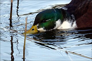
The tactile corpuscles of Grandry or Grandry corpuscles are mechanoreceptors found in the beak skin and oral mucosa of aquatic birds. They were first described by Grandry in 1869 in the bill skin of ducks and geese. Their general structure includes the flattened endings of an afferent nerve fiber sandwiched between two or more somewhat flattened sensory cells called Grandry cells, all surrounded by a layer of satellite cells and a partial capsule of collagen protein. Electrophysiological studies have shown that Grandry corpuscles function as rapidly adapting velocity detectors. In birds, Grandry and Merkel corpuscles share many morphological similarities, which has led to some confusion in the literature over their classification.
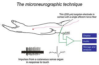
Microneurography is a neurophysiological method employed to visualize and record the traffic of nerve impulses that are conducted in peripheral nerves of waking human subjects. It can also be used in animal recordings. The method has been successfully employed to reveal functional properties of a number of neural systems, e.g. sensory systems related to touch, pain, and muscle sense as well as sympathetic activity controlling the constriction state of blood vessels. To study nerve impulses of an identified nerve, a fine tungsten needle microelectrode is inserted into the nerve and connected to a high input impedance differential amplifier. The exact position of the electrode tip within the nerve is then adjusted in minute steps until the electrode discriminates nerve impulses of interest. A unique feature and a significant strength of the microneurography method is that subjects are fully awake and able to cooperate in tests requiring mental attention, while impulses in a representative nerve fibre or set of nerve fibres are recorded, e.g. when cutaneous sense organs are stimulated or subjects perform voluntary precision movements.
Type II sensory fiber is a type of sensory fiber, the second of the two main groups of touch receptors. The responses of different type Aβ fibers to these stimuli can be subdivided based on their adaptation properties, traditionally into rapidly adapting (RA) or slowly adapting (SA) neurons. Type II sensory fibers are slowly-adapting (SA), meaning that even when there is no change in touch, they keep respond to stimuli and fire action potentials. In the body, Type II sensory fibers belong to pseudounipolar neurons. The most notable example are neurons with Merkel cell-neurite complexes on their dendrites and Ruffini endings. Under pathological conditions they may become hyper-excitable leading to stimuli that would usually elicit sensations of tactile touch causing pain. These changes are in part induced by PGE2 which is produced by COX1, and type II fibers with free nerve endings are likely to be the subdivision of fibers that carry out this function.
Eimer's organs are sensory organs in which the epidermis is modified to form bulbous papillae. First isolated by Theodor Eimer from the European mole in 1871, these organs are present in many moles, and are particularly common in the star-nosed mole, which bears 25,000 of them on its unique tentacled snout. The organs are formed from a stack of epidermal cells, which is innervated by nerve processes from myelinated fibers in the dermis, which form terminal swellings just below the outer keratinized layer of epidermis. They contain a Merkel cell-neurite complex in the epidermis and a lamellated corpuscle in the dermal connective tissue.
Mechanosensation is the transduction of mechanical stimuli into neural signals. Mechanosensation provides the basis for the senses of light touch, hearing, proprioception, and pain. Mechanoreceptors found in the skin, called cutaneous mechanoreceptors, are responsible for the sense of touch. Tiny cells in the inner ear, called hair cells, are responsible for hearing and balance. States of neuropathic pain, such as hyperalgesia and allodynia, are also directly related to mechanosensation. A wide array of elements are involved in the process of mechanosensation, many of which are still not fully understood.

In physiology, the somatosensory system is the network of neural structures in the brain and body that produce the perception of touch, as well as temperature (thermoception), body position (proprioception), and pain. It is a subset of the sensory nervous system, which also represents visual, auditory, olfactory, and gustatory stimuli.
Group A nerve fibers are one of the three classes of nerve fiber as generally classified by Erlanger and Gasser. The other two classes are the group B nerve fibers, and the group C nerve fibers. Group A are heavily myelinated, group B are moderately myelinated, and group C are unmyelinated.
Ellen Lumpkin is an American neuroscientist and professor of cell and developmental biology and neurobiology at the Helen Wills Neuroscience Institute at the University of California, Berkeley. She is also co-director of the MBL Advanced Training Course in Neurobiology, and adjunct associate professor of physiology and cellular biophysics and co-director of the Thompson Family Foundation Initiative in CIPN and Sensory Neuroscience at Columbia University. Lumpkin's group studies genes, cells and signals that mediate the sensation of touch. Lumpkin is most interested in the somatosensory system and how it gives feedback to the brain on sensations such as pain or touch. She is known for her significant contributions in somatosensory system research.

