The autonomic nervous system (ANS), formerly the vegetative nervous system, is a division of the peripheral nervous system that supplies smooth muscle and glands, and thus influences the function of internal organs. The autonomic nervous system is a control system that acts largely unconsciously and regulates bodily functions such as the heart rate, digestion, respiratory rate, pupillary response, urination, and sexual arousal. This system is the primary mechanism in control of the fight-or-flight response.

The facial nerve is the seventh cranial nerve, or simply CN VII. It emerges from the pons of the brainstem, controls the muscles of facial expression, and functions in the conveyance of taste sensations from the anterior two-thirds of the tongue. The nerves typically travels from the pons through the facial canal in the temporal bone and exits the skull at the stylomastoid foramen. It arises from the brainstem from an area posterior to the cranial nerve VI and anterior to cranial nerve VIII.

The glossopharyngeal nerve, known as the ninth cranial nerve, is a mixed nerve that carries afferent sensory and efferent motor information. It exits the brainstem out from the sides of the upper medulla, just rostral to the vagus nerve. The motor division of the glossopharyngeal nerve is derived from the basal plate of the embryonic medulla oblongata, while the sensory division originates from the cranial neural crest.

In human physiology, the lacrimal glands are paired, almond-shaped exocrine glands, one for each eye, that secrete the aqueous layer of the tear film. They are situated in the upper lateral region of each orbit, in the lacrimal fossa of the orbit formed by the frontal bone. Inflammation of the lacrimal glands is called dacryoadenitis. The lacrimal gland produces tears which then flow into canals that connect to the lacrimal sac. From that sac, the tears drain through the lacrimal duct into the nose.

The pterygopalatine ganglion is a parasympathetic ganglion found in the pterygopalatine fossa. It is largely innervated by the greater petrosal nerve ; and its axons project to the lacrimal glands and nasal mucosa. The flow of blood to the nasal mucosa, in particular the venous plexus of the conchae, is regulated by the pterygopalatine ganglion and heats or cools the air in the nose. It is one of four parasympathetic ganglia of the head and neck, the others being the submandibular ganglion, otic ganglion, and ciliary ganglion.

The ciliary ganglion is a parasympathetic ganglion located just behind the eye in the posterior orbit. It measures 1–2 millimeters in diameter and in humans contains approximately 2,500 neurons. The oculomotor nerve coming into the ganglion contains preganglionic axons from the Edinger-Westphal nucleus which form synapses with the ciliary neurons. The postganglionic axons run in the short ciliary nerves and innervate two eye muscles:

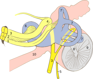
The greater (superficial) petrosal nerve is a nerve in the skull that branches from the facial nerve; it forms part of a chain of nerves that innervate the lacrimal gland. The fibers have synapses in the pterygopalatine ganglion.

The ophthalmic nerve is the first branch of the trigeminal nerve. The ophthalmic nerve is a sensory nerve mostly carrying general somatic afferent fibers that transmit sensory information to the CNS from structures of the eyeball, the skin of the upper face and anterior scalp, the lining of the upper part of the nasal cavity and air cells, and the meninges of the anterior cranial fossa. Some of ophthalmic nerve branches also convey parasympathetic fibers.

The geniculate ganglion is an L-shaped collection of fibers and pseudo-unipolar sensory neurons of the facial nerve located in the facial canal of the head. It receives fibers from the motor, sensory, and parasympathetic components of the facial nerve and sends fibers that will innervate the lacrimal glands, submandibular glands, sublingual glands, tongue, palate, pharynx, external auditory meatus, stapedius, posterior belly of the digastric muscle, stylohyoid muscle, and muscles of facial expression.

The lacrimal nerve is the smallest of the three branches of the ophthalmic division of the trigeminal nerve.

The zygomatic nerve is not to be confused with the zygomatic branches of the facial nerve.

The submandibular ganglion is part of the human autonomic nervous system. It is one of four parasympathetic ganglia of the head and neck..

In the autonomic nervous system, fibers from the CNS to the ganglion are known as preganglionic fibers. All preganglionic fibers, whether they are in the sympathetic division or in the parasympathetic division, are cholinergic and they are myelinated.

Parasympathetic ganglia are the autonomic ganglia of the parasympathetic nervous system. Most are small terminal ganglia or intramural ganglia, so named because they lie near or within (respectively) the organs they innervate. The exceptions are the four paired parasympathetic ganglia of the head and neck.

The nerve of the pterygoid canal is formed by the junction of the greater petrosal nerve and the deep petrosal nerve within the pterygoid canal containing the cartilaginous substance, which fills the foramen lacerum.

The intermediate nerve, nervus intermedius, nerve of Wrisberg or Glossopalatine nerve, is the part of the facial nerve located between the motor component of the facial nerve and the vestibulocochlear nerve. It contains the sensory and parasympathetic fibers of the facial nerve. Upon reaching the facial canal, it joins with the motor root of the facial nerve at the geniculate ganglion.

The salivatory nuclei are the superior salivatory nucleus, and the inferior salivatory nucleus that innervate the salivary glands. They are located in the pontine tegmentum in the brainstem. They both belong to the cranial nerve nuclei.

The sympathetic root of ciliary ganglion is one of three roots of the ciliary ganglion, a tissue mass behind the eye. It contains postganglionic sympathetic fibers whose cell bodies are located in the superior cervical ganglion. Their axons ascend with the internal carotid artery as a plexus of nerves, the carotid plexus. Sympathetic fibers innervating the eye separate from the carotid plexus within the cavernous sinus. They run forward through the superior orbital fissure and merge with the long ciliary nerves and the short ciliary nerves. Sympathetic fibers in the short ciliary nerves pass through the ciliary ganglion without forming synapses.

The parasympathetic root of ciliary ganglion provides parasympathetic innervation to the ciliary ganglion.


