Binaural fusion

Last updated

Binaural fusion or binaural integration is a cognitive process that involves the combination of different auditory information presented binaurally, or to each ear. In humans, this process is essential in understanding speech as one ear may pick up more information about the speech stimuli than the other.

Contents

The process of binaural fusion is important for computing the location of sound sources in the horizontal plane (sound localization), and it is important for sound segregation. [1] Sound segregation refers to the ability to identify acoustic components from one or more sound sources. [2] The binaural auditory system is highly dynamic and capable of rapidly adjusting tuning properties depending on the context in which sounds are heard. Each eardrum moves one-dimensionally; the auditory brain analyzes and compares movements of both eardrums to extract physical cues and synthesize auditory objects. [3]

When stimulation from a sound reaches the ear, the eardrum deflects in a mechanical fashion, and the three middle ear bones (ossicles) transmit the mechanical signal to the cochlea, where hair cells transform the mechanical signal into an electrical signal. The auditory nerve, also called the cochlear nerve, then transmits action potentials to the central auditory nervous system. [3]

In binaural fusion, inputs from both ears integrate and fuse to create a complete auditory picture at the brainstem. Therefore, the signals sent to the central auditory nervous system are representative of this complete picture, integrated information from both ears instead of a single ear.

The binaural squelch effect is a result of nuclei of the brainstem processing timing, amplitude, and spectral differences between the two ears. Sounds are integrated and then separated into auditory objects. For this effect to take place, neural integration from both sides is required. [4]

Anatomy

Transmissions from the SOC, in the pons of the brainstem, travel along the lateral lemniscus to the IC, located in the midbrain. Signals are then relayed to the thalamus and further ascending auditory pathway. Lateral lemniscus.PNG
Transmissions from the SOC, in the pons of the brainstem, travel along the lateral lemniscus to the IC, located in the midbrain. Signals are then relayed to the thalamus and further ascending auditory pathway.

As sound travels into the inner eardrum of vertebrate mammals, it encounters the hair cells that line the basilar membrane of the cochlea in the inner ear. [5] The cochlea receives auditory information to be binaurally integrated. At the cochlea, this information is converted into electrical impulses that travel by means of the cochlear nerve, which spans from the cochlea to the ventral cochlear nucleus, which is located in the pons of the brainstem. [6] The lateral lemniscus projects from the cochlear nucleus to the superior olivary complex (SOC), a set of brainstem nuclei that consists primarily of two nuclei, the medial superior olive (MSO) and the lateral superior olive (LSO), and is the major site of binaural fusion. The subdivision of the ventral cochlear nucleus that concerns binaural fusion is the anterior ventral cochlear nucleus (AVCN). [3] The AVCN consists of spherical bushy cells and globular bushy cells and can also transmit signals to the medial nucleus of the trapezoid body (MNTB), whose neuron projects to the MSO. Transmissions from the SOC travel to the inferior colliculus (IC) via the lateral lemniscus. At the level of the IC, binaural fusion is complete. The signal ascends to the thalamocortical system, and sensory inputs to the thalamus are then relayed to the primary auditory cortex. [3] [7] [8] [9]

Function

The ear functions to analyze and encode a sound’s dimensions. [10] Binaural fusion is responsible for avoiding the creation of multiple sound images from a sound source and its reflections. The advantages of this phenomenon are more noticeable in small rooms, decreasing as the reflective surfaces are placed farther from the listener. [11]

Central auditory system

The central auditory system converges inputs from both ears (inputs contain no explicit spatial information) onto single neurons within the brainstem. This system contains many subcortical sites that have integrative functions. The auditory nuclei collect, integrate, and analyze afferent supply, [10] the outcome is a representation of auditory space. [3] The subcortical auditory nuclei are responsible for extraction and analysis of dimensions of sounds. [10]

The integration of a sound stimulus is a result of analyzing frequency (pitch), intensity, and spatial localization of the sound source. [12] Once a sound source has been identified, the cells of lower auditory pathways are specialized to analyze physical sound parameters. [3] Summation is observed when the loudness of a sound from one stimulus is perceived as having been doubled when heard by both ears instead of only one. This process of summation is called binaural summation and is the result of different acoustics at each ear, depending on where sound is coming from. [4]

The cochlear nerve spans from the cochlea of the inner ear to the ventral cochlear nuclei located in the pons of the brainstem, relaying auditory signals to the superior olivary complex where it is to be binaurally integrated.

Medial superior olive and lateral superior olive

The MSO contains cells that function in comparing inputs from the left and right cochlear nuclei. [13] The tuning of neurons in the MSO favors low frequencies, whereas those in the LSO favor high frequencies. [14]

GABAB receptors in the LSO and MSO are involved in balance of excitatory and inhibitory inputs. The GABAB receptors are coupled to G proteins and provide a way of regulating synaptic efficacy. Specifically, GABAB receptors modulate excitatory and inhibitory inputs to the LSO. [3] Whether the GABAB receptor functions as excitatory or inhibitory for the postsynaptic neuron, depends on the exact location and action of the receptor. [1]

Sound localization

Sound localization is the ability to correctly identify the directional location of sounds. A sound stimulus localized in the horizontal plane is called azimuth; in the vertical plane it is referred to as elevation. The time, intensity, and spectral differences in the sound arriving at the two ears are used in localization. Localization of low frequency sounds is accomplished by analyzing interaural time difference (ITD). Localization of high frequency sounds is accomplished by analyzing interaural level difference (ILD). [4]

Mechanism

Binaural hearing

Action potentials originate in the hair cells of the cochlea and propagate to the brainstem; both the timing of these action potentials and the signal they transmit provide information to the SOC about the orientation of sound in space. The processing and propagation of action potentials is rapid, and therefore, information about the timing of the sounds that were heard, which is crucial to binaural processing, is conserved. [15] Each eardrum moves in one dimension, and the auditory brain analyzes and compares the movements of both eardrums in order to synthesize auditory objects. [3] This integration of information from both ears is the essence of binaural fusion. The binaural system of hearing involves sound localization in the horizontal plane, contrasting with the monaural system of hearing, which involves sound localization in the vertical plane. [3]

Superior olivary complex

The primary stage of binaural fusion, the processing of binaural signals, occurs at the SOC, where afferent fibers of the left and right auditory pathways first converge. This processing occurs because of the interaction of excitatory and inhibitory inputs in the LSO and MSO. [1] [3] [13] The SOC processes and integrates binaural information, in the form of ITD and ILD, entering the brainstem from the cochleae. This initial processing of ILD and ITD is regulated by GABAB receptors. [1]

ITD and ILD

The auditory space of binaural hearing is constructed based on the analysis of differences in two different binaural cues in the horizontal plane: sound level, or ILD, and arrival time at the two ears, or ITD, which allow for the comparison of the sound heard at each eardrum. [1] [3] ITD is processed in the MSO and results from sounds arriving earlier at one ear than the other; this occurs when the sound does not arise from directly in front or directly behind the hearer. ILD is processed in the LSO and results from the shadowing effect that is produced at the ear that is farther from the sound source. Outputs from the SOC are targeted to the dorsal nucleus of the lateral lemniscus as well as the IC. [3]

Lateral superior olive

LSO neurons are excited by inputs from one ear and inhibited by inputs from the other, and are therefore referred to as IE neurons. Excitatory inputs are received at the LSO from spherical bushy cells of the ipsilateral cochlear nucleus, which combine inputs coming from several auditory nerve fibers. Inhibitory inputs are received at the LSO from globular bushy cells of the contralateral cochlear nucleus. [3]

Medial superior olive

MSO neurons are excited bilaterally, meaning that they are excited by inputs from both ears, and they are therefore referred to as EE neurons. [3] Fibers from the left cochlear nucleus terminate on the left of MSO neurons, and fibers from the right cochlear nucleus terminate on the right of MSO neurons. [13] Excitatory inputs to the MSO from spherical bushy cells are mediated by glutamate, and inhibitory inputs to the MSO from globular bushy cells are mediated by glycine. MSO neurons extract ITD information from binaural inputs and resolve small differences in the time of arrival of sounds at each ear. [3] Outputs from the MSO and LSO are sent via the lateral lemniscus to the IC, which integrates the spatial localization of sound. In the IC, acoustic cues have been processed and filtered into separate streams, forming the basis of auditory object recognition. [3]

Binaural fusion abnormalities in autism

Current research is being performed on the dysfunction of binaural fusion in individuals with autism. The neurological disorder autism is associated with many symptoms of impaired brain function, including the degradation of hearing, both unilateral and bilateral. [16] Individuals with autism who experience hearing loss maintain symptoms such as difficulty listening to background noise and impairments in sound localization. Both the ability to distinguish particular speakers from background noise and the process of sound localization are key products of binaural fusion. They are particularly related to the proper function of the SOC, and there is increasing evidence that morphological abnormalities within the brainstem, namely in the SOC, of autistic individuals are a cause of the hearing difficulties. [17] The neurons of the MSO of individuals with autism display atypical anatomical features, including atypical cell shape and orientation of the cell body as well as stellate and fusiform formations. [18] Data also suggests that neurons of the LSO and MNTB contain distinct dysmorphology in autistic individuals, such as irregular stellate and fusiform shapes and a smaller than normal size. Moreover, a significant depletion of SOC neurons is seen in the brainstem of autistic individuals. All of these structures play a crucial role in the proper functioning of binaural fusion, so their dysmorphology may be at least partially responsible for the incidence of these auditory symptoms in autistic patients. [17]

Related Research Articles

<span class="mw-page-title-main">Cochlea</span> Snail-shaped part of inner ear involved in hearing

The cochlea is the part of the inner ear involved in hearing. It is a spiral-shaped cavity in the bony labyrinth, in humans making 2.75 turns around its axis, the modiolus. A core component of the cochlea is the Organ of Corti, the sensory organ of hearing, which is distributed along the partition separating the fluid chambers in the coiled tapered tube of the cochlea.

<span class="mw-page-title-main">Auditory system</span> Sensory system used for hearing

The auditory system is the sensory system for the sense of hearing. It includes both the sensory organs and the auditory parts of the sensory system.

<span class="mw-page-title-main">Lateral lemniscus</span>

The lateral lemniscus is a tract of axons in the brainstem that carries information about sound from the cochlear nucleus to various brainstem nuclei and ultimately the contralateral inferior colliculus of the midbrain. Three distinct, primarily inhibitory, cellular groups are located interspersed within these fibers, and are thus named the nuclei of the lateral lemniscus.

Sound localization is a listener's ability to identify the location or origin of a detected sound in direction and distance.

<span class="mw-page-title-main">Inferior colliculus</span> Midbrain structure involved in the auditory pathway.

The inferior colliculus (IC) is the principal midbrain nucleus of the auditory pathway and receives input from several peripheral brainstem nuclei in the auditory pathway, as well as inputs from the auditory cortex. The inferior colliculus has three subdivisions: the central nucleus, a dorsal cortex by which it is surrounded, and an external cortex which is located laterally. Its bimodal neurons are implicated in auditory-somatosensory interaction, receiving projections from somatosensory nuclei. This multisensory integration may underlie a filtering of self-effected sounds from vocalization, chewing, or respiration activities.

In physiology, tonotopy is the spatial arrangement of where sounds of different frequency are processed in the brain. Tones close to each other in terms of frequency are represented in topologically neighbouring regions in the brain. Tonotopic maps are a particular case of topographic organization, similar to retinotopy in the visual system.

<span class="mw-page-title-main">Medial geniculate nucleus</span>

The medial geniculate nucleus (MGN) or medial geniculate body (MGB) is part of the auditory thalamus and represents the thalamic relay between the inferior colliculus (IC) and the auditory cortex (AC). It is made up of a number of sub-nuclei that are distinguished by their neuronal morphology and density, by their afferent and efferent connections, and by the coding properties of their neurons. It is thought that the MGN influences the direction and maintenance of attention.

<span class="mw-page-title-main">Cochlear nerve</span> Nerve carrying auditory information from the inner ear to the brain

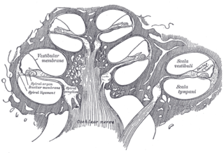
The cochlear nerve is one of two parts of the vestibulocochlear nerve, a cranial nerve present in amniotes, the other part being the vestibular nerve. The cochlear nerve carries auditory sensory information from the cochlea of the inner ear directly to the brain. The other portion of the vestibulocochlear nerve is the vestibular nerve, which carries spatial orientation information to the brain from the semicircular canals, also known as semicircular ducts.

<span class="mw-page-title-main">Cochlear nucleus</span> Two cranial nerve nuclei of the human brainstem

The cochlear nuclear (CN) complex comprises two cranial nerve nuclei in the human brainstem, the ventral cochlear nucleus (VCN) and the dorsal cochlear nucleus (DCN). The ventral cochlear nucleus is unlayered whereas the dorsal cochlear nucleus is layered. Auditory nerve fibers, fibers that travel through the auditory nerve carry information from the inner ear, the cochlea, on the same side of the head, to the nerve root in the ventral cochlear nucleus. At the nerve root the fibers branch to innervate the ventral cochlear nucleus and the deep layer of the dorsal cochlear nucleus. All acoustic information thus enters the brain through the cochlear nuclei, where the processing of acoustic information begins. The outputs from the cochlear nuclei are received in higher regions of the auditory brainstem.

<span class="mw-page-title-main">Dorsal cochlear nucleus</span>

The dorsal cochlear nucleus is a cortex-like structure on the dorso-lateral surface of the brainstem. Along with the ventral cochlear nucleus (VCN), it forms the cochlear nucleus (CN), where all auditory nerve fibers from the cochlea form their first synapses.

<span class="mw-page-title-main">Superior olivary complex</span> Collection of brainstem nuclei related to hearing

The superior olivary complex (SOC) or superior olive is a collection of brainstem nuclei that functions in multiple aspects of hearing and is an important component of the ascending and descending auditory pathways of the auditory system. The SOC is intimately related to the trapezoid body: most of the cell groups of the SOC are dorsal to this axon bundle while a number of cell groups are embedded in the trapezoid body. Overall, the SOC displays a significant interspecies variation, being largest in bats and rodents and smaller in primates.

<span class="mw-page-title-main">Interaural time difference</span> Difference in time that it takes a sound to travel between two ears

The interaural time difference when concerning humans or animals, is the difference in arrival time of a sound between two ears. It is important in the localization of sounds, as it provides a cue to the direction or angle of the sound source from the head. If a signal arrives at the head from one side, the signal has further to travel to reach the far ear than the near ear. This pathlength difference results in a time difference between the sound's arrivals at the ears, which is detected and aids the process of identifying the direction of sound source.

<span class="mw-page-title-main">Ventral cochlear nucleus</span>

In the ventral cochlear nucleus (VCN), auditory nerve fibers enter the brain via the nerve root in the VCN. The ventral cochlear nucleus is divided into the anterior ventral (anteroventral) cochlear nucleus (AVCN) and the posterior ventral (posteroventral) cochlear nucleus (PVCN). In the VCN, auditory nerve fibers bifurcate, the ascending branch innervates the AVCN and the descending branch innervates the PVCN and then continue to the dorsal cochlear nucleus. The orderly innervation by auditory nerve fibers gives the AVCN a tonotopic organization along the dorsoventral axis. Fibers that carry information from the apex of the cochlea that are tuned to low frequencies contact neurons in the ventral part of the AVCN; those that carry information from the base of the cochlea that are tuned to high frequencies contact neurons in the dorsal part of the AVCN. Several populations of neurons populate the AVCN. Bushy cells receive input from auditory nerve fibers through particularly large endings called end bulbs of Held. They contact stellate cells through more conventional boutons.

<span class="mw-page-title-main">Calyx of Held</span>

The Calyx of Held is a particularly large synapse in the mammalian auditory central nervous system, so named after Hans Held who first described it in his 1893 article Die centrale Gehörleitung because of its resemblance to the calyx of a flower. Globular bushy cells in the anteroventral cochlear nucleus (AVCN) send axons to the contralateral medial nucleus of the trapezoid body (MNTB), where they synapse via these calyces on MNTB principal cells. These principal cells then project to the ipsilateral lateral superior olive (LSO), where they inhibit postsynaptic neurons and provide a basis for interaural level detection (ILD), required for high frequency sound localization. This synapse has been described as the largest in the brain.

Coincidence detection is a neuronal process in which a neural circuit encodes information by detecting the occurrence of temporally close but spatially distributed input signals. Coincidence detectors influence neuronal information processing by reducing temporal jitter and spontaneous activity, allowing the creation of variable associations between separate neural events in memory. The study of coincidence detectors has been crucial in neuroscience with regards to understanding the formation of computational maps in the brain.

The olivocochlear system is a component of the auditory system involved with the descending control of the cochlea. Its nerve fibres, the olivocochlear bundle (OCB), form part of the vestibulocochlear nerve, and project from the superior olivary complex in the brainstem (pons) to the cochlea.

The neural encoding of sound is the representation of auditory sensation and perception in the nervous system. The complexities of contemporary neuroscience are continually redefined. Thus what is known of the auditory system has been continually changing. The encoding of sounds includes the transduction of sound waves into electrical impulses along auditory nerve fibers, and further processing in the brain.

Amblyaudia is a term coined by Dr. Deborah Moncrieff to characterize a specific pattern of performance from dichotic listening tests. Dichotic listening tests are widely used to assess individuals for binaural integration, a type of auditory processing skill. During the tests, individuals are asked to identify different words presented simultaneously to the two ears. Normal listeners can identify the words fairly well and show a small difference between the two ears with one ear slightly dominant over the other. For the majority of listeners, this small difference is referred to as a "right-ear advantage" because their right ear performs slightly better than their left ear. But some normal individuals produce a "left-ear advantage" during dichotic tests and others perform at equal levels in the two ears. Amblyaudia is diagnosed when the scores from the two ears are significantly different with the individual's dominant ear score much higher than the score in the non-dominant ear Researchers interested in understanding the neurophysiological underpinnings of amblyaudia consider it to be a brain based hearing disorder that may be inherited or that may result from auditory deprivation during critical periods of brain development. Individuals with amblyaudia have normal hearing sensitivity but have difficulty hearing in noisy environments like restaurants or classrooms. Even in quiet environments, individuals with amblyaudia may fail to understand what they are hearing, especially if the information is new or complicated. Amblyaudia can be conceptualized as the auditory analog of the better known central visual disorder amblyopia. The term “lazy ear” has been used to describe amblyaudia although it is currently not known whether it stems from deficits in the auditory periphery or from other parts of the auditory system in the brain, or both. A characteristic of amblyaudia is suppression of activity in the non-dominant auditory pathway by activity in the dominant pathway which may be genetically determined and which could also be exacerbated by conditions throughout early development.

<span class="mw-page-title-main">Sound localization in owls</span> Ability of owls to locate sounds in 3D space

Most owls are nocturnal or crepuscular birds of prey. Because they hunt at night, they must rely on non-visual senses. Experiments by Roger Payne have shown that owls are sensitive to the sounds made by their prey, not the heat or the smell. In fact, the sound cues are both necessary and sufficient for localization of mice from a distant location where they are perched. For this to work, the owls must be able to accurately localize both the azimuth and the elevation of the sound source.

Bushy cells are two types of second order neuron found in the anterior part of the ventral cochlear nucleus, the AVCN. They can be globular or spherical giving outputs to different parts of the superior olivary complex.

References

  1. 1 2 3 4 5 Grothe, Benedikt; Koch, Ursula (2011). "Dynamics of binaural processing in the mammalian sound localization pathway--the role of GABA(B) receptors". Hearing Research. 279 (1–2): 43–50. doi:10.1016/j.heares.2011.03.013. PMID   21447375. S2CID   7196476.
  2. Schwartz, Andrew; McDermott, Josh (2012). "Spatial cues alone produce inaccurate sound segregation: The effect of inter aural time differences". Journal of the Acoustical Society of America. 132 (1): 357–368. Bibcode:2012ASAJ..132..357S. doi:10.1121/1.4718637. PMC   3407160 . PMID   22779483.
  3. 1 2 3 4 5 6 7 8 9 10 11 12 13 14 15 16 Grothe, Benedikt; Pecka, Michael; McAlpine, David (2010). "Mechanisms of sound localization in mammals". Physiol Rev. 90 (3): 983–1012. doi:10.1152/physrev.00026.2009. PMID   20664077.
  4. 1 2 3 Tyler, R.S.; Dunn, C.C.; Witt, S.A.; Preece, J.P. (2003). "Update on bilateral cochlear implantation". Current Opinion in Otolaryngology & Head and Neck Surgery. 11 (5): 388–393. doi:10.1097/00020840-200310000-00014. PMID   14502072. S2CID   7209119.
  5. Lim, DJ (1980). "Cochlear anatomy related to cochlear micromechanics. A review". J. Acoust. Soc. Am. 67 (5): 1686–1695. Bibcode:1980ASAJ...67.1686L. doi:10.1121/1.384295. PMID   6768784.
  6. Moore, JK (2000). "Organization of the human superior olivary complex". Microsc Res Tech. 51 (4): 403–412. doi: 10.1002/1097-0029(20001115)51:4<403::AID-JEMT8>3.0.CO;2-Q . PMID   11071722. S2CID   10151612.
  7. Cant, Nell B; Benson, Christina G (2003). "Parallel auditory pathways: projection patterns of the different neuronal populations in the dorsal and ventral cochlear nuclei". Brain Research Bulletin. 60 (5–6): 457–474. doi:10.1016/s0361-9230(03)00050-9. PMID   12787867. S2CID   42563918.
  8. Herrero, Maria-Trinidad; Barcia, Carlos; Navarro, Juana Mari (2002). "Functional anatomy of thalamus and basal ganglia". Child's Nerv Syst. 18 (8): 386–404. doi:10.1007/s00381-002-0604-1. PMID   12192499. S2CID   8237423.
  9. Twefik, Ted L (2019-10-19). "Auditory System Anatomy".{{cite journal}}: Cite journal requires |journal= (help)
  10. 1 2 3 Masterton, R.B. (1992). "Role of the central auditory system in hearing: the new direction". Trends in Neurosciences. 15 (8): 280–285. doi:10.1016/0166-2236(92)90077-l. PMID   1384196. S2CID   4024835.
  11. Litovsky, R.; Colburn, H.; Yost, W. (1999). "The Precedence Effect". Journal of the Acoustical Society of America. 106 (4 Pt 1): 1633–1654. Bibcode:1999ASAJ..106.1633L. doi:10.1121/1.427914. PMID   10530009.
  12. Simon, E.; Perrot, E.; Mertens, P. (2009). "Functional anatomy of the cochlear nerve and the central auditory system". Neurochirurgie. 55 (2): 120–126. doi:10.1016/j.neuchi.2009.01.017. PMID   19304300.
  13. 1 2 3 Eldredge, D.H.; Miller, J.D. (1971). "Physiology of hearing". Annu. Rev. Physiol. 33: 281–310. doi:10.1146/annurev.ph.33.030171.001433. PMID   4951051.
  14. Guinan, JJ; Norris, BE; Guinan, SS (1972). "Single auditory units in the superior olivary complex II: Locations of unit categories and tonotopic organization". Int J Neurosci. 4 (4): 147‐166. doi:10.3109/00207457209164756.
  15. Forsythe, Ian D. "Excitatory and inhibitory transmission in the superior olivary complex" (PDF).
  16. Rosenhall, U; Nordin, V; Sandstrom, M (1999). "Autism and hearing loss". J Autism Dev Disord. 29 (5): 349–357. doi:10.1023/A:1023022709710. PMID   10587881. S2CID   18700224.
  17. 1 2 Kulesza Jr., Randy J.; Lukose, Richard; Stevens, Lisa Veith (2011). "Malformation of the human superior olive in autism spectrum disorders". Brain Research. 1367: 360–371. doi:10.1016/j.brainres.2010.10.015. PMID   20946889. S2CID   39753895.
  18. Kulesza, RJ; Mangunay, K (2008). "Morphological features of the medial superior olive in autism". Brain Res. 1200: 132–137. doi:10.1016/j.brainres.2008.01.009. PMID   18291353. S2CID   7388703.