Chilblains | |
---|---|
Other names | Pernio, perniones, perniosis [1] |
![]() | |
Toes inflamed by chilblains | |
Pronunciation | |
Specialty | Internal medicine, podiatry |
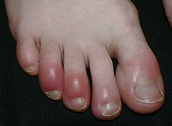
Chilblains, also known as pernio, is a medical condition in which damage occurs to capillary beds in the skin, most often in the hands or feet, when blood perfuses into the nearby tissue, resulting in redness, itching, inflammation, and possibly blisters. [2]
It occurs most frequently when predisposed individuals, predominantly women, [3] are exposed to cold and humidity. Ulcerated chilblains are referred to as kibes. Temperature-related chilblains can be prevented by keeping the feet and hands warm in cold weather and avoiding exposing these areas to extreme temperature changes. Once the diagnosis of chilblains is made, first-line treatment includes avoiding cold, damp environments and wearing gloves and warm socks. [3]
Chilblains can be idiopathic (spontaneous and unrelated to another disease), but similar symptoms may also be a manifestation of another serious medical condition that must be investigated. Related medical conditions include Raynaud syndrome, erythromelalgia, frostbite, and trench foot, as well as connective tissue diseases such as lupus or vasculitis. In infants affected by Aicardi–Goutières syndrome (a rare inherited condition which affects the nervous system) chilblain-like symptoms occur together with severe neurologic disturbances and unexplained fevers.
Dermatitis in extremities (toes, fingers, earlobes, nose, etc), including:
Chilblains caused by exposure to cold and humidity can usually heal within 7–14 days.
The word is a compound of Modern English chill ‘cold’ and the archaic word blain (now used only in the word in question), meaning ‘swelling’, ‘blister’ or ‘sore’ and derived from Old English bleġen, bleġene, having the same meaning. [10]
The medieval Bald's Leechbook recommended treating chilblains with a mixture of eggs, wine, and fennel root. [11] A modern-day home remedy is to put garlic on the chilblains. [12] While neither of these remedies has been supported by scientific research, vasodilating, rubiefacient and warming herbal remedies, both topical and internal are prescribed and efficacious.
Chilblain-like symptoms have also been linked to COVID-19. [13] [14] [15] COVID toes, as they are commonly known, [16] [17] [18] have mostly been reported in older children and adolescents, [19] [18] who often have not had other symptoms of COVID-19. [20] The symptoms are usually mild and disappear without treatment. [19] [18] Their cause is debated: it is uncertain whether COVID toes are a delayed consequence of the viral infection itself or are, at least partially, connected to environmental factors during the COVID-19 pandemic. [16] [17] [21] They may share some of the microscopic features of chilblains caused by lupus. [20] It has been suggested that in the absence of exposure to cold and damp, COVID-19 should be considered as a possible cause of chilblains. [20]
In a study at the dermatology department of Saint-Louis Hospital in Paris, researchers found that most of their study participants carried high levels of autoantibodies, proteins generated by the immune system that inadvertently attack the body's own tissues. Compared with healthy individuals, the participants showed high activity of proteins called type 1 interferons, which switch on pathogen-fighting genes in immune cells. [22]
A plantar wart is a wart occurring on the bottom of the foot or toes. Its color is typically similar to that of the skin. Small black dots often occur on the surface. One or more may occur in an area. They may result in pain with pressure such that walking is difficult.
Frostbite is a skin injury that occurs when someone is exposed to extremely low temperatures, causing the freezing of the skin or other tissues, commonly affecting the fingers, toes, nose, ears, cheeks and chin areas. Most often, frostbite occurs in the hands and feet. The initial symptoms are typically a feeling of cold and tingling or numbing. This may be followed by clumsiness with a white or bluish color to the skin. Swelling or blistering may occur following treatment. Complications may include hypothermia or compartment syndrome.
Raynaud syndrome, also known as Raynaud's phenomenon, is a medical condition in which the spasm of small arteries causes episodes of reduced blood flow to end arterioles. Typically the fingers, and, less commonly, the toes, are involved. Rarely, the nose, ears, nipples, or lips are affected. The episodes classically result in the affected part turning white and then blue. Often, numbness or pain occurs. As blood flow returns, the area turns red and burns. The episodes typically last minutes but can last several hours. The condition is named after the physician Auguste Gabriel Maurice Raynaud, who first described it in his doctoral thesis in 1862.
Rosacea is a long-term skin condition that typically affects the face. It results in redness, pimples, swelling, and small and superficial dilated blood vessels. Often, the nose, cheeks, forehead, and chin are most involved. A red, enlarged nose may occur in severe disease, a condition known as rhinophyma.
Athlete's foot, known medically as tinea pedis, is a common skin infection of the feet caused by a fungus. Signs and symptoms often include itching, scaling, cracking and redness. In rare cases the skin may blister. Athlete's foot fungus may infect any part of the foot, but most often grows between the toes. The next most common area is the bottom of the foot. The same fungus may also affect the nails or the hands. It is a member of the group of diseases known as tinea.
Calcinosis cutis is an uncommon condition marked by calcium buildup in the skin and subcutaneous tissues. Calcinosis cutis can range in intensity from little nodules in one area of the body to huge, crippling lesions affecting a vast portion of the body. Five kinds of the condition are typically distinguished: calciphylaxis, idiopathic calcification, iatrogenic calcification, dystrophic calcification, and metastatic calcification.
Pityriasis rosea is a type of skin rash. Classically, it begins with a single red and slightly scaly area known as a "herald patch". This is then followed, days to weeks later, by an eruption of many smaller scaly spots; pinkish with a red edge in people with light skin and greyish in darker skin. About 20% of cases show atypical deviations from this pattern. It usually lasts less than three months and goes away without treatment. Sometimes malaise or a fever may occur before the start of the rash or itchiness, but often there are few other symptoms.
Urticaria pigmentosa (also known as generalized eruption of cutaneous mastocytosis (childhood type) ) is the most common form of cutaneous mastocytosis. It is a rare disease caused by excessive numbers of mast cells in the skin that produce hives or lesions on the skin when irritated.
Lupus pernio is a chronic raised indurated (hardened) lesion of the skin, often purplish in color. It is seen on the nose, ears, cheeks, lips, and forehead. It is pathognomonic of sarcoidosis. The name "lupus pernio" is a misnomer, as microscopically this disease shows granulomatous infiltration and does not have features of either lupus nor pernio.
Erythema toxicum neonatorum is a common, non-threatening rash in newborns. It appears in 40-70% of newborns within the first week of life, and it typically improves within 1–2 weeks. It only occurs during the newborn period, but may appear slightly later in premature babies. The rash has a variable appearance. It typically includes blotchy red spots, often with overlying firm, yellow-white bumps or pus-filled boils. There may be only a few or many lesions. The lesions can appear almost anywhere on the body, and individual lesions may appear and disappear within hours. There are no other symptoms associated with erythema toxicum neonatorum, and the rash does not have any long-term effects on the skin. Erythema toxicum neonatorum is not harmful and does not require any treatment.
Proctalgia fugax, a variant of levator ani syndrome, is a severe, episodic pain in the regions of the rectum and anus. It can be caused by cramping of the levator ani muscle, particularly in the pubococcygeal part.
Hot tub folliculitis, also called Pseudomonalfolliculitis or Pseudomonas aeruginosa folliculitis, is a common type of folliculitis featuring inflammation of hair follicles and surrounding skin.
Chilblain lupus erythematosus was initially described by Hutchinson in 1888 as an uncommon manifestation of chronic cutaneous lupus erythematosus. Chilblain lupus erythematosus is characterized by a rash that primarily affects acral surfaces that are frequently exposed to cold temperatures, such as the toes, fingers, ears, and nose. The rash is defined by oedematous skin, nodules, and tender plaques with a purple discoloration.
Annular erythema of infancy(AEI) consists of self-limited eruptions of erythematous, annular to polycyclic patches and plaques. It is an idiopathic figurate erythema. Over several days, a single lesion disappears without leaving behind any scale or hyperpigmentation. Mostly affecting the trunk, face, and extremities, this rash has no symptoms. The diagnosis of AEI is made through a combination of histopathologic and clinical examinations. The disease first manifests in infancy, and if treatment is not received, the periodic eruptions usually stop after the first year of life.
Nakajo syndrome, also called nodular erythema with digital changes, is a rare autosomal recessive congenital disorder first reported in 1939 by A. Nakajo in the offspring of consanguineous parents. The syndrome can be characterized by erythema, loss of body fat in the upper part of the body, and disproportionately large eyes, ears, nose, lips, and fingers.
Senile pruritus is one of the most common conditions in the elderly or people over 65 years of age with an emerging itch that may be accompanied with changes in temperature and textural characteristics. In the elderly, xerosis, is the most common cause for an itch due to the degradation of the skin barrier over time. However, the cause of senile pruritus is not clearly known. Diagnosis is based on an elimination criteria during a full body examination that can be done by either a dermatologist or non-dermatologist physician.
Sucking blisters are a cutaneous condition on newborns that is present on their extremities at birth. This is not to be confused with sucking pads, which are similar in appearance. Sucking blisters are due to natural neonatal sucking reflexes on the affected areas in utero. These sucking blisters become present at birth when the sucking movements are very aggressive while in the uterus. These blisters are commonly found on the newborn's arms, fingers, or any body part they are able to suck on. It is a very rare condition, seen in less than 1% of newborns. It is difficult to diagnose because its appearance can be mistaken as other skin conditions. Diagnosis is usually done by eliminating other possible cutaneous conditions. This process of elimination includes examining blood culture, lesions, and inflammatory markers. This skin condition is usually self-inflicted, benign and can heal on its own. Sucking blisters do typically not require medical treatment and disappear within a week.
Cold injury is damage to the body from cold exposure, including hypothermia and several skin injuries. Cold-related skin injuries are categorized into freezing and nonfreezing cold injuries. Freezing cold injuries involve tissue damage when exposed to temperatures below freezing. Nonfreezing cold injuries involve tissue damage when exposed to temperatures often between 0-15 degrees Celsius for extended periods of time. While these injuries have disproportionally affected military members, recreational winter activities have also increased the risk and incidence within civilian populations. Additional risk factors include homelessness, inadequate or wet clothing, alcohol abuse or tobacco abuse, and pre-existing medical conditions that impair blood flow.
Non-freezing cold injuries (NFCI) is a class of tissue damage caused by sustained exposure to low temperature without actual freezing. There are several forms of NFCI, and the common names may refer to the circumstances in which they commonly occur or were first described, such as trench foot, which was named after its association with trench warfare. NFCI is caused by microvascular endothelial damage, stasis and vascular occlusion and is characterised by peripheral neuropathy. NFCI generally affects the hands or feet during exposure to temperatures just above freezing, often wet, and is typically found in soldiers.
Cutaneous manifestations of COVID-19 are characteristic signs or symptoms of the Coronavirus disease 2019 that occur in the skin. The American Academy of Dermatology reports that skin lesions such as morbilliform, pernio, urticaria, macular erythema, vesicular purpura, papulosquamous purpura and retiform purpura are seen in people with COVID-19. Pernio-like lesions were more common in mild disease while retiform purpura was seen only in critically ill patients. The major dermatologic patterns identified in individuals with COVID-19 are urticarial rash, confluent erythematous/morbilliform rash, papulovesicular exanthem, chilbain-like acral pattern, livedo reticularis and purpuric "vasculitic" pattern. Chilblains and Multisystem inflammatory syndrome in children are also cutaneous manifestations of COVID-19.
In acral chilblain-like lesions, a diffuse dense lymphoid infiltrate of the superficial and deep dermis, as well as hypodermis, with a prevalent perivascular pattern and signs of endothelial activation, are observed.