Cunninghamella bertholletiae

Last updated

Cunninghamella bertholletiae
Scientific classification
Kingdom:
Division:
Class:
Order:
Genus:
Species:
C. bertholletiae
Binomial name
Cunninghamella bertholletiae
Stadel (1911)

Cunninghamella bertholletiae is a species of zygomycetous fungi in the order Mucorales . [1] It is found globally, with increased prevalence in Mediterranean and subtropical climates. [2] It typically grows as a saprotroph and is found in a wide variety of substrates, including soil, fruits, vegetables, nuts, crops, and human and animal waste. [2] Although infections are still rare, C. betholletiae is emerging as an opportunistic human pathogen, predominantly in immunocompromised people, leukemia patients, and people with uncontrolled diabetes. [1] [2] [3] Cunninghamella bertholletiae infections are often highly invasive, and can be more difficult to treat with antifungal drugs than infections with other species of the Mucorales, making prompt and accurate recognition and diagnosis of mycoses caused by this fungus an important medical concern. [2] [3]

Contents

Growth and morphology

Cunninghamella bertholletiae grows as a mold. [3] Individual cells appear hyaline, but masses of fungi are darker in colour. [4] Colonies initially appear white, and become grey and powdery when they sporulate. [4] Cunninghamella bertholletiae displays very rapid growth on Sabouraud's agar (up to 20mm per day), which differentiates it from members of the Ascomycota and Basidiomycota. [2] However, culturing clinical materials infected by this species has been known to yield false negative results. [3] This species has very wide (10-20 μm), aseptate or partially septate hyphae, which contributes to a high capacity for cytoplasmic streaming. [2] Cytoplasmic streaming allows rapid diffusion of nutrients from a local nutrient source, which causes high growth rates and rapid nutrient depletion in culture or on limited substrates. [2] Like other members of the order Mucorales, C. bertholletiae is thermotolerant, [3] with a maximum growth temperature of 45-50˚C. [2] [4]

Hyphae branch at right angles and may appear twisted. [2] When growing in animal tissue, hyphae spread in all dimensions. [2] Cunninghamella bertholletiae produces spores in globose sporangia atop sporangiophores that are typically tall enough to be visible without a microscope. [2] Sporangiophores vary in length, and branch laterally to form concentric circles of shorter branches. [2] They lack the columella and apophysis present in sporangiophores of many other species of the Mucorales. [2] Due to the appearance of molds in this taxonomic order (a long stalk with a round, upward-pointing tip), members are often called "pin molds". [2] Unlike other members of the Mucorales, Cunninghamella species produce only one spore in each sporangium. [2] Sporangia form a halo around a central, round vesicle at the apex of a sporangiophore. [2] Spores are round to oval in shape and rough, with small spines or wart-like bumps. [4] The hyphae of C. bertholletiae may or may not produce rhizoids at the base of the sporangiophores. [2]

Physiology and reproduction

The formation of zygospores by two mating gametangia. Zygospore, Otto's Encyclopedia.jpg
The formation of zygospores by two mating gametangia.

As previously mentioned, C. bertholletiae grow hyphally and reproduce asexually via branching sporangiophores. Unlike in the case of dimorphic pathogenic fungi, [2] growth of C. bertholletiae is inhibited by cycloheximide. [2]

As a member of the Zygomycota, sexual reproduction in C. bertholletiae is through the formation of zygospores. [2] Specifically, in the case of C. bertholletiae, heterothallic mating occurs when hyphae of opposite mating types are stimulated by mutually-secreted pheromones to grow toward each other and differentiate into gametangia. [2] When they meet, these gametangia fuse (plasmogamy) and form a multinucleate, dikaryotic zygosporangium flanked by suspensor cells derived from the contributing hyphae. [2] Each zygosporangium produces one zygospore, which, after a dormant period of weeks to months, undergoes nuclear fusion (karyogamy) to produce a diploid nucleus. The diploid nucleus then undergoes meiosis and chromosomes recombine to produce recombinant progeny genomes. [2] A germosporangium forms, containing haploid spores, which are released into the environment to initiate the growth of a new mycelium. [2]

Cunninghamella betholletiae is not used widely in industry, but it is applied in industrial bioconversion to produce polyunsaturated acids. [2]

Habitat and ecology

Cunninghamella bertholletiae is found globally as a fruit and vegetable pathogen, as well as a cause of fruit and vegetable wastage due to rotting. However, it is more common in Mediterranean and subtropical zones than in temperate zones, and can grow at higher temperatures. [4] Its usual life cycle involves saprotrophy, and it is commonly found on dung, [5] rotting vegetables, fruit, nuts and seeds, soil, compost, sewage, and peat. Cunnginhamella bertholletiae can cause significant infections in agricultural crops. Hosts include plants in the genera Daucus , Gossypium and Tetragonia . [5] Cunninghamella bertholletiae can also be an opportunistic pathogen of both humans and animals, mainly in immunocompromised hosts. [2] [3] It can be transmitted between ecological niches via water and air. [2] The vast majority of the time, human infection is through airborne spores, although infections of deep wounds and medical devices can also occur through water contamination. [2] [3]

Human disease

Role in human disease

Cunninghamella bertholletiae is by far the most predominant opportunistic human pathogen of the genus Cunninghamella . [1] [2] Infections with this fungus are classified as opportunistic zygomycoses [3] or mucormycoses, [2] and risk factors for infection are similar for other mucormycoses, including diabetic ketoacidosis, and immunosuppression from chemotherapy, organ transplantation, and malnutrition. [3] Leukemia is a particularly high risk factor. [1] HIV-associated cases have been reported, but serious cases are more often seen in leukemia patients. [1] Disseminated infections have also been noted in renal and hepatic transplant patients. [1] Infection often occurs through traumatic introductions into the body (i.e. through a wound). [2] [3]

Cunninghamella bertholletiae can infect a wide variety of human tissue types, [3] exhibits hyphal growth in the body [6] and is angioinvasive. [2] Like other Mucorales, under appropriate host conditions, it can grow very aggressively and destroy tissue structure. [3] Typically, initial pathology is from thrombosis and infarction. [7] Common classes of mucormycoses include pulmonary, rhinocerebral (particularly when invasion into the vasculature of the brain is involved), multi-organ, cutaneous, and gastrointestinal (primarily in premature babies and malnourished children). Rhinocerebral infection and gastrointestinal cases are most immediately life-threatening. [3] Pulmonary infections, as well as disseminated infections with pulmonary origins, are most common for C. bertholletiae, which has been identified in 7% of mucormycosis cases globally, and 3.2% of cases in the United States. [2]

Diagnosis and treatment

A case of periorbiatal mucormycosis, a sign of highly invasive rhinocerebral C. bertholletiae infection. Periorbital fungal infection known as mucormycosis, or phycomycosis PHIL 2831 lores.jpg
A case of periorbiatal mucormycosis, a sign of highly invasive rhinocerebral C. bertholletiae infection.

Although C. bertholletiae is only responsible for a small percentage of mucormycoses, it is cited as having the worst prognosis of the Mucorales. [8] There are few identified cases per year, but C. bertholletiae infections and other mucormycoses are increasing in prevalence in North America, possibly due to growing populations of aging and immunosuppressed people. [2] [3]

Vascular invasion and tissue necrosis, often with black discharge, are good indicators of infection with Mucorales. [9] Cunninghamella bertholletiae can also grow at higher temperatures, which can be helpful in testing contaminated surfaces to differentiate between benign and pathogenic fungi. [7] Infections from the six different taxonomic families of Mucorales have virtually indistinguishable clinical courses. [2] [3] Furthermore, the difficulty of culturing C. bertholletiae and other species within Mucorales from tissue samples [2] makes laboratory analysis necessary to determine the causative organism of a mucormycosis. [9] Polymerase chain reaction-based sequencing of fungal isolates is preferred as a reliable diagnostic tool due to possible difficulty of isolating C. bertholletiae from patients in culture. [3] However, preliminary antifungal treatment should never be delayed if C. bertholletiae infection is suspected, as infections can often cause rapid and invasive tissue damage. [3] Genetic differences within the species C. bertholletiae can also be important determinants of pathogenicity and virulence. [8] Recently, DNA barcoding of the internal transcribed spacer (ITS) region of C. bertholletiae ribosomal DNA was performed to improve upon current diagnostic techniques, providing more accurate and detailed between- and within-species discrimination compared to traditional analysis of colony colour and morphology, maximum growth temperature, and reproductive characteristics. [8]

Because of its fast growth and invasiveness, treatment for C. bertholletiae infection can be expected to often require surgery in addition to antifungal treatment. [3] Immediate surgery is especially important in the case of rhinocerebral infection, in order to avoid dissemination into the vasculature of the brain and to avoid permanent optic nerve damage. [3] Surgical debridement is a common treatment. [3] Bacterial superinfection of debrided tissues after treatment can therefore be a significant problem. [7] Antifungal drugs that are used successfully against C. bertholletiae infection include amphotericin B, itraconazole, voriconazole and posaconazole. [4] However, compared to other Mucorales species, C. bertholletiae has decreased responsiveness to some antifungals that are commonly prescribed to treat mucormycoses, and samples should be tested for individual antibiotic susceptibility if possible. [2] Lipid formulations of amphotericin B are preferred for treatment of C. berthollettiae, because the high dosage required to treat infection can have significant toxic effects when administered in traditional formulations. [3] Relapse after antifungal treatment and surgery is rare if a patient's clinical course initially improves during therapy. [7]

Special case: ketoacidotic diabetes and iron availability

In cases of uncontrolled diabetes, where ketoacidosis is present and glucose levels are above 12mM, C. bertholletiae infection is promoted and can be highly invasive. [3] This may related to effects of low pH on iron acquisition, as low pH decreases affinity of transferrin for iron, freeing iron from its usual sequestration in blood and making if more available for fungal exploitation. [3] Because a primary factor in patient susceptibility to C. bertholletiae is increased iron availability, any condition that increases blood iron availability creates an increased risk of infection. [2] Patients undergoing iron chelation therapy with deferoxamine are also at risk of infection, because this treatment also increases accessible iron for C. bertholletiae. Furthermore, C. bertholletiae was identified as the causative agent of a fatal case of rhinocerebral mucormycosis in a patient with hemochromatosis, [10] expanding the recognized risk factors for infection.

Related Research Articles

Zygomycosis

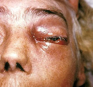
Zygomycosis is the broadest term to refer to infections caused by bread mold fungi of the zygomycota phylum. However, because zygomycota has been identified as polyphyletic, and is not included in modern fungal classification systems, the diseases that zygomycosis can refer to are better called by their specific names: mucormycosis, phycomycosis and basidiobolomycosis. These rare yet serious and potentially life-threatening fungal infections usually affect the face or oropharyngeal cavity. Zygomycosis type infections are most often caused by common fungi found in soil and decaying vegetation. While most individuals are exposed to the fungi on a regular basis, those with immune disorders (immunocompromised) are more prone to fungal infection. These types of infections are also common after natural disasters, such as tornadoes or earthquakes, where people have open wounds that have become filled with soil or vegetative matter.

Mucorales Order of fungi

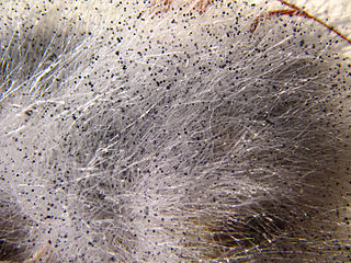
The Mucorales is the largest and best studied order of zygomycete fungi. Members of this order are sometimes called pin molds. The term mucormycosis is now preferred for infections caused by molds belonging to the order Mucorales.

<i>Mucor</i> Genus of fungi

Mucor is a microbial genus of approximately 40 species of moulds commonly found in soil, digestive systems, plant surfaces, some cheeses like tomme de savoie, rotten vegetable matter and iron oxide residue in the biosorption process.

<i>Aspergillus terreus</i> Species of fungus

Aspergillus terreus, also known as Aspergillus terrestris, is a fungus (mold) found worldwide in soil. Although thought to be strictly asexual until recently, A. terreus is now known to be capable of sexual reproduction. This saprotrophic fungus is prevalent in warmer climates such as tropical and subtropical regions. Aside from being located in soil, A. terreus has also been found in habitats such as decomposing vegetation and dust. A. terreus is commonly used in industry to produce important organic acids, such as itaconic acid and cis-aconitic acid, as well as enzymes, like xylanase. It was also the initial source for the drug mevinolin (lovastatin), a drug for lowering serum cholesterol.

Exophiala jeanselmei is a saprotrophic fungus in the family Herpotrichiellaceae. Four varieties have been discovered: Exophiala jeanselmei var. heteromorpha, E. jeanselmei var. lecanii-corni, E. jeanselmei var. jeanselmei, and E. jeanselmei var. castellanii. Other species in the genus Exophiala such as E. dermatitidis and E. spinifera have been reported to have similar annellidic conidiogenesis and may therefore be difficult to differentiate.

Mucormycosis

Mucormycosis is any fungal infection caused by fungi in the order Mucorales. Generally, species in the Mucor,Rhizopus,Absidia, and Cunninghamella genera are most often implicated.

<i>Pseudallescheria boydii</i> Species of fungus

Pseudallescheria boydii is a species of fungus classified in the Ascomycota. It is associated with some forms of eumycetoma, maduromycosis and pseudallescheriasis. Typically found in stagnant and polluted water, it has been implicated in the infection of immunocompromised and near-drowned pneumonia patients. Its asexual (anamorphic) form is Scedosporium apiospermum. Treatment of infections with P. boydii is complicated by its resistance to many of the standard antifungal agents normally used to treat infections by filamentous fungi.

Cunninghamellaceae Family of fungi

The Cunninghamellaceae are a family of fungi in the order Mucorales.

Saksenaea vasiformis is an infectious fungus associated with cutaneous or subcutaneous lesions following trauma. It causes opportunistic infections as the entry of the fungus is through open spaces of cutaneous barrier ranging in severity from mild to severe or fatal. It is found in soils worldwide, but is considered as a rare human pathogen since only 38 cases were reported as of 2012. Saksenaea vasiformis usually fails to sporulate on the routine culture media, creating a challenge for early diagnosis, which is essential for a good prognosis. Infections are usually treated using a combination of amphotericin B and surgery. Saksenaea vasiformis is one of the few fungi known to cause necrotizing fasciitis or "flesh-eating disease".

Rhizomucor pusillus is a species of Rhizomucor. It can cause disease in humans. R. pusillus is a grey mycelium fungi most commonly found in compost piles. Yellow-brown spores grow on a stalk to reproduce more fungal cells.

Fungal folliculitis Inflammation of hair follicles due to fungal infection

Majocchi's granuloma is a skin condition characterized by deep, pustular plaques, and is a form of tinea corporis. It is a localized form of fungal folliculitis. Lesions often have a pink and scaly central component with pustules or folliculocentric papules at the periphery. The name comes from Professor Domenico Majocchi, who discovered the disorder in 1883. Majocchi was a professor of dermatology at the University of Parma and later the University of Bologna. The most common dermatophyte is called Trichophyton rubrum.

<i>Apophysomyces variabilis</i> Species of fungus

Apophysomyces variabilis is an emerging fungal pathogen that can cause serious and sometimes fatal infection in humans. This fungus is a soil-dwelling saprobe with tropical to subtropical distribution. It is a zygomycete that causes mucormycosis, an infection in humans brought about by fungi in the order Mucorales. Infectious cases have been reported globally in locations including the Americas, Southeast Asia, India, and Australia. Apophysomyces variabilis infections are not transmissible from person to person.

Coniochaeta hoffmannii, also known as Lecythophora hoffmannii, is an ascomycete fungus that grows commonly in soil. It has also been categorized as a soft-rot fungus capable of bringing the surface layer of timber into a state of decay, even when safeguarded with preservatives. Additionally, it has pathogenic properties, although it causes serious infection only in rare cases. A plant pathogen lacking a known sexual state, C. hoffmannii has been classified as a "dematiaceous fungus" despite its contradictory lack of pigmentation; both in vivo and in vitro, there is no correlation between its appearance and its classification.

<i>Lichtheimia corymbifera</i> Species of fungus

Lichtheimia corymbifera is a thermophilic fungus in the phylum Zygomycota. It normally lives as a saprotrophic mold, but can also be an opportunistic pathogen known to cause pulmonary, CNS, rhinocerebral, or cutaneous infections in animals and humans with impaired immunity.

<i>Cunninghamella echinulata</i> Species of fungus

Cunninghamella echinulata is a fungal species in the genus Cunninghamella. It is an asexually reproducing fungus and a mesophile, preferring intermediate temperature ranges. C. echinulata is a common air contaminant, and is currently of interest to the biotechnology industry due to its ability to synthesize γ-linolenic acid as well as its capacity to bioconcentrate metals. This species is a soil saprotroph that forms rhizoids, preferring soils enriched in nitrogen, phosphorus and potassium. It has been reported occasionally an agent of mucormycosis following the inhalation of fungal spores. Czapek's agar is a suitable growth medium for the propagation of C. echinulata.

<i>Rhizopus stolonifer</i> Species of fungus

Rhizopus stolonifer is commonly known as black bread mold. It is a member of Zygomycota and considered the most important species in the genus Rhizopus. It is one of the most common fungi in the world and has a global distribution although it is most commonly found in tropical and subtropical regions. It is a common agent of decomposition of stored foods. Like other members of the genus Rhizopus, R. stolonifer grows rapidly, mostly in indoor environments.

Sarocladium kiliense is a saprobic fungus that is occasionally encountered as a opportunistic pathogen of humans, particularly immunocompromised and individuals. The fungus is frequently found in soil and has been linked with skin and systemic infections. This species is also known to cause disease in the green alga, Cladophora glomerata as well as various fruit and vegetable crops grown in warmer climates.

Microascus manginii is a filamentous fungal species in the genus Microascus. It produces both sexual (teleomorph) and asexual (anamorph) reproductive stages known as M. manginii and Scopulariopsis candida, respectively. Several synonyms appear in the literature because of taxonomic revisions and re-isolation of the species by different researchers. M. manginii is saprotrophic and commonly inhabits soil, indoor environments and decaying plant material. It is distinguishable from closely related species by its light colored and heart-shaped ascospores used for sexual reproduction. Scopulariopsis candida has been identified as the cause of some invasive infections, often in immunocompromised hosts, but is not considered a common human pathogen. There is concern about amphotericin B resistance in S. candida.

Arthrographis kalrae is an ascomycetous fungus responsible for human nail infections described in 1938 by Cochet as A. langeronii. A. kalrae is considered a weak pathogen of animals including human restricted to the outermost keratinized layers of tissue. Infections caused by this species are normally responsive to commonly used antifungal drugs with only very rare exceptions.

Mycotypha microspora, also known as Microtypha microspora, is a filamentous fungus in the division Zygomycota. It was discovered in a Citrus aurantium peel in 1932 by E. Aline Fenner, who proposed a new genus Mycotypha to accommodate it. Mycotypha africana, which is another species in the genus Mycotypha, is closely related to M. microspora. The fungus has subsequently been isolated from both outdoor and indoor settings around the world, and is typically found in soil and dung. The species rarely causes infections in humans, but has recently been involved in the clinical manifestation of the life-threatening disease mucormycosis.

References

  1. 1 2 3 4 5 6 "Cunninghamella Bertholletiae". Mycobank. Retrieved 11 November 2015.
  2. 1 2 3 4 5 6 7 8 9 10 11 12 13 14 15 16 17 18 19 20 21 22 23 24 25 26 27 28 29 30 31 32 33 34 35 36 37 38 Reiss, E; et al. (2011). Fundamental Medical Mycology. Hoboken, New Jersey: Wiley-Blackwell.
  3. 1 2 3 4 5 6 7 8 9 10 11 12 13 14 15 16 17 18 19 20 21 22 23 Kwon-Chung, K. June; Bennett, Joan E. (1992). Medical mycology. Philadelphia: Lea & Febiger. ISBN   0812114639.
  4. 1 2 3 4 5 6 Ellis, David. "Cunninhamella bertholletiae". Mycology Online. University of Adelaide. Archived from the original on 22 October 2015. Retrieved 18 October 2015.
  5. 1 2 Farr, David F.; Bills, Gerald F.; Chamuris, George P.; Rossman, Amy Y. (1989). Fungi on Plant Products in the United States. American Phytological Society.
  6. Honda, A.; Kamei, K.; Unno, H.; Hiroshima, K.; Kuriyama, T.; Miyaji, M. (1998). "A murine model of zygomycosis by Cunninghamella bertholletiae". Mycopathologia. 144 (3): 141–146. doi:10.1023/A:1007095831301. PMID   10531680. S2CID   6832679.
  7. 1 2 3 4 St-Germain, Guy; Summberbell, Richard (2011). Identifying Fungi: A Clinical Laboratory Handbook (2nd ed.). Belmont, CA: Star Publishing Company. ISBN   9780898631777.
  8. 1 2 3 Yu, J; Walther, G; Van Diepeningen, AD; Gerrits Van Den Ende, AH; Li, RY; Moussa, TA; Almaghrabi, OA; De Hoog, GS (1 February 2015). "DNA barcoding of clinically relevant Cunninghamella species". Medical Mycology. 53 (2): 99–106. doi: 10.1093/mmy/myu079 . PMID   25431472.
  9. 1 2 Mandell, Gerald L.; Bennett, John E.; Dolin, Raphael. Principles and Practice of Infectious Disease.
  10. Khan, Fida A.; Fisher, Melanie A.; Khakoo, Rashida A. (2007). "Association of hemochromatosis with infectious diseases: expanding spectrum". International Journal of Infectious Diseases. 11 (6): 482–487. doi: 10.1016/j.ijid.2007.04.007 . PMID   17600748.