Dural arteriovenous fistula

Last updated
Dural arteriovenous fistula
DAVF-SSS-IIIb.jpg
This dural arteriovenous fistula of the superior sagittal sinus drains into subarachnoid veins and is classified as Borden type IIIb.
Specialty
Symptoms
Complications Hemorrhagic stroke, cardiomegaly
Diagnostic method Medical imaging:
TreatmentArteriovenous fistula obliteration, including:

A dural arteriovenous fistula (DAVF) or malformation is an abnormal direct connection (fistula) between a meningeal artery and a meningeal vein or dural venous sinus.

Contents

Signs and symptoms

The most common signs/symptoms of DAVFs are: [1]

  1. Pulsatile tinnitus
  2. Occipital bruit
  3. Headache
  4. Visual impairment
  5. Papilledema

Pulsatile tinnitus is the most common symptom in patients, and it is associated with transverse-sigmoid sinus DAVFs. [1] Carotid-cavernous DAVFs, on the other hand, are more closely associated with pulsatile exophthalmos. DAVFs may also be asymptomatic (e.g. cavernous sinus DAVFs). [2]

Location

Most commonly found adjacent to dural sinuses in the following locations: [2]

  1. Transverse (lateral) sinus, [3] left-sided slightly more common than right [4]
  2. Intratentorial
  3. From the posterior cavernous sinus, usually draining to the transverse or sigmoid sinuses
  4. Vertebral artery (posterior meningeal branch) [2]

Causes

It is still unclear whether DAVFs are congenital or acquired. [5] Current evidence supports transverse-sigmoid sinus junction dural malformations are acquired defects, occurring in response to thrombosis and collateral revascularization of a venous sinus. [6] [1] [7]

Diagnosis

Cerebral angiography is the diagnostic standard. MRIs are typically normal but can identify venous hypertension as a result of arterial-venous shunting. [6]

Classification

Borden Classification

The Borden Classification of dural arteriovenous malformations or fistulas, groups into three types based upon their venous drainage: [8]

  1. Type I: dural arterial supply drains anterograde into venous sinus.
  2. Type II: dural arterial supply drains into venous sinus. High pressure in sinus results in both anterograde drainage and retrograde drainage via subarachnoid veins.
  3. Type III: dural arterial supply drains retrograde into subarachnoid veins.
Type I

Type I dural arteriovenous fistulas are supplied by meningeal arteries and drain into a meningeal vein or dural venous sinus. The flow within the draining vein or venous sinus is anterograde.

  1. Type Ia Archived 2007-03-12 at the Wayback Machine – simple dural arteriovenous fistulas have a single meningeal arterial supply
  2. Type Ib Archived 2007-03-12 at the Wayback Machine – more complex arteriovenous fistulas are supplied by multiple meningeal arteries

The distinction between Types Ia and Ib is somewhat specious as there is a rich system of meningeal arterial collaterals. Type I dural fistulas are often asymptomatic, do not have a high risk of bleeding and do not necessarily need to be treated.[ citation needed ]

Type II

The high pressure within a Type II Archived 2007-03-12 at the Wayback Machine dural AV fistula causes blood to flow in a retrograde fashion into subarachnoid veins which normally drain into the sinus. Typically this is because the sinus has outflow obstruction. Such draining veins form venous varices or aneurysms which can bleed. Type II fistulas need to be treated to prevent hemorrhage. The treatment may involve embolization of the draining sinus as well as clipping or embolization of the draining veins.

Type III

Type III dural AV fistulas drain directly into subarachnoid veins. [9] These veins can form aneurysms and bleed. Type III dural fistulas need to be treated to prevent hemorrhage. Treatment can be as simple as clipping the draining vein at the site of the dural sinus. If treatment involves embolization, it will only typically be effective if the glue traverses the actual fistula and enters, at least slightly, the draining vein.[ citation needed ]

The Cognard et al. Classification [10] correlates venous drainage patterns with increasingly aggressive neurological clinical course.

ClassificationLocation and clinical course
Type IConfined to sinus wall, typically after thrombosis.
Type IIIIa - confined to sinus with reflux (retrograde) into sinus but not cortical veins.

IIb - drains into sinus with reflux (retrograde) into cortical veins (10-20% hemorrhage).

Type IIIDrains direct into cortical veins (not into sinus) drainage (40% hemorrhage).
Type IVDrains direct into cortical veins (not into sinus) drainage with venous ectasia (65% hemorrhage).
Type VSpinal perimedullary venous drainage, associated with progressive myelopathy.

To simplify the above systems of DAVF classification, the two main factors that should be considered to determine aggressiveness of these lesions are:

  • DAVF that have bleed (as opposed to those that have not before)
  • DAVF resulting in cortical venous reflux

Treatment decisions are more complicated and require consultation with a neurosurgeon and team familiar with these lesions.

Treatment

Indications

Interventions

Embolization

One approach used for treatment is embolization. [11] A six-vessel angiogram is employed to determine the vascular supply to the fistula. [5] Detachable coils, liquid embolic agents like NBCA, and onyx, or combinations of both are injected into the blood vessel to occlude the DAVF. [2] Preoperative embolization can also be used to supplement surgery. [12]

Surgery

DAVFs are also managed surgically. The operative approach varies depending on the location of the lesion. [5]

Stereotactic radiosurgery

Stereotactic radiosurgery is used for obliterating DAVFs sometimes in conjunction with embolization or surgery, and is considered an important adjunct and sometimes a primary treatment method for non-aggressive DAVFs. [13] Use of this method, however, is limited as obliteration occurs over the course of up to 2–3 years after the delivery of radiation.[ citation needed ]

Epidemiology

10–15% of intracranial AV malformations are DAVFs. [4] There is a higher preponderance in females (61–66%), and typically patients are in their fourth or fifth decade of life. DAVFs are rarer in children. [2]

Research

External Manual Carotid Compression is Effective in Patients with Cavernous Sinus Dural Arteriovenous Fistulaetreatment. The patients were instructed to compress the carotid artery and jugular vein with the contralateral hand for ten seconds several times each hour (about 6 to 15 times per day).

See also

Related Research Articles

<span class="mw-page-title-main">Arteriovenous malformation</span> Vascular anomaly

An arteriovenous malformation (AVM) is an abnormal connection between arteries and veins, bypassing the capillary system. Usually congenital, this vascular anomaly is widely known because of its occurrence in the central nervous system, but can appear anywhere in the body. The symptoms of AVMs can range from none at all to intense pain or bleeding, and they can lead to other serious medical problems.

<span class="mw-page-title-main">Cerebral arteriovenous malformation</span> Medical condition

A cerebral arteriovenous malformation is an abnormal connection between the arteries and veins in the brain—specifically, an arteriovenous malformation in the cerebrum.

<span class="mw-page-title-main">Vein</span> Blood vessels that carry blood towards the heart

Veins are blood vessels in the circulatory system of humans and most other animals that carry blood towards the heart. Most veins carry deoxygenated blood from the tissues back to the heart; exceptions are those of the pulmonary and fetal circulations which carry oxygenated blood to the heart. In the systemic circulation, arteries carry oxygenated blood away from the heart, and veins return deoxygenated blood to the heart, in the deep veins.

<span class="mw-page-title-main">Dura mater</span> Outermost layer of the protective tissues around the central nervous system (meninges)

In neuroanatomy, dura mater is a thick membrane made of dense irregular connective tissue that surrounds the brain and spinal cord. It is the outermost of the three layers of membrane called the meninges that protect the central nervous system. The other two meningeal layers are the arachnoid mater and the pia mater. It envelops the arachnoid mater, which is responsible for keeping in the cerebrospinal fluid. It is derived primarily from the neural crest cell population, with postnatal contributions of the paraxial mesoderm.

<span class="mw-page-title-main">Great cerebral vein</span>

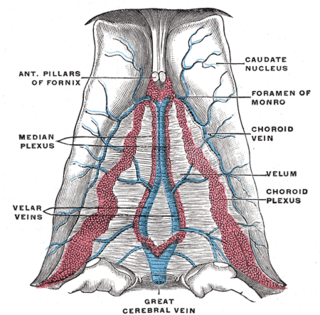
The great cerebral vein is one of the large blood vessels in the skull draining the cerebrum of the brain. It is also known as the vein of Galen, named for its discoverer, the Greek physician Galen.

<span class="mw-page-title-main">Intracranial hemorrhage</span> Hemorrhage, or bleeding, within the skull

Intracranial hemorrhage (ICH), also known as intracranial bleed, is bleeding within the skull. Subtypes are intracerebral bleeds, subarachnoid bleeds, epidural bleeds, and subdural bleeds.

<span class="mw-page-title-main">Cerebral angiography</span> Angiography that produces images of blood vessels in and around the brain

Cerebral angiography is a form of angiography which provides images of blood vessels in and around the brain, thereby allowing detection of abnormalities such as arteriovenous malformations and aneurysms. It was pioneered in 1927 by the Portuguese neurologist Egas Moniz at the University of Lisbon, who also helped develop thorotrast for use in the procedure.

<span class="mw-page-title-main">Arteriovenous fistula</span> Medical condition

An arteriovenous fistula is an abnormal connection or passageway between an artery and a vein. It may be congenital, surgically created for hemodialysis treatments, or acquired due to pathologic process, such as trauma or erosion of an arterial aneurysm.

<span class="mw-page-title-main">Carotid-cavernous fistula</span> Medical condition

A carotid-cavernous fistula results from an abnormal communication between the arterial and venous systems within the cavernous sinus in the skull. It is a type of arteriovenous fistula. As arterial blood under high pressure enters the cavernous sinus, the normal venous return to the cavernous sinus is impeded and this causes engorgement of the draining veins, manifesting most dramatically as a sudden engorgement and redness of the eye of the same side.

<span class="mw-page-title-main">Cavernous sinus</span> Sinus in the human head

The cavernous sinus within the human head is one of the dural venous sinuses creating a cavity called the lateral sellar compartment bordered by the temporal bone of the skull and the sphenoid bone, lateral to the sella turcica.

<span class="mw-page-title-main">Straight sinus</span> Venous sinus draining the brain

The straight sinus, also known as tentorial sinus or the sinus rectus, is an area within the skull beneath the brain. It receives blood from the inferior sagittal sinus and the great cerebral vein, and drains into the confluence of sinuses.

<span class="mw-page-title-main">Superior petrosal sinus</span> One of the dural venous sinuses located beneath the brain

The superior petrosal sinus is one of the dural venous sinuses located beneath the brain. It receives blood from the cavernous sinus and passes backward and laterally to drain into the transverse sinus. The sinus receives superior petrosal veins, some cerebellar veins, some inferior cerebral veins, and veins from the tympanic cavity. They may be affected by arteriovenous malformation or arteriovenous fistula, usually treated with surgery.

<span class="mw-page-title-main">Cerebellar veins</span> Veins that drain the cerebellum

The cerebellar veins are veins which drain the cerebellum. They consist of the superior cerebellar veins and the inferior cerebellar veins. The superior cerebellar veins drain to the straight sinus and the internal cerebral veins. The inferior cerebellar veins drain to the transverse sinus, the superior petrosal sinus, and the occipital sinus.
<span class="mw-page-title-main">Nasofrontal vein</span> Vein in the orbit around the eye

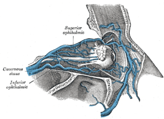
The nasofrontal vein is a vein in the orbit around the eye. It drains into the superior ophthalmic vein. It can be used for endovascular access to the cavernous sinus.

<span class="mw-page-title-main">Central retinal vein</span> Vein in the retina of the eye

The central retinal vein is a vein that drains the retina of the eye. It travels backwards through the centre of the optic nerve accompanied by the central retinal artery before exiting the optic nerve together with the central retinal artery to drain into either the superior ophthalmic vein or the cavernous sinus.

A vascular malformation is a blood vessel or lymph vessel abnormality. Vascular malformations are one of the classifications of vascular anomalies, the other grouping is vascular tumors. They may cause aesthetic problems as they have a growth cycle, and can continue to grow throughout life.

Vascular myelopathy refers to an abnormality of the spinal cord in regard to its blood supply. The blood supply is complicated and supplied by two major vessel groups: the posterior spinal arteries and the anterior spinal arteries—of which the Artery of Adamkiewicz is the largest. Both the posterior and anterior spinal arteries run the entire length of the spinal cord and receive anastomotic (conjoined) vessels in many places. The anterior spinal artery has a less efficient supply of blood and is therefore more susceptible to vascular disease. Whilst atherosclerosis of spinal arteries is rare, necrosis in the anterior artery can be caused by disease in vessels originating from the segmental arteries such as atheroma or aortic dissection.

<span class="mw-page-title-main">Vein of Galen aneurysmal malformations</span> Medical condition

Vein of Galen aneurysmal malformations(VGAMs) and Vein of Galen aneurysmal dilations (VGADs) are the most frequent arteriovenous malformations in infants and fetuses. A VGAM consists of a tangled mass of dilated vessels supplied by an enlarged artery. The malformation increases greatly in size with age, although the mechanism of the increase is unknown. Dilation of the great cerebral vein of Galen is a secondary result of the force of arterial blood either directly from an artery via an arteriovenous fistula or by way of a tributary vein that receives the blood directly from an artery. There is usually a venous anomaly downstream from the draining vein that, together with the high blood flow into the great cerebral vein of Galen causes its dilation. The right sided cardiac chambers and pulmonary arteries also develop mild to severe dilation.

Interventional neuroradiology (INR) also known as neurointerventional surgery (NIS), endovascular therapy (EVT), endovascular neurosurgery, and interventional neurology is a medical subspecialty of neurosurgery, neuroradiology, intervention radiology and neurology specializing in minimally invasive image-based technologies and procedures used in diagnosis and treatment of diseases of the head, neck, and spine.

Alexander Coon is an American neurosurgeon who is the Director of Endovascular and Cerebrovascular Neurosurgery at the Carondelet Neurological Institute of St. Joseph's and St. Mary's Hospitals in Tucson, Arizona. He was previously the Director of Endovascular Neurosurgery at the Johns Hopkins Hospital and an assistant professor of neurosurgery, Neurology, and Radiology at the Johns Hopkins Hospital. He is known for his work in cerebrovascular and endovascular neurosurgery and his research in neuroendovascular devices and clinical outcomes in the treatment of cerebral aneurysms, subarachnoid hemorrhage, and AVMs.

References

  1. 1 2 3 Sundt, Thoralf M.; Piepgras, David G. (1983-07-01). "The surgical approach to arteriovenous malformations of the lateral and sigmoid dural sinuses". Journal of Neurosurgery. 59 (1): 32–39. doi:10.3171/jns.1983.59.1.0032. ISSN   0022-3085. PMID   6864280.
  2. 1 2 3 4 5 6 Greenberg, Mark S. (2010-01-01). Handbook of neurosurgery. Greenberg Graphics. ISBN   978-1604063264. OCLC   892183792.
  3. Graeb, D. A.; Dolman, C. L. (1986-06-01). "Radiological and pathological aspects of dural arteriovenous fistulas. Case report". Journal of Neurosurgery. 64 (6): 962–967. doi:10.3171/jns.1986.64.6.0962. ISSN   0022-3085. PMID   3701446.
  4. 1 2 Arnautovic, Kenan I.; Krisht, Ali F. (July 1999). "Transverse-Sigmoid Sinus Dural Arteriovenous Malformations". Contemporary Neurosurgery. 21 (15): 1–6. doi:10.1097/00029679-199907020-00001.
  5. 1 2 3 Rahul, Jandial; R., Aizenberg, Michele; Y., Chen, Mike (2016-10-27). 100 case reviews in neurosurgery. Elsevier. ISBN   9780323356374. OCLC   966563367.{{cite book}}: CS1 maint: multiple names: authors list (link)
  6. 1 2 Polster, Sean P.; Zeineddine, Hussein A.; Baron, Joseph; Lee, Seon-Kyu; Awad, Issam A. (October 2018). "Patients with cranial dural arteriovenous fistulas may benefit from expanded hypercoagulability and cancer screening". Journal of Neurosurgery. 129 (4): 954–960. doi: 10.3171/2017.5.JNS17788 . ISSN   1933-0693. PMID   29148899.
  7. LaHue, Sara C.; Kim, Helen; Pawlikowska, Ludmila; Nelson, Jeffrey; Cooke, Daniel L.; Hetts, Steven W.; Singh, Vineeta (2018-04-01). "Frequency and characteristics associated with inherited thrombophilia in patients with intracranial dural arteriovenous fistula". Journal of Neurosurgery. 130 (4): 1346–1350. doi:10.3171/2017.10.JNS171987. ISSN   1933-0693. PMC   6173991 . PMID   29624150.
  8. Borden JA, Wu JK, Shucart WA (1995). "A proposed classification for spinal and cranial dural arteriovenous fistulous malformations and implications for treatment". Journal of Neurosurgery. 82 (2): 166–79. doi:10.3171/jns.1995.82.2.0166. PMID   7815143. S2CID   358930.
  9. "jonathanborden-md.com". Archived from the original on 2022-03-31. Retrieved 2007-12-22.
  10. Cognard, C.; Gobin, Y. P.; Pierot, L.; Bailly, A. L.; Houdart, E.; Casasco, A.; Chiras, J.; Merland, J. J. (1995). "Cerebral dural arteriovenous fistulas: clinical and angiographic correlation with a revised classification of venous drainage". Radiology. 194 (3): 671–80. doi:10.1148/radiology.194.3.7862961. PMID   7862961.
  11. Carlson AP, Taylor CL, Yonas H (2007). "Treatment of dural arteriovenous fistula using ethylene vinyl alcohol (onyx) arterial embolization as the primary modality: short-term results". Journal of Neurosurgery. 107 (6): 1120–5. doi:10.3171/JNS-07/12/1120. PMID   18077948. S2CID   27522333.
  12. Barnwell, Stanley L.; Halbach, Van V.; Higashida, Randall T.; Hieshima, Grant; Wilson, Charles B. (1989-09-01). "Complex dural arteriovenous fistulas". Journal of Neurosurgery. 71 (3): 352–358. doi:10.3171/jns.1989.71.3.0352. ISSN   0022-3085. PMID   2769386. S2CID   11425407.
  13. Lewis, Adam I.; Tomsick, Thomas A.; Tew, John M. (2009-05-28). "Management of tentorial dural arteriovenous malformations: transarterial embolization combined with stereotactic radiation or surgery". Journal of Neurosurgery. 15 (4): 851–859. doi:10.3171/jns.1994.81.6.0851. PMID   7965115.