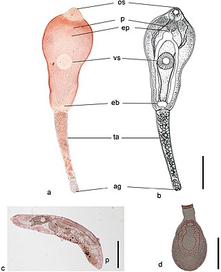
Trematoda is a class of flatworms known as flukes or trematodes. They are obligate internal parasites with a complex life cycle requiring at least two hosts. The intermediate host, in which asexual reproduction occurs, is usually a snail. The definitive host, where the flukes sexually reproduce, is a vertebrate. Infection by trematodes can cause disease in all five traditional vertebrate classes: mammals, birds, amphibians, reptiles, and fish.

Fasciola hepatica, also known as the common liver fluke or sheep liver fluke, is a parasitic trematode of the class Trematoda, phylum Platyhelminthes. It infects the livers of various mammals, including humans, and is transmitted by sheep and cattle to humans all over the world. The disease caused by the fluke is called fasciolosis or fascioliasis, which is a type of helminthiasis and has been classified as a neglected tropical disease. Fasciolosis is currently classified as a plant/food-borne trematode infection, often acquired through eating the parasite's metacercariae encysted on plants. F. hepatica, which is distributed worldwide, has been known as an important parasite of sheep and cattle for decades and causes significant economic losses in these livestock species, up to £23 million in the UK alone. Because of its relatively large size and economic importance, it has been the subject of many scientific investigations and may be the best-known of any trematode species. F. hepatica's closest relative is Fasciola gigantica. These two flukes are sister species; they share many morphological features and can mate with each other.

Trematodes are parasitic flatworms of the class Trematoda, specifically parasitic flukes with two suckers: one ventral and the other oral. Trematodes are covered by a tegument, that protects the organism from the environment by providing secretory and absorptive functions.

Paragonimus westermani is the most common species of lung fluke that infects humans, causing paragonimiasis. Human infections are most common in eastern Asia and in South America. Paragonimiasis may present as a sub-acute to chronic inflammatory disease of the lung. It was discovered by Coenraad Kerbert (1849–1927) in 1878.

Echinostoma is a genus of trematodes (flukes), which can infect both humans and other animals. These intestinal flukes have a three-host life cycle with snails or other aquatic organisms as intermediate hosts, and a variety of animals, including humans, as their definitive hosts.

Paragonimiasis is a food-borne parasitic disease caused by several species of lung flukes belonging to genus Paragonimus. Infection is acquired by eating crustaceans such as crabs and crayfishes which host the infective forms called metacercariae, or by eating raw or undercooked meat of mammals harboring the metacercariae from crustaceans.
Metagonimoides oregonensis is a trematode, or fluke worm, in the family Heterophyidae. This North American parasite is found primarily in the intestines of raccoons, American minks, frogs in the genus Rana, and freshwater snails in the genus Goniobasis. It was first described in 1931 by E. W. Price. The parasite has a large distribution, from Oregon to North Carolina. Adult flukes vary in host range and morphology dependent on the geographical location. This results in different life cycles, as well as intermediate hosts, across the United States. On the west coast, the intermediate host is freshwater snails (Goniobasis), while on the east coast the intermediate host is salamanders (Desmognathus). The parasites on the west coast are generally much larger than on the east coast. For example, the pharynx as well as the body of the parasite are distinctly larger in Oregon than in North Carolina. The reverse pattern is observed on the east coast for uterine eggs, which are larger on the west coast. In snails, there is also a higher rate of infection in female snails than in males. Research on the life history traits of the parasites have been performed with hamsters and frogs as model species.

Leucochloridium variae, the brown-banded broodsac, is a species of trematode whose life cycle involves the alternate parasitic infection of certain species of snail and bird. While there is no external evidence of the worm's existence within the bird host, the infection of the snail host is visible when its eye stalks become grotesquely engorged with the parasite's brood sacs. These brood sacks pulsate and move to imitate insect larva, attracting the parasite's next host, insectivore birds. The bird rips off the eye stalk and eats it, thus becoming infected. Later on, the parasite's eggs are dropped with the bird's feces. Similar life-histories are found in other species of the genus Leucochloridium, including Leucochloridium paradoxum.

Fasciolopsis is a genus of trematodes. They are also known as giant intestinal flukes.

Nanophyetus salmincola is a food-borne intestinal trematode parasite prevalent on the Pacific Northwest coast. The species may be the most common trematode endemic to the United States.

Echinostoma revolutum is a trematode parasite of which the adults can infect birds and mammals, including humans. In humans, it causes echinostomiasis.

Heterophyes heterophyes, or the intestinal fish fluke, was discovered by Theodor Maximaillian Bilharz in 1851. This parasite was found during an autopsy of an Egyptian mummy. H. heterophyes is found in the Middle East, West Europe and Africa. They use different species to complete their complex lifestyle. Humans and other mammals are the definitive host, first intermediate host are snails, and second intermediate are fish. Mammals that come in contact with the parasite are dogs, humans, and cats. Snails that are affected by this parasite are the Cerithideopsilla conica. Fish that come in contact with this parasite are Mugil cephalus, Tilapia milotica, Aphanius fasciatus, and Acanthgobius sp. Humans and mammals will come in contact with this parasite by the consumption of contaminated or raw fish. This parasite is one of the smallest endoparasite to infect humans. It can cause intestinal infection called heterophyiasis.
Echinostoma cinetorchis is a species of human intestinal fluke, a trematode in the family Echinostomatidae.
Megalodiscus temperatus is a Digenean in the phylum Platyhelminthes. This parasite belongs to the Cladorchiidae family and is a common parasite located in the urinary bladder and rectum of frogs. The primary host is frogs and the intermediate hosts of Megalodiscus temeperatus are freshwater snails in the genus Helisoma.

Philophthalmus gralli, commonly known as the Oriental avian eye fluke, parasitises the conjunctival sac of the eyes of many species of birds, including birds of the orders Galliformes and Anseriformes. In Brazil this parasite was reported in native Anseriformes species. It was first discovered by Mathis and Leger in 1910 in domestic chickens from Hanoi, Vietnam. Birds are definitive hosts and freshwater snail species are intermediate hosts. Human cases of philophthalmosis are rare, but have been previously reported in Europe, Asia, and America.
Alaria americana is a species of trematode in the family Diplostomidae. All Diplostomidae species infect carnivorous mammals by living in their small intestines as mature worms. A. americana is most frequently found in temperate regions, predominantly in northern North America. Its habit is extremely diverse, as the species occupies four different hosts throughout its lifetime. It thrives in areas close to water as water is needed for several developmental stages to occur. It has been isolated to a wide range of North American mammals as definitive hosts, including cattle, lynx, martens, skunks, bobcats, foxes, coyotes, and wolves.
Paramphistomum cervi, the type species of Paramphistomum, is a parasitic flat worm belonging to the class Trematoda. It is a tiny fluke mostly parasitising livestock ruminants, as well as some wild mammals. Uniquely, unlike most parasites, the adult worms are relatively harmless, but it is the developing juveniles that cause serious disease called paramphistomiasis, especially in cattle and sheep. Its symptoms include profuse diarrhoea, anaemia, lethargy, and often result in death if untreated.
Paramphistomum is a genus of parasitic flatworms belonging to the digenetic trematodes. It includes flukes which are mostly parasitising livestock ruminants, as well as some wild mammals. They are responsible for the serious disease called paramphistomiasis, also known as amphistomosis, especially in cattle and sheep. Its symptoms include profuse diarrhoea, anaemia, lethargy, and often result in death if untreated. They are found throughout the world, and most abundantly in livestock farming regions such as Australia, Asia, Africa, Eastern Europe, and Russia.
Echinostoma caproni is a species of 37-spined Egyptian echinostome. It is naturally found in Cameroon, Congo, Egypt, Madagascar, and Togo.

Metagonimus yokogawai, or the Yokogawa fluke, is a species of a trematode, or fluke worm, in the family Heterophyidae.


