Ferredoxins (from Latin ferrum: iron + redox, often abbreviated "fd") are iron–sulfur proteins that mediate electron transfer in a range of metabolic reactions. The term "ferredoxin" was coined by D.C. Wharton of the DuPont Co. and applied to the "iron protein" first purified in 1962 by Mortenson, Valentine, and Carnahan from the anaerobic bacterium Clostridium pasteurianum . [1] [2]
Another redox protein, isolated from spinach chloroplasts, was termed "chloroplast ferredoxin". [3] The chloroplast ferredoxin is involved in both cyclic and non-cyclic photophosphorylation reactions of photosynthesis. In non-cyclic photophosphorylation, ferredoxin is the last electron acceptor thus reducing the enzyme NADP+ reductase. It accepts electrons produced from sunlight-excited chlorophyll and transfers them to the enzyme ferredoxin: NADP+ oxidoreductase EC 1.18.1.2.
Ferredoxins are small proteins containing iron and sulfur atoms organized as iron–sulfur clusters. These biological "capacitors" can accept or discharge electrons, with the effect of a change in the oxidation state of the iron atoms between +2 and +3. In this way, ferredoxin acts as an electron transfer agent in biological redox reactions.
Other bioinorganic electron transport systems include rubredoxins, cytochromes, blue copper proteins, and the structurally related Rieske proteins.
Ferredoxins can be classified according to the nature of their iron–sulfur clusters and by sequence similarity.
Ferredoxins typically carry out a single electron transfer.
However a few bacterial ferredoxins (of the 2[4Fe4S] type) have two iron sulfur clusters and can carry out two electron transfer reactions. Depending on the sequence of the protein, the two transfers can have nearly identical reduction potentials or they may be significantly different. [4] [5]
Ferredoxins are one of the most reducing biological electron carriers. They typically have a mid point potential of -420 mV. [6] The reduction potential of a substance in the cell will differ from its midpoint potential depending on the concentrations of its reduced and oxidized forms. For a one electron reaction, the potential changes by around 60 mV for each power of ten change in the ratio of the concentration. For example, if the ferredoxin pool is around 95% reduced, the reduction potential will be around -500 mV. [7] In comparison, other biological reactions mostly have less reducing potentials: for example the primary biosynthetic reductant of the cell, NADPH has a cellular redox potential of -370 mV (E
0 = -320 mV).
Depending on the sequence of the supporting protein ferredoxins have reduction potential from around -500 mV [6] [8] to -340 mV. [9] A single cell can have multiple types of ferredoxins where each type is tuned to optimally carry out different reactions. [10]
The highly reducing ferredoxins are reduced either by using another strong reducing agent, or by using some source of energy to "boost" electrons from less reducing sources to the ferredoxin. [11]
Reactions that reduce Fd include the oxidation of aldehydes to acids like the glyceraldehyde to glycerate reaction (-580 mV), the carbon monoxide dehydrogenase reaction (-520 mV), and the 2-oxoacid:Fd Oxidoreductase reactions (-500 mV) [12] [8] like the reaction carried out by pyruvate synthase. [7]
Ferredoxin can also be reduced by using NADH (-320 mV) or H
2 (-414 mV), but these processes are coupled to the consumption of the membrane potential to power the "boosting" of electrons to the higher energy state. [6] The Rnf complex is a widespread membrane protein in bacteria that reversibly transfers electrons between NADH and ferredoxin while pumping Na+
or H+
ions across the cell membrane. The chemiosmotic potential of the membrane is consumed to power the unfavorable reduction of Fd
ox by NADH. This reaction is an essential source of Fd−
red in many autotrophic organisms. If the cell is growing on substrates that provide excess Fd−
red, the Rnf complex can transfer these electrons to NAD+
and store the resultant energy in the membrane potential. [13] The energy converting hydrogenases (Ech) are a family of enzymes that reversibly couple the transfer of electrons between Fd and H
2 while pumping H+
ions across the membrane to balance the energy difference. [14]
The unfavourable reduction of Fd from a less reducing electron donor can be coupled simultaneously with the favourable reduction of an oxidizing agent through an electron bifurcation reaction. [6] An example of the electron bifurcation reaction is the generation of Fd−
red for nitrogen fixation in certain aerobic diazotrophs. Typically, in oxidative phosphorylation the transfer of electrons from NADH to ubiquinone (Q) is coupled to charging the proton motive force. In Azotobacter the energy released by transferring one electron from NADH to Q is used to simultaneously boost the transfer of one electron from NADH to Fd. [15] [16]
Some ferredoxins have a sufficiently high redox potential that they can be directly reduced by NADPH. One such ferredoxin is adrenoxin (-274 mV) which takes part in the biosynthesis of many mammalian steroids. [17] The ferredoxin Fd3 in the roots of plants that reduces nitrate and sulfite has a midpoint potential of -337 mV and is also reduced by NADPH. [10]
2Fe-2S iron-sulfur cluster binding domain | |||||||||
---|---|---|---|---|---|---|---|---|---|
Identifiers | |||||||||
Symbol | Fer2 | ||||||||
Pfam | PF00111 | ||||||||
Pfam clan | CL0486 | ||||||||
InterPro | IPR001041 | ||||||||
PROSITE | PDOC00642 | ||||||||
SCOP2 | 3fxc / SCOPe / SUPFAM | ||||||||
OPM protein | 1kf6 | ||||||||
|
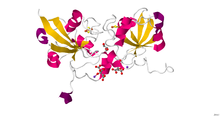
Members of the 2Fe–2S ferredoxin superfamily (InterPro : IPR036010 ) have a general core structure consisting of beta(2)-alpha-beta(2), which includes putidaredoxin, terpredoxin, and adrenodoxin. [18] [19] [20] [21] They are proteins of around one hundred amino acids with four conserved cysteine residues to which the 2Fe–2S cluster is ligated. This conserved region is also found as a domain in various metabolic enzymes and in multidomain proteins, such as aldehyde oxidoreductase (N-terminal), xanthine oxidase (N-terminal), phthalate dioxygenase reductase (C-terminal), succinate dehydrogenase iron–sulphur protein (N-terminal), and methane monooxygenase reductase (N-terminal).
One group of ferredoxins, originally found in chloroplast membranes, has been termed "chloroplast-type" or "plant-type" (InterPro : IPR010241 ). Its active center is a [Fe2S2] cluster, where the iron atoms are tetrahedrally coordinated both by inorganic sulfur atoms and by sulfurs of four conserved cysteine (Cys) residues.
In chloroplasts, Fe2S2 ferredoxins function as electron carriers in the photosynthetic electron transport chain and as electron donors to various cellular proteins, such as glutamate synthase, nitrite reductase, sulfite reductase, and the cyclase of chlorophyll biosynthesis. [22] Since the cyclase is a ferredoxin dependent enzyme this may provide a mechanism for coordination between photosynthesis and the chloroplasts need for chlorophyll by linking chlorophyll biosynthesis to the photosynthetic electron transport chain. In hydroxylating bacterial dioxygenase systems, they serve as intermediate electron-transfer carriers between reductase flavoproteins and oxygenase.
The Fe2S2 ferredoxin from Clostridium pasteurianum (Cp2FeFd; P07324 ) has been recognized as distinct protein family on the basis of its amino acid sequence, spectroscopic properties of its iron–sulfur cluster and the unique ligand swapping ability of two cysteine ligands to the [Fe2S2] cluster. Although the physiological role of this ferredoxin remains unclear, a strong and specific interaction of Cp2FeFd with the molybdenum-iron protein of nitrogenase has been revealed. Homologous ferredoxins from Azotobacter vinelandii (Av2FeFdI; P82802 ) and Aquifex aeolicus (AaFd; O66511 ) have been characterized. The crystal structure of AaFd has been solved. AaFd exists as a dimer. The structure of AaFd monomer is different from other Fe2S2 ferredoxins. The fold belongs to the α+β class, with first four β-strands and two α-helices adopting a variant of the thioredoxin fold. [23] UniProt categorizes these as the "2Fe2S Shethna-type ferredoxin" family. [24]
ferredoxin 1 | |||||||
---|---|---|---|---|---|---|---|
Identifiers | |||||||
Symbol | FDX1 | ||||||
Alt. symbols | FDX | ||||||
NCBI gene | 2230 | ||||||
HGNC | 3638 | ||||||
OMIM | 103260 | ||||||
RefSeq | NM_004109 | ||||||
UniProt | P10109 | ||||||
Other data | |||||||
Locus | Chr. 11 q22.3 | ||||||
|
Adrenodoxin (adrenal ferredoxin; InterPro : IPR001055 ), putidaredoxin, and terpredoxin make up a family of soluble Fe2S2 proteins that act as single electron carriers, mainly found in eukaryotic mitochondria and Pseudomonadota. The human variant of adrenodoxin is referred to as ferredoxin-1 and ferredoxin-2. In mitochondrial monooxygenase systems, adrenodoxin transfers an electron from NADPH:adrenodoxin reductase to membrane-bound cytochrome P450. In bacteria, putidaredoxin and terpredoxin transfer electrons between corresponding NADH-dependent ferredoxin reductases and soluble P450s. [26] [27] The exact functions of other members of this family are not known, although Escherichia coli Fdx is shown to be involved in biogenesis of Fe–S clusters. [28] Despite low sequence similarity between adrenodoxin-type and plant-type ferredoxins, the two classes have a similar folding topology.
Ferredoxin-1 in humans participates in the synthesis of thyroid hormones. It also transfers electrons from adrenodoxin reductase to CYP11A1, a CYP450 enzyme responsible for cholesterol side chain cleavage. FDX-1 has the capability to bind to metals and proteins. [29] Ferredoxin-2 participates in heme A and iron–sulphur protein synthesis. [30]
The [Fe4S4] ferredoxins may be further subdivided into low-potential (bacterial-type) and high-potential (HiPIP) ferredoxins.
Low- and high-potential ferredoxins are related by the following redox scheme:
The formal oxidation numbers of the iron ions can be [2Fe3+, 2Fe2+] or [1Fe3+, 3Fe2+] in low-potential ferredoxins. The oxidation numbers of the iron ions in high-potential ferredoxins can be [3Fe3+, 1Fe2+] or [2Fe3+, 2Fe2+].
3Fe-4S binding domain | |||||||||||
---|---|---|---|---|---|---|---|---|---|---|---|
Identifiers | |||||||||||
Symbol | Fer4 | ||||||||||
Pfam | PF00037 | ||||||||||
InterPro | IPR001450 | ||||||||||
PROSITE | PDOC00176 | ||||||||||
SCOP2 | 5fd1 / SCOPe / SUPFAM | ||||||||||
OPM protein | 1kqf | ||||||||||
|
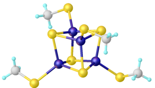
A group of Fe4S4 ferredoxins, originally found in bacteria, has been termed "bacterial-type". Bacterial-type ferredoxins may in turn be subdivided into further groups, based on their sequence properties. Most contain at least one conserved domain, including four cysteine residues that bind to a [Fe4S4] cluster. In Pyrococcus furiosus Fe4S4 ferredoxin, one of the conserved Cys residues is substituted with aspartic acid.
During the evolution of bacterial-type ferredoxins, intrasequence gene duplication, transposition and fusion events occurred, resulting in the appearance of proteins with multiple iron–sulfur centers. In some bacterial ferredoxins, one of the duplicated domains has lost one or more of the four conserved Cys residues. These domains have either lost their iron–sulfur binding property or bind to a [Fe3S4] cluster instead of a [Fe4S4] cluster [31] and dicluster-type. [32]
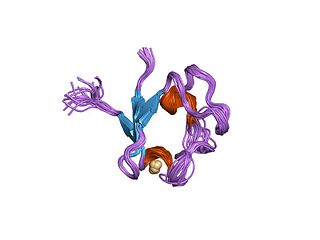
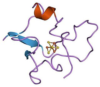
3-D structures are known for a number of monocluster and dicluster bacterial-type ferredoxins. The fold belongs to the α+β class, with 2-7 α-helices and four β-strands forming a barrel-like structure, and an extruded loop containing three "proximal" Cys ligands of the iron–sulfur cluster.
High potential iron–sulfur proteins (HiPIPs) form a unique family of Fe4S4 ferredoxins that function in anaerobic electron transport chains. Some HiPIPs have a redox potential higher than any other known iron–sulfur protein (e.g., HiPIP from Rhodopila globiformis has a redox potential of ca. -450 mV). Several HiPIPs have so far been characterized structurally, their folds belonging to the α+β class. As in other bacterial ferredoxins, the [Fe4S4] unit forms a cubane-type cluster and is ligated to the protein via four Cys residues.
Oxidative phosphorylation or electron transport-linked phosphorylation or terminal oxidation is the metabolic pathway in which cells use enzymes to oxidize nutrients, thereby releasing chemical energy in order to produce adenosine triphosphate (ATP). In eukaryotes, this takes place inside mitochondria. Almost all aerobic organisms carry out oxidative phosphorylation. This pathway is so pervasive because it releases more energy than alternative fermentation processes such as anaerobic glycolysis.
Iron–sulfur clusters are molecular ensembles of iron and sulfide. They are most often discussed in the context of the biological role for iron–sulfur proteins, which are pervasive. Many Fe–S clusters are known in the area of organometallic chemistry and as precursors to synthetic analogues of the biological clusters. It is supposed that the last universal common ancestor had many iron-sulfur clusters.
Photosystem I is one of two photosystems in the photosynthetic light reactions of algae, plants, and cyanobacteria. Photosystem I is an integral membrane protein complex that uses light energy to catalyze the transfer of electrons across the thylakoid membrane from plastocyanin to ferredoxin. Ultimately, the electrons that are transferred by Photosystem I are used to produce the moderate-energy hydrogen carrier NADPH. The photon energy absorbed by Photosystem I also produces a proton-motive force that is used to generate ATP. PSI is composed of more than 110 cofactors, significantly more than Photosystem II.
Iron–sulfur proteins are proteins characterized by the presence of iron–sulfur clusters containing sulfide-linked di-, tri-, and tetrairon centers in variable oxidation states. Iron–sulfur clusters are found in a variety of metalloproteins, such as the ferredoxins, as well as NADH dehydrogenase, hydrogenases, coenzyme Q – cytochrome c reductase, succinate – coenzyme Q reductase and nitrogenase. Iron–sulfur clusters are best known for their role in the oxidation-reduction reactions of electron transport in mitochondria and chloroplasts. Both Complex I and Complex II of oxidative phosphorylation have multiple Fe–S clusters. They have many other functions including catalysis as illustrated by aconitase, generation of radicals as illustrated by SAM-dependent enzymes, and as sulfur donors in the biosynthesis of lipoic acid and biotin. Additionally, some Fe–S proteins regulate gene expression. Fe–S proteins are vulnerable to attack by biogenic nitric oxide, forming dinitrosyl iron complexes. In most Fe–S proteins, the terminal ligands on Fe are thiolate, but exceptions exist.
The cytochrome b6f complex (plastoquinol/plastocyanin reductase or plastoquinol/plastocyanin oxidoreductase; EC 7.1.1.6) is an enzyme found in the thylakoid membrane in chloroplasts of plants, cyanobacteria, and green algae, that catalyzes the transfer of electrons from plastoquinol to plastocyanin:
Rieske proteins are iron–sulfur protein (ISP) components of cytochrome bc1 complexes and cytochrome b6f complexes and are responsible for electron transfer in some biological systems. John S. Rieske and co-workers first discovered the protein and in 1964 isolated an acetylated form of the bovine mitochondrial protein. In 1979, Trumpower's team isolated the "oxidation factor" from bovine mitochondria and showed it was a reconstitutively-active form of the Rieske iron-sulfur protein.
It is a unique [2Fe-2S] cluster in that one of the two Fe atoms is coordinated by two histidine residues rather than two cysteine residues. They have since been found in plants, animals, and bacteria with widely ranging electron reduction potentials from -150 to +400 mV.
Aromatic-ring-hydroxylating dioxygenases (ARHD) incorporate two atoms of dioxygen (O2) into their substrates in the dihydroxylation reaction. The product is (substituted) cis-1,2-dihydroxycyclohexadiene, which is subsequently converted to (substituted) benzene glycol by a cis-diol dehydrogenase.
Outer sphere refers to an electron transfer (ET) event that occurs between chemical species that remain separate and intact before, during, and after the ET event. In contrast, for inner sphere electron transfer the participating redox sites undergoing ET become connected by a chemical bridge. Because the ET in outer sphere electron transfer occurs between two non-connected species, the electron is forced to move through space from one redox center to the other.
A photosynthetic reaction center is a complex of several proteins, biological pigments, and other co-factors that together execute the primary energy conversion reactions of photosynthesis. Molecular excitations, either originating directly from sunlight or transferred as excitation energy via light-harvesting antenna systems, give rise to electron transfer reactions along the path of a series of protein-bound co-factors. These co-factors are light-absorbing molecules (also named chromophores or pigments) such as chlorophyll and pheophytin, as well as quinones. The energy of the photon is used to excite an electron of a pigment. The free energy created is then used, via a chain of nearby electron acceptors, for a transfer of hydrogen atoms (as protons and electrons) from H2O or hydrogen sulfide towards carbon dioxide, eventually producing glucose. These electron transfer steps ultimately result in the conversion of the energy of photons to chemical energy.
Any enzyme system that includes cytochrome P450 protein or domain can be called a P450-containing system.
Rubredoxins are a class of low-molecular-weight iron-containing proteins found in sulfur-metabolizing bacteria and archaea. Sometimes rubredoxins are classified as iron-sulfur proteins; however, in contrast to iron-sulfur proteins, rubredoxins do not contain inorganic sulfide. Like cytochromes, ferredoxins and Rieske proteins, rubredoxins are thought to participate in electron transfer in biological systems. Recent work in bacteria and algae have led to the hypothesis that some rubredoxins may instead have a role in delivering iron to metalloproteins.
P700, or photosystem I primary donor, is a molecular dimer of chlorophyll a associated with the reaction-center of photosystem I in plants, algae, and cyanobacteria.
In enzymology, carbon monoxide dehydrogenase (CODH) (EC 1.2.7.4) is an enzyme that catalyzes the chemical reaction
The enzyme sirohydrochlorin ferrochelatase (EC 4.99.1.4) catalyzes the following reaction:
Adrenal ferredoxin is a protein that in humans is encoded by the FDX1 gene. In addition to the expressed gene at this chromosomal locus (11q22), there are pseudogenes located on chromosomes 20 and 21.
High potential iron-sulfur proteins (HIPIP) are a class of iron-sulfur proteins. They are ferredoxins that participate in electron transfer in photosynthetic bacteria as well as in Paracoccus denitrificans.
Light-dependent reactions are certain photochemical reactions involved in photosynthesis, the main process by which plants acquire energy. There are two light dependent reactions: the first occurs at photosystem II (PSII) and the second occurs at photosystem I (PSI).
Ferredoxin-thioredoxin reductase EC 1.8.7.2, systematic name ferredoxin:thioredoxin disulfide oxidoreductase, is a [4Fe-4S] protein that plays an important role in the ferredoxin/thioredoxin regulatory chain. It catalyzes the following reaction:
The fnr gene of Escherichia coli encodes a transcriptional activator (FNR) which is required for the expression of a number of genes involved in anaerobic respiratory pathways. The FNR protein of E. coli is an oxygen – responsive transcriptional regulator required for the switch from aerobic to anaerobic metabolism.
"Type III mutants, originally frdB, were designated fnr because they were defective in fumarate and nitrate reduction and impaired in their ability to produce gas." - Lambden and Guest, 1976 Journal of General Microbiology97, 145-160
In enzymology, an aldehyde ferredoxin oxidoreductase (EC 1.2.7.5) is an enzyme that catalyzes the chemical reaction