Gestational choriocarcinoma

Last updated

Gestational choriocarcinoma is a form of gestational trophoblastic neoplasia, which is a type of gestational trophoblastic disease (GTD), that can occur during pregnancy. It is a rare disease where the trophoblast, a layer of cells surrounding the blastocyst, undergoes abnormal developments, leading to trophoblastic tumors. The choriocarcinoma can metastasize to other organs, including the lungs, kidney, and liver. [1] The amount and degree of choriocarcinoma spread to other parts of the body can vary greatly from person to person. [2]

Contents

Gestational choriocarcinoma can happen during and after any type of pregnancy event, though risk of the disease is higher in and after complete or partial molar pregnancies. [3] Risk of disease may also be higher in those experiencing pregnancy at younger or older ages that average, such as below 15 years old or above 45 years old. [4] Those with gestational choriocarcinoma may experience abnormal vaginal bleeding, abdominal pain, and have high levels of human chorionic gonadotropin (hCG), in addition to history of molar pregnancy or other metastatic cancer. A combination of history, symptoms, human chorionic gonadotropin levels, and imaging can be used in the diagnosis process, with ultrasonography being commonly used to image. [5] Approximately 50% of those with gestational choriocarcinoma have experienced molar pregnancy, approximately 25% developed the disease after a regular, term pregnancy, and other situations have included history of ectopic pregnancy, where the pregnancy does not occur in the uterus. [6]

Guidelines from the Federation of Gynecologists and Obstetricians (FIGO), Royal College of Obstetricians and Gynaecologists (RCOG), European Society for Medical Oncology (ESMO), and Royal Australian and New Zealand College of Obstetricians and Gynaecologists (RANZCOG) exist to evaluate risk and treatment of the disease. There are generally three levels of risk: low risk, high risk, and ultrahigh risk. [6] The primary form of treatment is chemotherapy with one or more agents. Duration can go upwards of six weeks following the return of human chorionic gonadotropin levels to the normal range. Depending on the risk of gestational trophoblastic disease (GTD) development, such as in certain people with mole pregnancies, chemotherapy has been used in a preventative manner in the past. However, this type of use is now advised against due to risk of toxicity and resistance to agents that may be needed in future treatment of the disease, on top of medical costs. [2] Guidelines also detail options for salvage therapies in the situation of resistance to preferred choice of chemotherapy and surgical procedures possible in the situation of drug-resistant tumors. [6]

Signs and symptoms

Vaginal bleeding is a common symptom of gestational choriocarcinoma. In the event the gestational choriocarcinoma has already spread to other parts of the body, bleeding events in other organs may be the first symptom. [3] If the choriocarcinoma has metastasized to the lungs, one of the most common organs of metastasis, then symptoms may include abnormally quick breathing, coughing, and chest pain. [7] However, it is also possible for there to be none of these symptoms and only a difference in biomarkers in an individual instead. [4]

Uterine enlargement as seen in an ultrasound can be a presenting sign of gestational choriocarcinoma. Transvaginal ultrasounds may also be used in screening for the disease, with the positron emission tomography (PET) scan, computed tomography (CT), and magnetic resonance imaging (MRI) in locations such as the abdomen, pelvis, and brain being other imaging options. Chest x-rays may also be used to see if gestational choriocarcinoma has potentially spread to the lungs. [6]

Human chorionic gonadotropin is a hormone produced by the trophoblast and can be measured in both urine and blood. Another option of measuring this biomarker level is through lumbar puncture, which can show fluid-to-serum ratio of human chorionic gonadotropic and reflect whether the disease has potentially spread to the central nervous system. [7] [6] Elevated or rising human chorionic gonadotropin can be a sign of gestational choriocarcinoma, though it is also used as a biomarker in other types of gestational trophoblastic diseases (GTD). It is useful not only for diagnosis but also for monitoring disease progression, treatment response, and potential of recurring gestational choriocarcinoma. [4]

Causes

Gestational choriocarcinoma (GC) is the most aggressive form of trophoblastic tumor; it most commonly arises from certain fertilization defects, such as molar pregnancy, which results in increased level of growth factors and uncontrolled proliferation of trophoblasts in the uterus. [8] Statistics from clinical cases have shown that GC is associated with any pregnancy events, with 50% of GC arise from hydatidiform moles, 25% from gestation, and 25% from abortion or tubal pregnancy. [9]

Risk factors

The precise underlying causes of this disease remain incompletely understood, primarily due to its rarity and clinical challenges in distinguishing it from invasive mole, another type of trophoblastic tumor that shares similarities with gestational choriocarcinoma (GC). [9] Despite these complexities, several risk factors have been identified and reported to be associated with gestational choriocarcinoma.

Pathogenesis

At the cellular level, any abnormalities in the placental trophoblastic proliferation could potentially contribute to molar pregnancy and tumor development. [9] There are three different types of trophoblasts: cytotrophoblasts, syncytiotrophoblasts, and intermediate trophoblasts. [11] Each of them possesses distinct specialized functions to support a developing embryo. Cytotrophoblasts are the germinating cells that forms the chorion villi and can be differentiated into syncytiotrophoblasts which produce hCG and can intrude to the inner muscle layer of the uterus to help embed the developing embryo, and intermediate trophoblasts which spread throughout the chorion villi to support maternal-fetal circulation. [9] [11] Malignant cellular transformation of all of these three trophoblastic cells contribute to the development of gestational choriocarcinoma. [12] Common pathological features of GC include abnormal trophoblastic enlargement, loss of trophoblastic specialized features and functions, absence of villi in the placenta, abnormal bleeding, and necrosis. [3]

Many efforts have been made to try to understand the mechanism of how non-malignant mole could become invasive. It is suspected that activation of certain oncogenes (such as up-regulations of MDM2, c-ERB2, and BLC2) and inactivation of tumor suppressor genes (such as up-regulations of p53, p21) were involved in the processes of genetic changes in this malignant transformation. [10] But trophoblastic cells, by nature, are highly active in cell division, the increased activities of those genes are also necessary to maintain their normal cell function. [10] Therefore, it is still unclear how significantly these genetic changes are in the pathogenesis of gestational choriocarcinoma.

Recent research in the role of long non-coding RNAs (lncRNAs) in the GC development is believed to bring hope in this field. [13] Long non-coding RNAs are groups of RNAs that do not code for protein expression and are usually over 200 nucleotides long; they are increasingly recognized to have essential role in many aspects of cellular function, like transcriptional regulation, sub-cellular protein localization, and epigenetic remodeling. [13] To date, several types of lncRNAs are reported to have role in GC pathogenesis, which are metastasis-associated lung adenocarcinoma transcript 1 (MALAT1), H19, maternally expressed gene 3 (MEG3), prostate cancer antigen 3 (PCA3), the long intergenic non-coding RNA 00261 (LINC00261), etc. [13] However, as many of these studies are still in the preliminary stages, more investigation is needed to fully understand the underlying mechanism. [13]

Diagnosis

Common characteristic manifestations of gestational choriocarcinoma include irregular vaginal bleeding and hydatidiform moles. A hydatidiform mole is a red hemorrhagic mass with various sizes in the uterus. [14] Often, diagnosis is presumptive. It is based on clinical findings and the identification of a malignant trophoblast. One prevalent symptom is vaginal bleeding after a pregnancy, abortion, or hydatidiform mole. [15] In the presence of choriocarcinoma, a pregnancy test will be positive even if there is no embryo/fetus. [15]

A confirmed diagnosis usually happens after the disease has progressed to a late clinical stage. This is partly due to the wide spectrum and rare clinical presentations of the disease. [16] Individuals presented with the disease are mostly in their reproductive years. [3]

Similar to other non-gestational tumors, gestational choriocarcinoma can be reflected via an elevated level in serum hCG concentration. [7] Besides the change in serum hCG levels, the diagnosis of gestational choriocarcinoma also focuses on the presence of metastases after any types of pregnancy events. [9] Based on the International Federation of Gynecology and Obstetrics (FIGO)'s updated guidelines on gestational trophoblastic disease management, the diagnostic criteria of a post-gestational trophoblastic neoplasia (GTN) is as follows: [4]

  1. Over a period of 3 weeks or longer (day 1, 7, 14, 21), there are 4 or more plateaued hCG levels.
  2. Over a period of 2 weeks or longer (day 1, 7, 14), there are 3 consecutive weekly measurements of rising hCG levels.
  3. A histological diagnosis of choriocarcinoma.

There are currently different guidelines available in terms of diagnosing and recommending treatment options for gestational trophoblastic neoplasia (GTN). The 4 guidelines identified are: the Royal College of Obstetricians and Gynecologists (RCOG), the International Federation of Gynecology and Obstetrics (FIGO), the European Society for Medical Oncology (ESMO), and the Royal Australian and New Zealand College of Obstetricians and Gynecologists (RANZCOG).[ citation needed ] The differences in GTN diagnosis between the guidelines are as follows: [6]

Comparison chart of available guidelines on gestational trophoblastic neoplasia (GTN) diagnosis criteria
RCOGESMOFIGORANZCOG
Clinical presentationxx
FIGO Criteria (*see above)xxxx
Urine hCG levelx
Post-molar hCG levelxx

To differentiate gestational choriocarcinoma from other tumors such as lung or brain cancers, a genetic test is usually completed on top of a pathological diagnosis. [7] DNA genotyping is a powerful tool that helps with the differentiation. The technique can accurately determine the time that it takes to develop the observed tumor and the type of index gestation, which includes term pregnancy, molar gestation, or non-molar abortion. [17]

Treatment

Due to the susceptibility of choriocarcinomas to chemotherapy, it is the first line treatment for this disease. [18] Treatment course depends on the FIGO Scoring System for GTN, which has various prognostic factors, and categorizes based on low risk with a score of 0-6 and high risk with a score of 7 and above. [18]

Low risk

Individuals with low risk GTN are usually treated with single agent chemotherapy, such as methotrexate and folinic acid (MTX/FA) or actinomycin D (ActD). [18] Other treatment options for those with low risk include hysterectomy and surgical removal of the tumor if possible. [18]

High risk

Those with high risk GTN receive a multi-agent chemotherapy regimen, usually consisting of a weekly rotation of etoposide, methotrexate, and actinomycin D given one week and cyclophosphamide and vincristine given the following week, with variations to treatment occurring on a patient-specific basis. [18]  In individuals who relapse, secondary chemotherapy is needed where treatment may vary from the original regimen. [19] The treatment includes a platinum-etopside combination given with other chemotherapy medications, such as methotrexate and actinomycin D (EMA-EP), bleomycin (BEP), or ifosfamide (VIP, ICE). [19] Additional treatment also consists of surgery and radiotherapy if deemed appropriate for the individual. [19] This condition can also result in resistance to the medications used to treat it and failure of their success in the individuals. [20] If this occurs, individuals usually undergo a more intensive medication regimen than what was originally given to them. [20]

Immune checkpoint inhibitors

New treatment options designed to reduce the risk of resistance are currently being researched, such as immune checkpoint inhibitors. [20] Studies of gestational choriocarcinoma demonstrate an increased expression of the ligand PD-L1 and its corresponding receptor PD-1, leading to drug research and development into medications that can block the PD-1 receptor, such as Pembrolizumab. [20] In individuals requiring more intensive chemotherapy options, using an immune checkpoint inhibitor may be preferable to long-term use of multi-agent chemotherapy to prevent prolonged toxicity to the body. [20]

FIGO Scoring System for GTN [18]
Prognostic Factor0124
Age<40≥40
Previous PregnancyHydatidiform moleAbortionTerm
Months since last pregnancyLess than 44 to 67-12>12
Pre-treatment hCG (IU/mL)<103103 to 104>104 to 105>105
Largest tumor size, including uterus<3 cm3–4 cm≥5 cm
Site of spreadLungSpleen or kidneyGI TractBrain, liver
Number of metastases1 to 45-8>8
Previous Failed chemotherapySingle Drug≥2

Prognosis

The survival rate following treatment with chemotherapy is approximately at least 90%. [4] If gestational choriocarcinoma has spread to the liver in an individual, survival rate may be lower. [7] Overall survival rate is also higher when management of gestational choriocarcinoma occurs in a setting with physicians familiar with the condition. Earlier diagnosis and treatment intervention can lead to greater success in preserving potential of future pregnancy in those seeking it. [2]

Return of human chorionic gonadotropic levels to within the bounds of normal values lowers the chance of recurring gestational choriocarcinoma. These levels can be checked monthly as follow-up. For chemotherapy, patients should wait a minimum of one year following chemotherapy before they plan to have a child to minimize the chance of miscarriage. [6]

Related Research Articles

<span class="mw-page-title-main">Miscarriage</span> Natural death and expulsion of an embryo or fetus before its independent survival

Miscarriage, also known in medical terms as a spontaneous abortion, is the death and expulsion of an embryo or fetus before it is able to survive independently. The term miscarriage is sometimes used to refer to all forms of pregnancy loss and pregnancy with abortive outcome before 20 weeks of gestation.

<span class="mw-page-title-main">Amniocentesis</span> Sampling of amniotic fluid done mainly to detect fetal chromosomal abnormalities

Amniocentesis is a medical procedure used primarily in the prenatal diagnosis of genetic conditions. It has other uses such as in the assessment of infection and fetal lung maturity. Prenatal diagnostic testing, which includes amniocentesis, is necessary to conclusively diagnose the majority of genetic disorders, with amniocentesis being the gold-standard procedure after 15 weeks' gestation.

<span class="mw-page-title-main">Human chorionic gonadotropin</span> Hormone

Human chorionic gonadotropin (hCG) is a hormone for the maternal recognition of pregnancy produced by trophoblast cells that are surrounding a growing embryo, which eventually forms the placenta after implantation. The presence of hCG is detected in some pregnancy tests. Some cancerous tumors produce this hormone; therefore, elevated levels measured when the patient is not pregnant may lead to a cancer diagnosis and, if high enough, paraneoplastic syndromes, however, it is not known whether this production is a contributing cause, or an effect of carcinogenesis. The pituitary analog of hCG, known as luteinizing hormone (LH), is produced in the pituitary gland of males and females of all ages.

<span class="mw-page-title-main">Pregnancy test</span> A test based on scientific data to determine if a female is pregnant

A pregnancy test is used to determine whether a female is pregnant or not. The two primary methods are testing for the female pregnancy hormone in blood or urine using a pregnancy test kit, and scanning with ultrasonography. Testing blood for hCG results in the earliest detection of pregnancy. Almost all pregnant women will have a positive urine pregnancy test one week after the first day of a missed menstrual period.

<span class="mw-page-title-main">Trophoblast</span> Early embryonic structure that gives rise to the placenta

The trophoblast is the outer layer of cells of the blastocyst. Trophoblasts are present four days after fertilization in humans. They provide nutrients to the embryo and develop into a large part of the placenta. They form during the first stage of pregnancy and are the first cells to differentiate from the fertilized egg to become extraembryonic structures that do not directly contribute to the embryo. After blastulation, the trophoblast is contiguous with the ectoderm of the embryo and is referred to as the trophectoderm. After the first differentiation, the cells in the human embryo lose their totipotency because they can no longer form a trophoblast. They become pluripotent stem cells.

<span class="mw-page-title-main">Molar pregnancy</span> Abnormal form of pregnancy (human disorder)

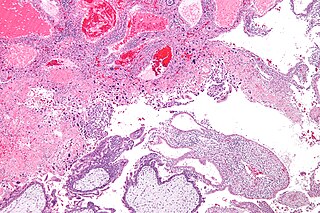
A molar pregnancy, also known as a hydatidiform mole, is an abnormal form of pregnancy in which a non-viable fertilized egg implants in the uterus. It falls under the category of gestational trophoblastic diseases and was previously known as a hydatidiform mole. During a molar pregnancy, the uterus contains a growing mass characterized by swollen chorionic villi, resembling clusters of grapes. The occurrence of a molar pregnancy can be attributed to the fertilized egg lacking an original maternal nucleus. As a result, the products of conception may or may not contain fetal tissue. These molar pregnancies are categorized into two types: partial moles and complete moles, where the term 'mole' simply denotes a clump of growing tissue or a ‘growth'.

<span class="mw-page-title-main">Choriocarcinoma</span> Malignant, trophoblastic cancer, usually of the placenta

Choriocarcinoma is a malignant, trophoblastic cancer, usually of the placenta. It is characterized by early hematogenous spread to the lungs. It belongs to the malignant end of the spectrum in gestational trophoblastic disease (GTD). It is also classified as a germ cell tumor and may arise in the testis or ovary.

<span class="mw-page-title-main">Gestational trophoblastic disease</span> Pregnancy-related tumours

Gestational trophoblastic disease (GTD) is a term used for a group of pregnancy-related tumours. These tumours are rare, and they appear when cells in the womb start to proliferate uncontrollably. The cells that form gestational trophoblastic tumours are called trophoblasts and come from tissue that grows to form the placenta during pregnancy.

<span class="mw-page-title-main">Seminoma</span> Medical condition

A seminoma is a germ cell tumor of the testicle or, more rarely, the mediastinum or other extra-gonadal locations. It is a malignant neoplasm and is one of the most treatable and curable cancers, with a survival rate above 95% if discovered in early stages.

<span class="mw-page-title-main">Dactinomycin</span> Chemical compound

Dactinomycin, also known as actinomycin D, is a chemotherapy medication used to treat a number of types of cancer. This includes Wilms tumor, rhabdomyosarcoma, Ewing's sarcoma, trophoblastic neoplasm, testicular cancer, and certain types of ovarian cancer. It is given by injection into a vein.

<span class="mw-page-title-main">Trophoblastic neoplasm</span> Medical condition

Gestational trophoblastic neoplasia (GTN) is group of rare diseases related to pregnancy and included in gestational trophoblastic disease (GTD) in which abnormal trophoblast cells grow in the uterus. GTN can be classified into benign and malignant lesions. Benign lesions include placental site nodule and hydatidiform moles while malignant lesions have four subtypes including invasive mole, gestational choriocarcinoma, placental site trophoblastic tumor (PSTT) and epithelioid trophoblastic tumor (ETT). The choriocarcinoma has 2 significant subtypes including gestational and non-gestational and they are differentiated by their different biological feature and prognosis. Signs and symptoms of GTN will appear vary from person to person and depending upon the type of the disease. They may include uterine bleeding not related to menstruation, pain or pressure in pelvis, large uterus and high blood pressure during pregnancy. The cause of this disease is unknown but the identification of the tumor based on total beta-human chorionic gonadotropin (β-hCG) in the serum.

<span class="mw-page-title-main">Placenta accreta spectrum</span> Medical condition

Placenta accreta occurs when all or part of the placenta attaches abnormally to the myometrium. Three grades of abnormal placental attachment are defined according to the depth of attachment and invasion into the muscular layers of the uterus:

  1. Accreta – chorionic villi attached to the myometrium, rather than being restricted within the decidua basalis.
  2. Increta – chorionic villi invaded into the myometrium.
  3. Percreta – chorionic villi invaded through the perimetrium.
<span class="mw-page-title-main">Cytotrophoblast</span>

"Cytotrophoblast" is the name given to both the inner layer of the trophoblast or the cells that live there. It is interior to the syncytiotrophoblast and external to the wall of the blastocyst in a developing embryo.

<span class="mw-page-title-main">Placental disease</span> Medical condition

A placental disease is any disease, disorder, or pathology of the placenta.

<span class="mw-page-title-main">Interstitial pregnancy</span> Medical condition

An interstitial pregnancy is a uterine but ectopic pregnancy; the pregnancy is located outside the uterine cavity in that part of the fallopian tube that penetrates the muscular layer of the uterus. The term cornual pregnancy is sometimes used as a synonym, but remains ambiguous as it is also applied to indicate the presence of a pregnancy located within the cavity in one of the two upper "horns" of a bicornuate uterus. Interstitial pregnancies have a higher mortality than ectopics in general.

<span class="mw-page-title-main">Placental site trophoblastic tumor</span> Medical condition

Placental site trophoblastic tumor is a form of gestational trophoblastic disease, which is thought to arise from intermediate trophoblast.

Theca lutein cyst is a type of bilateral functional ovarian cyst filled with clear, straw-colored fluid. These cysts result from exaggerated physiological stimulation due to elevated levels of beta-human chorionic gonadotropin (beta-hCG) or hypersensitivity to beta-hCG. On ultrasound and MRI, theca lutein cysts appear in multiples on ovaries that are enlarged.

<span class="mw-page-title-main">Min Chiu Li</span> American physician

Min Chiu Li was a Chinese-American oncologist and cancer researcher. Li was the first scientist to use chemotherapy to cure widely metastatic, malignant cancer.

Early pregnancy bleeding refers to vaginal bleeding before 14 weeks of gestational age. If the bleeding is significant, hemorrhagic shock may occur. Concern for shock is increased in those who have loss of consciousness, chest pain, shortness of breath, or shoulder pain.

Epithelioid trophoblastic tumor (ETT) is a gestational trophoblastic disease with about 110 case reports in the literature. It is a trophoblastic tumor of neoplastic chorionic type associated with the intermediate trophoblast.

References

  1. Sharami SR, Saffarieh E (March 2020). "A review on management of gestational trophoblastic neoplasia". Journal of Family Medicine and Primary Care. 9 (3): 1287–1295. doi: 10.4103/jfmpc.jfmpc_876_19 . PMC   7266251 . PMID   32509606.
  2. 1 2 3 Braga A, Mora P, de Melo AC, Nogueira-Rodrigues A, Amim-Junior J, Rezende-Filho J, Seckl MJ (February 2019). "Challenges in the diagnosis and treatment of gestational trophoblastic neoplasia worldwide". World Journal of Clinical Oncology. 10 (2): 28–37. doi: 10.5306/wjco.v10.i2.28 . PMC   6390119 . PMID   30815369.
  3. 1 2 3 4 Hui P (January 2019). "Gestational Trophoblastic Tumors: A Timely Review of Diagnostic Pathology". Archives of Pathology & Laboratory Medicine. 143 (1): 65–74. doi: 10.5858/arpa.2018-0234-RA . PMID   30407075. S2CID   53213943.
  4. 1 2 3 4 5 Ngan HY, Seckl MJ, Berkowitz RS, Xiang Y, Golfier F, Sekharan PK, et al. (October 2018). "Update on the diagnosis and management of gestational trophoblastic disease". International Journal of Gynaecology and Obstetrics. 143 (Suppl 2): 79–85. doi: 10.1002/ijgo.12615 . PMID   30306586. S2CID   52958887.
  5. Shaaban AM, Rezvani M, Haroun RR, Kennedy AM, Elsayes KM, Olpin JD, et al. (2017). "Gestational Trophoblastic Disease: Clinical and Imaging Features". Radiographics. 37 (2): 681–700. doi:10.1148/rg.2017160140. PMID   28287945.
  6. 1 2 3 4 5 6 7 Tsakiridis I, Giouleka S, Kalogiannidis I, Mamopoulos A, Athanasiadis A, Dagklis T (December 2020). "Diagnosis and Management of Gestational Trophoblastic Disease: A Comparative Review of National and International Guidelines". Obstetrical & Gynecological Survey. 75 (12): 747–756. doi:10.1097/OGX.0000000000000848. PMID   33369685. S2CID   229380333.
  7. 1 2 3 4 5 Lok C, Frijstein M, van Trommel N (July 2021). "Clinical presentation and diagnosis of Gestational Trophoblastic Disease". Best Practice & Research. Clinical Obstetrics & Gynaecology. 74: 42–52. doi:10.1016/j.bpobgyn.2020.12.001. PMID   33422446. S2CID   231577251.
  8. Ning F, Hou H, Morse AN, Lash GE (2019). "Understanding and management of gestational trophoblastic disease". F1000Research. 8: F1000 Faculty Rev–428. doi: 10.12688/f1000research.14953.1 . PMC   6464061 . PMID   31001418.
  9. 1 2 3 4 5 Lurain JR (December 2010). "Gestational trophoblastic disease I: epidemiology, pathology, clinical presentation and diagnosis of gestational trophoblastic disease, and management of hydatidiform mole". American Journal of Obstetrics and Gynecology. 203 (6): 531–539. doi:10.1016/j.ajog.2010.06.073. PMID   20728069.
  10. 1 2 3 4 5 6 Altieri A, Franceschi S, Ferlay J, Smith J, La Vecchia C (November 2003). "Epidemiology and aetiology of gestational trophoblastic diseases". The Lancet. Oncology. 4 (11): 670–678. doi:10.1016/S1470-2045(03)01245-2. PMID   14602247.
  11. 1 2 3 Steigrad SJ (December 2003). "Epidemiology of gestational trophoblastic diseases". Best Practice & Research. Clinical Obstetrics & Gynaecology. 17 (6): 837–847. doi:10.1016/s1521-6934(03)00049-x. PMID   14614884.
  12. Silva AL, Monteiro KD, Sun SY, Borbely AU (December 2021). "Gestational trophoblastic neoplasia: Novelties and challenges". Placenta. Trophoblast Research - Volume 39. 116: 38–42. doi: 10.1016/j.placenta.2021.02.013 . PMID   33685753. S2CID   232161179.
  13. 1 2 3 4 Di Fiore R, Suleiman S, Felix A, O'Toole SA, O'Leary JJ, Ward MP, et al. (June 2021). "An Overview of the Role of Long Non-Coding RNAs in Human Choriocarcinoma". International Journal of Molecular Sciences. 22 (12): 6506. doi: 10.3390/ijms22126506 . PMC   8235025 . PMID   34204445.
  14. Buza N (June 2022). "Gestational Trophoblastic Disease: Contemporary Diagnostic Approach". Surgical Pathology Clinics. 15 (2): 197–218. doi:10.1016/j.path.2022.02.002. PMID   35715158. S2CID   248959673.
  15. 1 2 Serno J, Zeppernick F, Jäkel J, Schrading S, Maass N, Meinhold-Heerlein I, Bauerschlag DO (2012). "Primary pulmonary choriocarcinoma: case report and review of the literature". Gynecologic and Obstetric Investigation. 74 (2): 171–176. doi:10.1159/000336784. PMID   22738859. S2CID   27897722.
  16. Mangla M, Singla D, Kaur H, Sharma S (February 2017). "Unusual clinical presentations of choriocarcinoma: A systematic review of case reports". Taiwanese Journal of Obstetrics & Gynecology. 56 (1): 1–8. doi: 10.1016/j.tjog.2015.05.011 . PMID   28254207.
  17. Buza N, Hui P (September 2021). "Genotyping diagnosis of gestational trophoblastic disease: frontiers in precision medicine". Modern Pathology. 34 (9): 1658–1672. doi: 10.1038/s41379-021-00831-9 . PMID   34088998.
  18. 1 2 3 4 5 6 Clark JJ, Slater S, Seckl MJ (February 2021). "Treatment of gestational trophoblastic disease in the 2020s". Current Opinion in Obstetrics & Gynecology. 33 (1): 7–12. doi:10.1097/GCO.0000000000000674. PMC   7116872 . PMID   33337613.
  19. 1 2 3 Lurain JR, Nejad B (May 2005). "Secondary chemotherapy for high-risk gestational trophoblastic neoplasia". Gynecologic Oncology. 97 (2): 618–623. doi: 10.1016/j.ygyno.2005.02.004 . PMID   15863169.
  20. 1 2 3 4 5 Winter MC (July 2021). "Treatment of low-risk gestational trophoblastic neoplasia". Best Practice & Research. Clinical Obstetrics & Gynaecology. 74: 67–80. doi:10.1016/j.bpobgyn.2021.01.006. PMID   33741258. S2CID   232302437.