Idiopathic Ulnar neuropathy at the elbow

Last updated

Idiopathic Ulnar neuropathy at the elbow
Anatomy of Ulnar nerve.JPG
Anatomy of ulnar nerve
Specialty Neurology   OOjs UI icon edit-ltr-progressive.svg

Idiopathic Ulnar neuropathy at the elbow (aka Ulnar nerve entrapment or Cubital tunnel syndrome) is a condition where pressure on the ulnar nerve as it passes through the cubital tunnel causes nerve dysfunction (neuropathy). The symptoms of neuropathy are paresthesia (tingling) and numbness (loss of sensibility) primarily affecting the little finger and ring finger of the hand. Ulnar neuropathy can progress to weakness and atrophy of the muscles in the hand (interossei and small and ring finger lumbricals). Symptoms can be alleviated by attempts to keep the elbow from flexing while sleeping, such as sticking one's arm in the pillow case, so the pillow restricts flexion.

Contents

Signs and symptoms

In general, ulnar neuropathy will result in symptoms in a specific anatomic distribution, affecting the little finger, the ulnar half of the ring finger, and the intrinsic muscles of the hand.

The specific symptoms experienced in the characteristic distribution depend on the specific location of ulnar nerve compression. The hallmark symptoms of ulnar neuropathy at the elbow is paresthesia (tingling). This can progress to a loss of sensibility. Muscle weakness is usually experienced as a loss of dexterity.

Ulnar neuropathy at the wrist is associated with variable symptoms, as the ulnar nerve separates near the hand into distinct motor and sensory branches.

In cubital tunnel syndrome (ulnar neuropathy at the elbow), sensory and motor symptoms tend to occur in a certain sequence. Initially, there may be intermittent paresthesia and loss of sensibility of the small and ulnar half of the ring fingers. Next is constant numbness (loss of sensibility). The final stage is intrinsic hand muscle atrophy and weakness.

In contrast, when ulnar neuropathy occurs at the wrist (Guyon canal syndrome), motor symptoms predominate. There may be an ulnar claw hand from imbalance between the muscles innervated by the ulnar nerve in the forearm (which are functioning normally) and those in the hand (which are weak). The back of the hand will have normal sensation. [1]

Diagnosis

Ulnar neuropathy at the cubital tunnel is diagnosed based on characteristic symptoms and signs. Intermittent or static numbness in the small finger and ulnar half of the ring finger, weakness or atrophy of the first dorsal interosseous, positive Tinel sign over the ulnar nerve proximal to the cubital tunnel, and positive elbow flexion test (elicitation of paresthesia in the small and ring finger with sustained elbow flexion) establish the diagnosis. The diagnosis can be confirmed using electrophysiological tests: nerve conduction velocity and electromyography.

Imaging studies are not routinely used. Ultrasound or MRI may reveal enlargement of the ulnar nerve proximal to the cubital tunnel. Variations in anatomy such as the anconeus epitrochlearis muscle are common and their relationship to ulnar neuropathy is uncertain. [2]

Differential diagnosis

Trauma can cause symptoms of ulnar neuropathy. The symptoms are transient after blunt trauma and constant after a laceration.

It is theorized that dislocation of the ulnar nerve anteriorly over the medial epicondyle can result in ulnar neuropathy, but this is not established by experimental evidence.

Ulnar nerve dislocation is a common variation of normal and has not been experimentally associated with ulnar neuropathy.

Median neuropathy at the carpal tunnel (the symptoms of signs of which are carpal tunnel syndrome) is typically characterized by numbness in the thumb, index, middle, and half of the ring finger. Because of variable cross over between the median and ulnar nerve, as well as patient imprecise experience of and report of the symptoms, median neuropathy at the carpal tunnel can be considered among people with intermittent paresthesia of the small and ring fingers.

Classification

McGowan classified idiopathic ulnar neuropathy at the elbow as follows: i) Mild (intermittent paresthesia); ii) moderate (intermittent paresthesia and measurable weakness); and iii) severe (constant paresthesia and measurable weakness).

Dellon and Goldberg modified the classification to subdivide grade 2 neuropathy into grade 2A and 2B on the basis of the extent of motor compromise. The modified classification is as follows: Type 1 Subjective sensory symptoms without objective loss of two-point sensibility or muscular atrophy; Type 2A Sensory symptoms and weakness on pinch and grip without atrophy of intrinsic muscles; Type 2B Sensory symptoms and atrophy and intrinsic muscle strength less than 3 out of 5 on the Medical Research Council scale; Type 3 Profound muscular atrophy and sensory disturbance.

Cubital tunnel syndrome

The most common location of ulnar nerve impingement at the elbow is within the cubital tunnel, and is known as cubital tunnel syndrome. [3] [4] The tunnel is formed by the medial epicondyle of the humerus, the olecranon process of the ulna and the tendinous arch joining the humeral and ulnar heads of the flexor carpi ulnaris muscle. [5] While most cases of injury are minor and resolve spontaneously with time, chronic compression or repetitive trauma may cause more persistent problems. Commonly cited scenarios include:

Compression of the ulnar nerve at the medial elbow may occasionally be caused by an epitrocheloanconeus muscle, an anatomical variant. [6]

Ulnar tunnel syndrome

Ulnar nerve impingement along an anatomical space in the wrist called the ulnar canal is known as ulnar tunnel syndrome (or Guyon canal's syndrome). [7] Recognized causes of ulnar nerve impingement at this location include local trauma, fractures, ganglion cysts, [8] and classically avid cyclists who experience repetitive trauma against bicycle handlebars. [9] This form of ulnar neuropathy comprises two work-related syndromes: so-called "hypothenar hammer syndrome," seen in workers who repetitively use a hammer, and "occupational neuritis" due to hard, repetitive compression against a desk surface. [8] This syndrome can be categorized into three zones based on the localization of the ulnar nerve within the Guyon's canal. [10]

Prevention

Cubital tunnel syndrome may be prevented or reduced by maintaining good posture and proper use of the elbow and arms, such as wearing an arm splint while sleeping to maintain the arm is in a straight position instead of keeping the elbow tightly bent. [4] [11] A recent example of this is popularization of the concept of cell phone elbow and game hand. [11]

Treatment

The most effective treatment for cubital tunnel syndrome is surgical decompression. The most safe and effective operation is in-situ decompression +/- medial epicondylectomy. [12]

For pain symptoms, medications such as NSAID, amitriptyline, or vitamin B6 supplementation may help although there is no evidence to support this claim.[ citation needed ]

Mild symptoms may first be treated non-operatively, with the following:[ citation needed ]

It is important to identify positions and activities that aggravate symptoms and to find ways to avoid them. [4] For example, if the person experiences symptoms when holding a telephone up to the head, then the use of a telephone headset will provide immediate symptomatic relief and reduce the likelihood of further damage and inflammation to the nerve. For cubital tunnel syndrome, it is recommended to avoid repetitive elbow flexion and also avoiding prolonged elbow flexion during sleep, as this position puts stress of the ulnar nerve. [13]

Cubital tunnel decompression surgery involves an incision posteromedial to the medial epicondyle which helps avoid the medial antebrachial cutaneous nerve branches. The ulnar nerve is identified and released from its fascia proximally and distally up to the flexor carpi ulnaris heads. After release, flexion and extension of the arm are performed to ensure there is no subluxation of the ulnar nerve. [14]

Prognosis

Following surgery, on average, 85% of patients report an improvement in their symptoms [12]

Most patients diagnosed with cubital tunnel syndrome have advanced disease (atrophy, static numbness, weakness) that might reflect permanent nerve damage that will not recover after surgery. [15] When diagnosed prior to atrophy, weakness or static numbness, the disease can be arrested with treatment. Mild and intermittent symptoms often resolve spontaneously. [4]

Epidemiology

People with diabetes mellitus are at higher risk for any kind of peripheral neuropathy, including ulnar nerve entrapments. [4]

Cubital tunnel syndrome is more common in people who spend long periods of time with their elbows bent, such as when holding a telephone to the head. [4] Flexing the elbow while the arm is pressed against a hard surface, such as leaning against the edge of a table, is a significant risk factor. [4] The use of vibrating tools at work or other causes of repetitive activities increase the risk, including throwing a baseball. [4]

Damage to or deformity of the elbow joint increases the risk of cubital tunnel syndrome. [4] Additionally, people who have other nerve entrapments elsewhere in the arm and shoulder are at higher risk for ulnar nerve entrapment. [4] There is some evidence that soft tissue compression of the nerve pathway in the shoulder by a bra strap over many years can cause symptoms of ulnar neuropathy, especially in very large-breasted women.[ citation needed ]

See also

Related Research Articles

<span class="mw-page-title-main">Carpal tunnel syndrome</span> Medical condition

Carpal tunnel syndrome (CTS) is the collection of symptoms and signs associated with nerve compression of the median nerve at the carpal tunnel. Most CTS is related to idiopathic compression of the median nerve as it travels through the wrist at the carpal tunnel. Idiopathic means that there is no other disease process contributing to pressure on the nerve. As with most structural issues, it occurs in both hands, and the strongest risk factor is genetics.

<span class="mw-page-title-main">Radial nerve</span> Nerve in the human body that supplies the posterior portion of the upper limb

The radial nerve is a nerve in the human body that supplies the posterior portion of the upper limb. It innervates the medial and lateral heads of the triceps brachii muscle of the arm, as well as all 12 muscles in the posterior osteofascial compartment of the forearm and the associated joints and overlying skin.

<span class="mw-page-title-main">Median nerve</span> Nerve of the upper limb

The median nerve is a nerve in humans and other animals in the upper limb. It is one of the five main nerves originating from the brachial plexus.

<span class="mw-page-title-main">Ulnar nerve</span> Nerve which runs near the ulna bone

In human anatomy, the ulnar nerve is a nerve that runs near the ulna bone. The ulnar collateral ligament of elbow joint is in relation with the ulnar nerve. The nerve is the largest in the human body unprotected by muscle or bone, so injury is common. This nerve is directly connected to the little finger, and the adjacent half of the ring finger, innervating the palmar aspect of these fingers, including both front and back of the tips, perhaps as far back as the fingernail beds.

<span class="mw-page-title-main">Wrist drop</span> Medical condition

Wrist drop is a medical condition in which the wrist and the fingers cannot extend at the metacarpophalangeal joints. The wrist remains partially flexed due to an opposing action of flexor muscles of the forearm. As a result, the extensor muscles in the posterior compartment remain paralyzed.

The pronator teres is a muscle that, along with the pronator quadratus, serves to pronate the forearm. It has two origins, at the medial humeral supracondylar ridge and the ulnar tuberosity, and inserts near the middle of the radius.

<span class="mw-page-title-main">Flexor retinaculum of the hand</span> Thickened fascia over the carpal tunnel

The flexor retinaculum is a fibrous band on the palmar side of the hand near the wrist. It arches over the carpal bones of the hands, covering them and forming the carpal tunnel.

<span class="mw-page-title-main">Ulnar canal</span>

The ulnar canal or ulnar tunnel (also known as Guyon's canal or tunnel) is a semi-rigid longitudinal canal in the wrist that allows passage of the ulnar artery and ulnar nerve into the hand. The roof of the canal is made up of the superficial palmar carpal ligament, while the deeper flexor retinaculum and hypothenar muscles comprise the floor. The space is medially bounded by the pisiform and pisohamate ligament more proximally, and laterally bounded by the hook of the hamate more distally. It is approximately 4 cm long, beginning proximally at the transverse carpal ligament and ending at the aponeurotic arch of the hypothenar muscles.

<span class="mw-page-title-main">Anterior interosseous nerve</span>

The anterior interosseous nerve is a branch of the median nerve that supplies the deep muscles on the anterior of the forearm, except the ulnar (medial) half of the flexor digitorum profundus. Its nerve roots come from C8 and T1.

<span class="mw-page-title-main">Ulnar tunnel syndrome</span> Medical condition

Ulnar tunnel syndrome, also known as Guyon's canal syndrome or Handlebar palsy, is ulnar neuropathy at the wrist where it passes through the Guyon canal. The most common presentation is a palsy of the deep motor branch of the ulnar nerve causing weakness of the interosseous muscles. Many are associated with a ganglion cyst pressing on the ulnar nerve, but most are idiopathic. Long distance bicycle rides are associated with transient alterations in ulnar nerve function. Sensory loss in the ring and small fingers is usually due to ulnar nerve entrapment at the cubital tunnel near the elbow, which is known as cubital tunnel syndrome, although it can uncommonly be due to compression at the wrist.

<span class="mw-page-title-main">Carpal tunnel</span> Structure of human wrist

In the human body, the carpal tunnel or carpal canal is a flattened body cavity on the flexor (palmar/volar) side of the wrist, bounded by the carpal bones and flexor retinaculum. It forms the passageway that transmits the median nerve and the tendons of the extrinsic flexor muscles of the hand from the forearm to the hand. There are described cases of the anatomical variant median artery occurrence.

<span class="mw-page-title-main">Ulnar neuropathy</span> Medical condition

Ulnar neuropathy is a disorder involving the ulnar nerve. Ulnar neuropathy may be caused by entrapment of the ulnar nerve with resultant numbness and tingling. It may also cause weakness or paralysis of the muscles supplied by the nerve.

<span class="mw-page-title-main">Elbow</span> Joint between the upper and lower parts of the arm

The elbow is the region between the upper arm and the forearm that surrounds the elbow joint. The elbow includes prominent landmarks such as the olecranon, the cubital fossa, and the lateral and the medial epicondyles of the humerus. The elbow joint is a hinge joint between the arm and the forearm; more specifically between the humerus in the upper arm and the radius and ulna in the forearm which allows the forearm and hand to be moved towards and away from the body. The term elbow is specifically used for humans and other primates, and in other vertebrates it is not used. In those cases, forelimb plus joint is used.

Anterior interosseous syndrome is a medical condition in which damage to the anterior interosseous nerve (AIN), a distal motor and sensory branch of the median nerve, classically with severe weakness of the pincer movement of the thumb and index finger, and can cause transient pain in the wrist.

<span class="mw-page-title-main">Pronator teres syndrome</span> Medical condition

Pronator teres syndrome is a compression neuropathy of the median nerve at the elbow. It is rare compared to compression at the wrist or isolated injury of the anterior interosseous branch of the median nerve.

Wartenberg's sign is a neurological sign consisting of involuntary abduction of the fifth (little) finger, caused by unopposed action of the extensor digiti minimi.

<span class="mw-page-title-main">Median nerve palsy</span> Medical condition

Injuries to the arm, forearm or wrist area can lead to various nerve disorders. One such disorder is median nerve palsy. The median nerve controls the majority of the muscles in the forearm. It controls abduction of the thumb, flexion of hand at wrist, flexion of digital phalanx of the fingers, is the sensory nerve for the first three fingers, etc. Because of this major role of the median nerve, it is also called the eye of the hand. If the median nerve is damaged, the ability to abduct and oppose the thumb may be lost due to paralysis of the thenar muscles. Various other symptoms can occur which may be repaired through surgery and tendon transfers. Tendon transfers have been very successful in restoring motor function and improving functional outcomes in patients with median nerve palsy.

<span class="mw-page-title-main">Radial nerve dysfunction</span> Medical condition

Radial nerve dysfunction is a problem associated with the radial nerve resulting from injury consisting of acute trauma to the radial nerve. The damage has sensory consequences, as it interferes with the radial nerve's innervation of the skin of the posterior forearm, lateral three digits, and the dorsal surface of the lateral side of the palm. The damage also has motor consequences, as it interferes with the radial nerve's innervation of the muscles associated with the extension at the elbow, wrist, and fingers, as well the supination of the forearm. This type of injury can be difficult to localize, but relatively common, as many ordinary occurrences can lead to the injury and resulting mononeuropathy. One out of every ten patients with radial nerve dysfunction do so because of a fractured humerus.

<span class="mw-page-title-main">Osborne's ligament</span> Connective tissue in the body

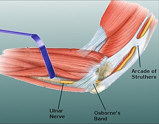
Osborne's ligament, also Osborne's band, Osborne's fascia, Osborne's arcade, arcuate ligament of Osborne, or the cubital tunnel retinaculum, refers to either the connective tissue which spans the humeral and ulnar heads of the flexor carpi ulnaris (FCU) or another distinct tissue located between the olecranon process of the ulna and the medial epicondyle of the humerus. It is named after Geoffrey Vaughan Osborne, a British orthopedic surgeon, who described the eponymous tissue in 1957.

Nerve glide, also known as nerve flossing or nerve stretching, is an exercise that stretches nerves. It facilitates the smooth and regular movement of peripheral nerves in the body. It allows the nerve to glide freely along with the movement of the joint and relax the nerve from compression. Nerve gliding cannot proceed with injuries or inflammations as the nerve is trapped by the tissue surrounding the nerve near the joint. Thus, nerve gliding exercise is widely used in rehabilitation programs and during the post-surgical period.

References

  1. Aguiar, Paulo Henrique; Bor-Seng-Shu, Edson; Gomes-Pinto, Fernando; Almeida- Leme, Ricardo Jose de; Freitas, Alexandre Bruno R.; Martins, Roberto S.; Nakagawa, Edison S.; Tedesco-Marchese, Antonio J. (March 2001). "Surgical management of Guyon's canal syndrome, an ulnar nerve entrapment at the wrist: report of two cases". Arquivos de Neuro-Psiquiatria. 59 (1): 106–111. doi: 10.1590/S0004-282X2001000100022 . PMID   11299442.
  2. Miller TT, Reinus WR (September 2010). "Nerve entrapment syndromes of the elbow, forearm, and wrist". American Journal of Roentgenology. 195 (3): 585–94. doi:10.2214/AJR.10.4817. PMID   20729434.
  3. Thakker, Arjuna; Gupta, Vinay Kumar; Gupta, Keshav Kumar (December 2020). "The Anatomy, Presentation and Management Options of Cubital Tunnel Syndrome". The Journal of Hand Surgery Asian-Pacific Volume. 25 (4): 393–401. doi:10.1142/S2424835520400032. ISSN   2424-8363. PMID   33115358. S2CID   226051048.
  4. 1 2 3 4 5 6 7 8 9 10 Cutts, S. (2007). "Cubital tunnel syndrome". Postgraduate Medical Journal. 83 (975): 28–31. doi:10.1136/pgmj.2006.047456. PMC   2599973 . PMID   17267675.
  5. Moore, Keith L. (2010). Clinically Oriented Anatomy 6th Ed. Baltimore, MD: Lippincott, Williams and Wilkins. p. 770. ISBN   978-07817-7525-0.
  6. Erdem Bagatur, A.; Yalcin, Mehmet Burak; Ozer, Utku Erdem (1 September 2016). "Anconeus Epitrochlearis Muscle Causing Ulnar Neuropathy at the Elbow: Clinical and Neurophysiological Differential Diagnosis". Orthopedics. 39 (5): e988–991. doi:10.3928/01477447-20160623-11. ISSN   1938-2367. PMID   27398787.
  7. "Guyon's Canal Syndrome". Archived from the original on 5 September 2015. Retrieved 17 September 2009.
  8. 1 2 Shea JD, McClain EJ (1969). "Ulnar-nerve compression syndromes at and below the wrist". J Bone Joint Surg Am. 51 (6): 1095–1103. doi:10.2106/00004623-196951060-00004. PMID   5805411.
  9. Patterson JM, Jaggars MM, Boyer MI (2003). "Ulnar and median nerve palsy in long-distance cyclists. A prospective study". Am J Sports Med. 31 (4): 585–589. doi:10.1177/03635465030310041801. PMID   12860549. S2CID   22497516.
  10. Aleksenko, Dmitri; Varacallo, Matthew (2023), "Guyon Canal Syndrome", StatPearls, Treasure Island (FL): StatPearls Publishing, PMID   28613717 , retrieved 24 August 2023
  11. 1 2 Thomas, Jennifer (2 June 2009). "'Cell Phone Elbow' -- A New Ill for the Wired Age". HealthDay News. Archived from the original on 5 April 2017. Retrieved 2 June 2009.
  12. 1 2 Wade, Ryckie G.; Griffiths, Timothy T.; Flather, Robert; Burr, Nicholas E.; Teo, Mario; Bourke, Grainne (24 November 2020). "Safety and Outcomes of Different Surgical Techniques for Cubital Tunnel Decompression: A Systematic Review and Network Meta-analysis". JAMA Network Open. 3 (11): e2024352. doi: 10.1001/jamanetworkopen.2020.24352 . PMC   7686867 . PMID   33231636.
  13. Guardia, Charles F (24 August 2014). "Ulnar Neuropathy Treatment & Management: Non-surgical therapy". Medscape.
  14. , Ilyas A, Herman Z. Cubital Tunnel Release. J Med Ins. 2017;2017(206.4) doi:https://jomi.com/article/206.4
  15. Mallette, Paige; Zhao, Meijuan; Zurakowski, David; Ring, David (2007). "Muscle Atrophy at Diagnosis of Carpal and Cubital Tunnel Syndrome". The Journal of Hand Surgery. 32 (6): 855–8. doi:10.1016/j.jhsa.2007.03.009. PMID   17606066.