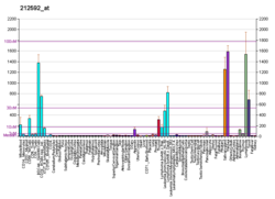
The Joining (J) chain is a protein component that links monomers of antibodies IgM and IgA to form polymeric antibodies capable of secretion. [5] The J chain is well conserved in the animal kingdom, but its specific functions are yet to be fully understood. It is a 137 residue polypeptide, [6] encoded by the IGJ gene. [7] [8] [9]
The J chain is a glycoprotein of molecular weight 15 kDa. Its secondary structure remains undetermined but is believed to adopt either a single β-barrel or two-domain folded structure with standard immunoglobulin domains. [10] The J chain's primary structure is unusually acidic having a high content of negatively charged amino acids. [11] It has 8 cysteine residues, 6 of which are involved in intramolecular disulfide bonds while the remaining two function to bind the Fc tailpiece regions of IgA or IgM antibodies, the α chain and μ chain respectively. An N-linked carbohydrate resulting from N-glycosylation is also essential in the protein's incorporation to antibody polymers. [12] There is no known protein family with significant homology to the J chain. [13]
The J chain regulates the multimerization of IgM and IgA in mammals. When expressed in cells, it favors the formation of a pentameric IgM and an IgA dimer. IgM pentamers are most commonly found with a single J chain, but some studies have seen as many as 4 J chains associated to a single IgM pentamer.
The J chain is incorporated late in the formation of IgM polymers and thermodynamically favors the formation of pentamers as opposed to hexamers. [12] In J chain-knockout (KO) mice, the hexameric IgM polymer dominates. [14] These J chain negative IgM hexamers are 15-20 times more effective at activating complement than J chain positive IgM pentamers. [15] However, J chain-KO mice have been shown have low concentrations of hexameric IgM and a deficiency in complement activation, suggesting additional in vivo regulatory mechanisms. [16] Another consequence of pentameric IgM reduced complement activation is its allowance of J chain positive pIgM to bind antigen without causing excessive damage to epithelial membranes through complement activation. [17]
The J chain facilitates IgA dimerization by linking two monomer secretory tails. Structurally, the J chain joins two antibody monomers asymmetrically by forming intermolecular disulfide bonds and bringing hydrophobic β-sandwiches on each molecule together. [18] This multimerization mechanism involves chaperone proteins including binding immunoglobulin protein (BiP) and MZB1 each sequentially recruiting distinct factors of the polymerized antibody. [19]
Mucosal membrane antibody secretion from the basal membrane to apical epithelial cells is facilitated by the polymeric Ig receptor (pIgR). A basal protein of the pIgR known as secretory component (SC) recognizes Ig ready for secretion. [20] The binding between the secretory component and secretory Ig is facilitated by the antibody's J chain which makes physical contact with the secretory component in order to change the transporter's conformation to an open state. [21] The complex is then transcytosed and the secretory component proteolytically cleaved from the receptor releasing the antibody to the apical side of the epithelial cell and to the lumen at large. This mechanism is thought to be largely conserved between the secretion of IgM and IgA. [19]
J chain was originally believed to only be expressed in antibody-secreting plasma cells, however, the J chain has been seen to be expressed in earlier stages of B cell differentiation prior to Ig expression. [22] J chain expression is believed to occur in the early stages of lymphoid cell differentiation as it is expressed in both B and T cell precursors. As cells develop, it seems that expression of the μ-chain becomes necessary for J chain synthesis. [23]
The J chain gene is transcriptionally regulated through canonical Pax5 repression. [24] As Pax5 is a common transcriptional regulator, the J chain is still expressed in plasma cells that secrete monomeric antibodies. In such cells it is believed to provide no function and is quickly degraded. [19] In plasma cells that secrete monomeric IgA, a Pax5-independent mechanism is likely to prevent IgA dimerization. [25]
The J chain is likely to have evolutionarily arisen in early jaw-boned vertebrates. [26] Groups of bony fish including teleosts have since lost J chain expression. [13] Xenopus are able to polymerize mucosal IgX in the absence of J chain, perhaps due to a loss of the conserved cysteine residues that link the J chain and Ig secretory tail. [27]
Sharks do not express IgA and thus use J chain expression solely for the polymerization of IgM. [28] This makes sharks an intriguing model organism in studying J chain regulation and polymerization without the confounding variables of mucosal secretion. [29]
An antibody (Ab), also known as an immunoglobulin (Ig), is a large, Y-shaped protein used by the immune system to identify and neutralize foreign objects such as pathogenic bacteria and viruses. The antibody recognizes a unique molecule of the pathogen, called an antigen. Each tip of the "Y" of an antibody contains a paratope that is specific for one particular epitope on an antigen, allowing these two structures to bind together with precision. Using this binding mechanism, an antibody can tag a microbe or an infected cell for attack by other parts of the immune system, or can neutralize it directly.
Immunoglobulin A is an antibody that plays a role in the immune function of mucous membranes. The amount of IgA produced in association with mucosal membranes is greater than all other types of antibody combined. In absolute terms, between three and five grams are secreted into the intestinal lumen each day. This represents up to 15% of total immunoglobulins produced throughout the body.
Immunoglobulin D (IgD) is an antibody isotype that makes up about 1% of proteins in the plasma membranes of immature B-lymphocytes where it is usually co-expressed with another cell surface antibody called IgM. IgD is also produced in a secreted form that is found in very small amounts in blood serum, representing 0.25% of immunoglobulins in serum. The relative molecular mass and half-life of secreted IgD is 185 kDa and 2.8 days, respectively. Secreted IgD is produced as a monomeric antibody with two heavy chains of the delta (δ) class, and two Ig light chains.
Immunoglobulin E (IgE) is a type of antibody that has been found only in mammals. IgE is synthesised by plasma cells. Monomers of IgE consist of two heavy chains and two light chains, with the ε chain containing four Ig-like constant domains (Cε1–Cε4). IgE is thought to be an important part of the immune response against infection by certain parasitic worms, including Schistosoma mansoni, Trichinella spiralis, and Fasciola hepatica. IgE is also utilized during immune defense against certain protozoan parasites such as Plasmodium falciparum. IgE may have evolved as a defense to protect against venoms.
Immunoglobulin M (IgM) is one of several isotypes of antibody that are produced by vertebrates. IgM is the largest antibody, and it is the first antibody to appear in the response to initial exposure to an antigen. In humans and other mammals that have been studied, plasmablasts residing in the spleen are the main source for specific IgM production.
Plasma cells, also called plasma B cells or effector B cells, are white blood cells that originate in the lymphoid organs as B lymphocytes and secrete large quantities of proteins called antibodies in response to being presented specific substances called antigens. These antibodies are transported from the plasma cells by the blood plasma and the lymphatic system to the site of the target antigen, where they initiate its neutralization or destruction. B cells differentiate into plasma cells that produce antibody molecules closely modeled after the receptors of the precursor B cell.
In immunology, a memory B cell (MBC) is a type of B lymphocyte that forms part of the adaptive immune system. These cells develop within germinal centers of the secondary lymphoid organs. Memory B cells circulate in the blood stream in a quiescent state, sometimes for decades. Their function is to memorize the characteristics of the antigen that activated their parent B cell during initial infection such that if the memory B cell later encounters the same antigen, it triggers an accelerated and robust secondary immune response. Memory B cells have B cell receptors (BCRs) on their cell membrane, identical to the one on their parent cell, that allow them to recognize antigen and mount a specific antibody response.
The immunoglobulin heavy chain (IgH) is the large polypeptide subunit of an antibody (immunoglobulin). In human genome, the IgH gene loci are on chromosome 14.
Protein A is a 42 kDa surface protein originally found in the cell wall of the bacteria Staphylococcus aureus. It is encoded by the spa gene and its regulation is controlled by DNA topology, cellular osmolarity, and a two-component system called ArlS-ArlR. It has found use in biochemical research because of its ability to bind immunoglobulins. It is composed of five homologous Ig-binding domains that fold into a three-helix bundle. Each domain is able to bind proteins from many mammalian species, most notably IgGs. It binds the heavy chain within the Fc region of most immunoglobulins and also within the Fab region in the case of the human VH3 family. Through these interactions in serum, where IgG molecules are bound in the wrong orientation, the bacteria disrupts opsonization and phagocytosis.
The neonatal Fc receptor is a protein that in humans is encoded by the FCGRT gene. It is an IgG Fc receptor which is similar in structure to the MHC class I molecule and also associates with beta-2-microglobulin. In rodents, FcRn was originally identified as the receptor that transports maternal immunoglobulin G (IgG) from mother to neonatal offspring via mother's milk, leading to its name as the neonatal Fc receptor. In humans, FcRn is present in the placenta where it transports mother's IgG to the growing fetus. FcRn has also been shown to play a role in regulating IgG and serum albumin turnover. Neonatal Fc receptor expression is up-regulated by the proinflammatory cytokine, TNF-α, and down-regulated by IFN-γ.
In immunology, antibodies are classified into several types called isotypes or classes. The variable (V) regions near the tip of the antibody can differ from molecule to molecule in countless ways, allowing it to specifically target an antigen . In contrast, the constant (C) regions only occur in a few variants, which define the antibody's class. Antibodies of different classes activate distinct effector mechanisms in response to an antigen . They appear at different stages of an immune response, differ in structural features, and in their location around the body.
Polymeric immunoglobulin receptor (pIgR) is a transmembrane protein that in humans is encoded by the PIGR gene. It is an Fc receptor which facilitates the transcytosis of the soluble polymeric isoforms of immunoglobulin A and immunoglobulin M (pIg) and immune complexes. pIgRs are mainly located on the epithelial lining of mucosal surfaces of the gastrointestinal tract. The composition of the receptor is complex, including 6 immunoglobulin-like domains, a transmembrane region, and an intracellular domain. pIgR expression is under the strong regulation of cytokines, hormones, and pathogenic stimuli.
ICOS ligand is a protein that in humans is encoded by the ICOSLG gene located at chromosome 21. ICOSLG has also been designated as CD275.
Ig mu chain C region is a protein that in humans is encoded by the IGHM gene.
Cluster of Differentiation 276 (CD276) or B7 Homolog 3 (B7-H3) is a human protein encoded by the CD276 gene.
Immunoglobulin iota chain is a protein that in humans is encoded by the VPREB1 gene. VPREB1 has also recently been designated CD179A.
Fc fragment of IgG receptor IIb is a low affinity inhibitory receptor for the Fc region of immunoglobulin gamma (IgG). FCGR2B participates in the phagocytosis of immune complexes and in the regulation of antibody production by B lymphocytes.
Leukocyte immunoglobulin-like receptor subfamily A member 3 (LILR-A3) also known as CD85 antigen-like family member E (CD85e), immunoglobulin-like transcript 6 (ILT-6), and leukocyte immunoglobulin-like receptor 4 (LIR-4) is a protein that in humans is encoded by the LILRA3 gene located within the leukocyte receptor complex on chromosome 19q13.4. Unlike many of its family, LILRA3 lacks a transmembrane domain. The function of LILRA3 is currently unknown; however, it is highly homologous to other LILR genes, and can bind human leukocyte antigen (HLA) class I. Therefore, if secreted, the LILRA3 might impair interactions of membrane-bound LILRs with their HLA ligands, thus modulating immune reactions and influencing susceptibility to disease.
Cluster of differentiation CD79A also known as B-cell antigen receptor complex-associated protein alpha chain and MB-1 membrane glycoprotein, is a protein that in humans is encoded by the CD79A gene.
Fc fragment of IgA receptor (FCAR) is a human gene that codes for the transmembrane receptor FcαRI, also known as CD89. FcαRI binds the heavy-chain constant region of Immunoglobulin A (IgA) antibodies. FcαRI is present on the cell surface of myeloid lineage cells, including neutrophils, monocytes, macrophages, and eosinophils, though it is notably absent from intestinal macrophages and does not appear on mast cells. FcαRI plays a role in both pro- and anti-inflammatory responses depending on the state of IgA bound. Inside-out signaling primes FcαRI in order for it to bind its ligand, while outside-in signaling caused by ligand binding depends on FcαRI association with the Fc receptor gamma chain.