Penile fracture

Last updated

Penile Fracture
Specialty Urology
Symptoms Immediate pain and swelling of the penis, rapid erection loss, discoloration of the penile shaft [1]
Complications Gangrene, erectile dysfunction, Peyronie's disease
CausesBlunt trauma to an erect penis
Risk factors Sexual intercourse, masturbation
Diagnostic method Based on symptoms, ultrasound
Differential diagnosis Testicular torsion
Treatment Emergency surgery
Prognosis ~10–50% of people develop erectile dysfunction or Peyronie's disease [2]
Frequency~1 per 175,000 men per year [3]

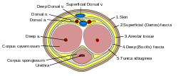
Penile fracture is rupture of one or both of the tunica albuginea , the fibrous coverings that envelop the penis's corpora cavernosa . It is caused by rapid blunt force to an erect penis, usually during vaginal intercourse, or aggressive masturbation. [4] It sometimes also involves partial or complete rupture of the urethra or injury to the dorsal nerves, veins and arteries. [5]

Contents

Signs and symptoms

A popping or cracking sound, significant pain, swelling, immediate loss of erection leading to flaccidity, and skin hematoma of various sizes are commonly associated with the sexual event. [4] [6]

Causes

Bending penis in doggy style position Penisfracture - risk.png
Bending penis in doggy style position
Cross-section of the human penis Penis cross section.svg
Cross-section of the human penis

Penile fracture is a relatively uncommon clinical condition. [7] Vaginal intercourse and aggressive masturbation are the most common causes. [4] A 2014 study of accident and emergency records at three hospitals in Campinas, Brazil, showed that woman on top positions caused the greatest risk with the missionary position being the safest. The research conjectured that when the receptive partner is on top, they usually control the movement and are not able to interrupt movement when the penis suffers a misaligned penetration. Conversely, when the penetrative partner is controlling the movement, they have better chances of stopping in response to pain from misalignment, minimizing harm. [7]

The practice of taqaandan (also taghaandan) also puts men at risk of penile fracture. Taqaandan, which comes from a Kurdish word meaning "to click", involves bending the top part of the erect penis while holding the lower part of the shaft in place, until a click is heard and felt. Taqaandan is said to be painless and has been compared to cracking one's knuckles, but the practice of taqaandan has led to an increase in the prevalence of penile fractures in western Iran. [8] Taqaandan may be performed to achieve detumescence. [9]

Diagnosis

Imaging studies

Ultrasound examination is able to depict the tunica albuginea tear in the majority of cases (as a hypoechoic discontinuity in the normally echogenic tunica). In a study on 25 patients, Zare Mehrjardi et al. concluded that ultrasound is unable to find the tear just when it is located at the penile base. In their study magnetic resonance imaging (MRI) accurately diagnosed all of the tears (as a discontinuity in the normally low signal tunica on both T1- and T2-weighted sequences). They concluded that ultrasound should be considered as the initial imaging method, and MRI can be helpful in cases that ultrasound does not depict any tear but clinical suspicions for fracture are still high. In the same study, authors investigated accuracy of ultrasound and MRI for determining the tear location (mapping of fracture) in order to perform a tailored surgical repair. MRI was more accurate than ultrasound for this purpose, but ultrasound mapping was well correlated with surgical results in cases where the tear was clearly visualized on ultrasound exam. [10] The advantage of ultrasound in the diagnosis of penile fracture is unrivaled when its noninvasive, cost-effective, and nonionising nature are considered. [11]

Penile trauma can result from a blunt or penetrating injury, the latter being rarely investigated by imaging methods, almost always requiring immediate surgical exploration. In the erect penis, trauma results from stretching and narrowing of the tunica albuginea, which can undergo segmental rupture of one or both of the corpora cavernosa, constituting a penile fracture. [2]

In the ultrasound examination, a lesion of the tunica albuginea presents as an interruption in (loss of continuity of) the echoic line representing it (Figure 4). Small, moderate, or broad hematomas demonstrate the extent of that discontinuity. Intracavernous hematomas, sometimes without the presence of a tunica albuginea fracture, can be observed when there is a lesion of the smooth muscle of the trabeculae surrounding the sinusoid spaces or the subtunical venular plexus. [2]

In 10–15% of penile traumas, there can be an accompanying urethral lesion. When blood is observed in the urethral meatus, contrast-enhanced evaluation of the urethra is necessary. In cases in which the ultrasound findings are inconclusive, the use of magnetic resonance imaging can facilitate the diagnosis and is recommended by various authors. [2]

Treatment

Penile fracture is a medical emergency, and emergency surgical repair is the usual treatment. Delay in seeking treatment increases the complication rate. Non-surgical approaches result in 10–50% complication rates including erectile dysfunction, permanent penile curvature, damage to the urethra and pain during sexual intercourse, while operatively treated patients experience an 11% complication rate. [4] [12]

In some cases, retrograde urethrogram may be performed to rule out concurrent urethral injury. [12]

In the United States, the case of Doe v. Moe , 63 Mass. App. Ct. 516, 827 N.E.2d 240 (2005), tested liability for a penile fracture injury caused during sexual intercourse. The court declined to find duty as between two consensual adults. The plaintiff in this case, a man who suffered a fractured penis, complained that the defendant, his ex-girlfriend, had caused his injury while she was on top of him during sexual intercourse. The court ruled in her favor, determining that her conduct was neither legally wanton nor reckless.

Related Research Articles

<span class="mw-page-title-main">Urethra</span> Tube that connects the urinary bladder to the external urethral orifice

The urethra is a tube that connects the mammalian urinary bladder to the urinary meatus. Male and female placental mammals release urine through the urethra during urination, but males also release semen through the urethra during ejaculation.

<span class="mw-page-title-main">Peyronie's disease</span> Medical condition

Peyronie's disease is a connective tissue disorder involving the growth of fibrous plaques in the soft tissue of the penis. Specifically, scar tissue forms in the tunica albuginea, the thick sheath of tissue surrounding the corpora cavernosa, causing pain, abnormal curvature, erectile dysfunction, indentation, loss of girth and shortening.

<span class="mw-page-title-main">Urethral stricture</span> Medical condition

A urethral stricture is a narrowing of the urethra, the tube connected to the bladder that allows the passing of urine. The narrowing reduces the flow of urine and makes it more difficult or even painful to empty the bladder.

<span class="mw-page-title-main">Hypospadias</span> Medical condition

Hypospadias is a common variation in fetal development of the penis in which the urethra does not open from its usual location on the head of the penis. It is the second-most common birth defect of the male reproductive system, affecting about one of every 250 males at birth. Roughly 90% of cases are the less serious distal hypospadias, in which the urethral opening is on or near the head of the penis (glans). The remainder have proximal hypospadias, in which the meatus is all the way back on the shaft of the penis, near or within the scrotum. Shiny tissue that typically forms the urethra instead extends from the meatus to the tip of the glans; this tissue is called the urethral plate.

<span class="mw-page-title-main">Chordee</span> Medical condition

Chordee is a condition in which the head of the penis curves downward or upward, at the junction of the head and shaft of the penis. The curvature is usually most obvious during erection, but resistance to straightening is often apparent in the flaccid state as well. In many cases but not all, chordee is associated with hypospadias. This is not the same condition as Peyronie's disease, which involves curvature of the shaft of the penis most commonly due to injury during adult life.

<span class="mw-page-title-main">Urethral sounding</span> Use of a probe on the urethra

Urethral sounding is the practice of inserting objects into the urethra for sexual gratification. Urethral dilatation is a urological procedure that uses probes called sounds to enlarge the inside diameter of the urethra and locate obstructions in the urethra, or as a treatment for urethral strictures.

<span class="mw-page-title-main">Bladder exstrophy</span> Medical condition

Bladder exstrophy is a congenital anomaly that exists along the spectrum of the exstrophy-epispadias complex, and most notably involves protrusion of the urinary bladder through a defect in the abdominal wall. Its presentation is variable, often including abnormalities of the bony pelvis, pelvic floor, and genitalia. The underlying embryologic mechanism leading to bladder exstrophy is unknown, though it is thought to be in part due to failed reinforcement of the cloacal membrane by underlying mesoderm. Exstrophy means the inversion of a hollow organ.

<span class="mw-page-title-main">Radical retropubic prostatectomy</span>

Radical retropubic prostatectomy is a surgical procedure in which the prostate gland is removed through an incision in the abdomen. It is most often used to treat individuals who have early prostate cancer. Radical retropubic prostatectomy can be performed under general, spinal, or epidural anesthesia and requires blood transfusion less than one-fifth of the time. Radical retropubic prostatectomy is associated with complications such as urinary incontinence and impotence, but these outcomes are related to a combination of individual patient anatomy, surgical technique, and the experience and skill of the surgeon.

<span class="mw-page-title-main">Penile implant</span> Medical device

A penile implant is an implanted device intended for the treatment of erectile dysfunction, Peyronie's disease, ischemic priapism, deformity and any traumatic injury of the penis, and for phalloplasty or metoidioplasty, including in gender-affirming surgery. Men also opt for penile implants for aesthetic purposes. Men's satisfaction and sexual function is influenced by discomfort over genital size which leads to seek surgical and non-surgical solutions for penis alteration. Although there are many distinct types of implants, most fall into one of two categories: malleable and inflatable transplants.

Testicular rupture is a rip or tear in the tunica albuginea resulting in extrusion of the testicular contents, including the seminiferous tubules. It is a rare complication of testicular trauma, and can result from blunt or penetrating trauma, although blunt trauma is more likely to cause rupture.

Urethroplasty is the surgical repair of an injury or defect within the walls of the urethra. Trauma, iatrogenic injury and infections are the most common causes of urethral injury/defect requiring repair. Urethroplasty is regarded as the gold standard treatment for urethral strictures and offers better outcomes in terms of recurrence rates than dilatations and urethrotomies. It is probably the only useful modality of treatment for long and complex strictures though recurrence rates are higher for this difficult treatment group.

<span class="mw-page-title-main">Retrograde urethrogram</span> Medical imaging of the urethra

A retrograde urethrography is a routine radiologic procedure used to image the integrity of the urethra. Hence a retrograde urethrogram is essential for diagnosis of urethral injury, or urethral stricture.

<span class="mw-page-title-main">Diphallia</span> Genital medical condition

Diphallia, penile duplication (PD), diphallic terata, or diphallasparatus is an extremely rare developmental abnormality in which a male is born with two penises. The first reported case was by Johannes Jacob Wecker in 1609. Its occurrence is 1 in 5.5 million boys in the United States.

<span class="mw-page-title-main">Mechanics of human sexuality</span> Biomechanics of human sexual intercourse

The mechanics of human sexuality or mechanics of sex, or more formally the biomechanics of human sexuality, is the study of the mechanics related to human sexual activity. Examples of topics include the biomechanical study of the strength of vaginal tissues and the biomechanics of male erectile function. The mechanics of sex under limit circumstances, such as sexual activity at zero-gravity in outer space, are also being studied.

Bulbar urethral necrosis is a problem that can occur after a pelvic fracture associated urethral distraction defect (PFUDD).

<span class="mw-page-title-main">Webbed penis</span> Medical condition

Webbed penis also known as buried or concealed penis is an acquired or congenital condition in which the scrotal skin extends onto the ventral penile shaft. The penile shaft is buried in the scrotum or tethered to the scrotal midline by a fold or web of skin. The urethra and erectile bodies are usually normal. Webbed penis is usually asymptomatic, but the cosmetic appearance is often unacceptable. This condition may be corrected by surgical techniques.

<span class="mw-page-title-main">Septum glandis</span> Part of the human glans penis

The septum glandis, also septum of the glans, refers to the fibrous partition of the ventral aspect of the glans penis that separates the two glans wings in the ventral midline. The septum extends from the urethral meatus through the glanular urethra and ends in the tunica albuginea of the human penis. Externally it is attached to the frenulum which extends lower on the neck of the penis.

Genital trauma is trauma to the genitalia.

A penile injury is a medical emergency that afflicts the penis. Common injuries include fracture, avulsion injury, strangulation, entrapment, and amputation.

Penile ulltrasonography is medical ultrasonography of the penis. Ultrasound is an excellent method for the study of the penis, such as indicated in trauma, priapism, erectile dysfunction or suspected Peyronie's disease.

References

  1. "Penis fracture: Is it possible?". Mayo Clinic .
  2. 1 2 3 4 5 6 Originally copied from:
    Fernandes, Maitê Aline Vieira; Souza, Luis Ronan Marquez Ferreira de; Cartafina, Luciano Pousa (2018). "Ultrasound evaluation of the penis". Radiologia Brasileira. 51 (4): 257–261. doi:10.1590/0100-3984.2016.0152. ISSN   1678-7099. PMC   6124582 . PMID   30202130.
    CC-BY license
  3. Amer, Tarik; Wilson, Rebekah; Chlosta, Piotr; Albuheissi, Salah; Qazi, Hasan; Fraser, Michael; Aboumarzouk, Omar M. (2016). "Penile Fracture: A Meta-Analysis". Urologia Internationalis. 96 (3): 315–329. doi: 10.1159/000444884 . PMID   26953932. S2CID   8901660.
  4. 1 2 3 4 Greenberg's Text-Atlas of Emergency Medicine. Lippincott Williams & Wilkins. 22 November 2004. p. 318. ISBN   978-0-7817-4586-4 . Retrieved 15 October 2012.
  5. Haas CA, Brown SL, Spirnak JP (April 1999). "Penile fracture and testicular rupture". World J Urol. 17 (2): 101–6. doi:10.1007/s003450050114. PMID   10367369. S2CID   27097515.
  6. Stein DM, Santucci RA (July 2015). "An update on urotrauma". Current Opinion in Urology. 25 (4): 323–30. doi:10.1097/MOU.0000000000000184. PMID   26049876. S2CID   26994715.
  7. 1 2 Reis, Leonardo O.; Cartapatti, Marcelo; Marmiroli, Rafael; Oliveira Júnior, Eduardo Jeronimo de; Saade, Ricardo Destro; Fregonesi, Adriano (2014). "Mechanisms Predisposing Penile Fracture and Long-Term Outcomes on Erectile and Voiding Functions". Advances in Urology. 2014: 1–4. doi: 10.1155/2014/768158 . PMC   4005103 . PMID   24822062.
  8. Nuzzo, Regina (9 February 2009). "Preventing penile fractures and Peyronie's disease - latimes.com". Los Angeles Times. Archived from the original on 14 February 2009.
  9. Zargooshi J (August 2000). "Penile fracture in Kermanshah, Iran: report of 172 cases". J. Urol. 164 (2): 364–6. doi:10.1016/s0022-5347(05)67361-2. PMID   10893586.
  10. Zare Mehrjardi, Mohammad; Darabi, Mohsen; Bagheri, Seyed Morteza; Kamali, Koosha; Bijan, Bijan (June 2017). "The role of ultrasound (US) and magnetic resonance imaging (MRI) in penile fracture mapping for modified surgical repair". International Urology and Nephrology. 49 (6): 937–945. doi:10.1007/s11255-017-1550-x. ISSN   1573-2584. PMID   28258528. S2CID   4094189.
  11. Napier, Donna (2018). "The role of ultrasound in the diagnosis of penile fracture". Sonography. 6: 15–23. doi: 10.1002/sono.12167 . ISSN   2054-6750.
  12. 1 2 Andrew B. Peitzman; Michael Rhodes; C. William Schwab; Donald M Yealy; Timothy C Fabian (1 September 2007). The trauma manual: trauma and acute care surgery. Lippincott Williams & Wilkins. pp. 305–. ISBN   978-0-7817-6275-5 . Retrieved 18 April 2010.

Further reading