Plasmablastic lymphoma

Last updated
Plasmablast, Wright stain Plasmablast, Wright stain.png
Plasmablast, Wright stain

Plasmablastic lymphoma (PBL) is a type of large B-cell lymphoma recognized by the World Health Organization (WHO) in 2017 as belonging to a subgroup of lymphomas termed lymphoid neoplasms with plasmablastic differentiation. The other lymphoid neoplasms within this subgroup are: plasmablastic plasma cell lymphoma (or the plasmacytoma variant of this disease); primary effusion lymphoma that is Kaposi's sarcoma-associated herpesvirus positive or Kaposi's sarcoma-associated Herpesvirus negative; anaplastic lymphoma kinase-positive large B-cell lymphoma; and human herpesvirus 8-positive diffuse large B-cell lymphoma, not otherwise specified. All of these lymphomas are malignancies of plasmablasts, i.e. B-cells that have differentiated into plasmablasts but because of their malignant nature: fail to differentiate further into mature plasma cells; proliferate excessively; and accumulate in and injure various tissues and organs. [1]

Contents

The lymphomas in the lymphoid neoplasms with plasmablastic differentiation sub-group that are not PBL have sometimes been incorrectly considered to be variants of PBL. Each of the lymphomas in this subgroup of malignancies have distinctive clinical, morphological, and abnormal gene features. However, key features of these lymphomas sometime overlap with other lymphomas including those that are in this sub-group. In consequence, correctly diagnosing these lymphomas has been challenging. [2] Nonetheless, it is particularly important to diagnose them correctly because they can have very different prognoses and treatments than the lymphomas which they resemble. [1]

Plasmablastic lymphomas are aggressive and rare malignancies that usually respond poorly to chemotherapy and carry a very poor prognosis. They occur predominantly in males who have HIV/AIDS, had a solid organ transplant, or are immunosuppressed in other ways; ~5% of all individuals with PBL appear to be immunocompetent, i.e. to have no apparent defect in their immune system. [2] The malignant plasmablasts in more than half the cases of PBL are infected with a potentially cancer-causing virus, Epstein–Barr virus (EBV), and rare cases of PBL appear due to the plasmablastic transformation of a preexisting low-grade B-cell lymphoma. [3] One variant of PBL, sometimes termed plasmablastic lymphoma of the elderly, has a significantly better prognosis than most other cases of PBL. [4] The development of this variant appears due, at least in part, to immunosenescence, i.e. the immunodeficiency occurring in old age. [3]

Presentation

Plasmablastic lymphoma lesions are most commonly rapidly growing, soft tissue masses [5] that may be ulcerating, bleeding, and/or painful. [6] In a recent (2020) review of published cases, individuals presenting with PBD were typically middle-aged or elderly (range 1–88 years; median age 58 tears) males (~73% of cases). [7] Only a few cases have been reported in pediatric cases. [6] The PDL lesions occurred most commonly in lymph nodes (~23% of cases), the gastrointestinal tract (~18%), bone marrow (16%), and oral cavity (12%). [7] Less frequently involved tissues include the skin, genitourinary tract, [7] paranasal sinuses, lung, and bones. [5] While cases of PBL may present as a primary oral, [8] or, very rarely a skin [3] or lymph node [8] disease, most individuals present with a widespread stage III or IV disease which in ~40% of cases, is accompanied by systemic B-symptoms such as fever, night sweats, and recent weight loss. [2] Some 48%-63% of PBL cases occur in individuals with HIV/AIDS; ~80% of these HIV/AIDS-afflicted individuals have EBV+ disease whereas only ~50% of PBL individuals that do not have HIV/AIDS are EBV-positive. [2] Individuals with PBL often present with a history of being immunosuppressed due to prior organ transplantation, immunosuppressive drug treatment, or other causes. [9] This is particularly the case for individuals with HIV/AIDS-negative disease. [7] Individuals who develop PBL following organ transplantation are EBV/AIDS-positive in >85% of cases. [7] Most post-transplant and HIV/AIDS-positive patients have an extremely aggressive disease. However, patients whose major contributing factor to PBL-development is EBV-positivity often present with, and continue to have, a significantly less aggressive disease than other patients with PBL. [5] It is similarly clear that, on average, individuals who are elderly (>68 years) [4] or have HIV/AIDS-negative disease [7] likewise present with, and continue to have, a significantly less aggressive cancer.

Pathophysiology

In addition to the immunodeficiency-causing viral disease, HIV/AIDS (which is an AIDS-defining clinical condition [5] ), recent studies have diagnosed PBL in individuals who have one or more other causes for immunodeficiency. [7] These causes include prior organ transplantation; immunosuppressive drugs; autoimmune and chronic inflammatory diseases (e.g. hepatitis C, [3] rheumatoid arthritis, Graves' disease, Giant-cell arteritis, sarcoidosis, and severe psoriasis [10] ); and immunosenescence due to age (e.g. >60 years). Rare cases of PDL have also occurred as a transformation of a low grade B-cell malignancy such as chronic lymphocytic leukemia/small lymphocytic lymphoma and follicular lymphoma. [11] Studies also find that 60-75% of individuals diagnosed with PBL have Epstein–Barr virus-infected plasmablasts. [1] EBV infects ~95% of the world's population to cause no symptoms, minor non-specific symptoms, or infectious mononucleosis. The virus then enters a latency phase in which infected individuals become lifetime asymptomatic carriers of the virus in a set of their B-cells. Some weeks, months, years, or decades thereafter, a very small fraction of these carriers, particularly those with an immunodeficiency, develop any one of various EBV-associated benign or malignant diseases, including, in extremely rare cases, Epstein–Barr virus-positive plasmablastic lymphoma. [12] The virus in infected plasmablastic cells appears to be in its latency I phase; consequently, these infected cells express EBV products such as EBER nuclear RNAs and BART microRNAs. These RNAs promote infected cells to proliferate, avoid attack by the host's immune system's cytotoxic T-cells, and, possibly, block the infected cells' apoptosis (i.e. programmed cell death) response to injury. [12]

The predisposing conditions described in the previous paragraph can serve to enhance the ability of the plasmablasts in PBL to: avoid the host's immune surveillance; survive for prolonged periods, grow excessively, and acquire pro-malignant gene abnormalities. Some of the gene abnormalities found in PBL include: 1) increased expression of the MYC proto-oncogene due to its rearrangement with an antibody gene by genetic recombination or, less commonly, other causes (Myc protein, the product of this gene, enhances cell proliferation, inhibits apoptosis, and promotes malignancy); 2) loss in the expression of the PRDM1 gene whose product, PRDM1/BLMP1 protein, represses the expression of Myc protein; [13] ) 3) frequent duplications in certain areas of chromosomes 1, 7, 11, and 22 (these duplications are similar to those often seen in diffuse large cell lymphoma); [8] 4) reduced expression of at least 13 genes that are involved in B-cell responses to signaling agents. [1] 5) increased expression of genes which promote the maturation of B-cells toward plasma cells (e.g. CD38, CD138, IR4/MUM1, XBP1, IL21R , and, as just indicated, PRDM1); and 6) reduced expression of genes characteristic of B-cells (e.g. CD20 and PAX5 ). [2]

Diagnosis

Microscopic examination of involved PBD masses and infiltrates generally reveals diffuse proliferations of immunoblast-like cells with prominent features of plasma cells, i.e. plasmablastic cells. [2] Immunostaining of these cells indicate that they lack B-cell marker proteins (e.g. CD20 and PAX5 [in ~10% of cases CD20 may be expressed at very low levels [2] ]) but rather express plasma cell marker proteins (e.g. CD38, CD138, IR4/MUM1, XBP1, IL21R, and/or PRDM1). The abnormalities in gene structures and expressions reported in the Pathophysiology section, particularly rearrangement and/or over expression of the MYC proto-oncogene, may also be apparent in these cells. The presence of HIV/AIDS or other causes of immuno-incompetence (see previous section), a history of having a low-grade lymphoma, and/or the presence of EVB+ plasmablasts in the disease's lesions would support the diagnosis of PBL. [1]

Differential diagnosis

Various lymphomas can exhibit the microscopic appearance, including plasmablastic cells, and presentation of PBL. These lymphomas can usually be differentiated from PBL by further examinations of the plasmablasts for various marker proteins and determining other factors that favor the diagnosis of these lymphomas rather than PBL, [14] as indicated in the following descriptions.

Anaplastic lymphoma kinase-positive large B-cell lymphoma

Unlike PBL, the plasmablastic cells in anaplastic lymphoma kinase-positive large B-cell lymphoma strongly express the product of the ACVRL1 gene, i.e. activin receptor-like kinase 1 (ALK1) and are not infected with EBV and therefore do not express this virus's EBER or BART RNAs. [8]

Human herpesvirus 8-positive diffuse large B-cell lymphoma, not otherwise specified

Unlike PBL, the plasmablastic cells in human herpesvirus 8-positive diffuse large B-cell lymphoma, not otherwise specified express products of herpesvirus 8 (also termed Kaposi sarcoma virus) such as LANA-1 protein. Also unlike PBL, these plasmablastic cells do not express CD30, CD138, CD79a, [1] or a clonal IgM antibody and usually are not EBV-infected and therefore usually do not express this virus's EBER or BART RNAs. [8]

Primary effusion lymphoma

In contrast to PBL, the plasmablastic cells in primary effusion lymphomas, whether HHV8-positive or HHV8-negative, usually strongly express CD45 [8] and in HHV8 cases express HHV8 proteins such as the LANA-1 protein. Primary effusion lymphoma, HH8-negative also differs from PBL in that its plasmablastic cells frequently express certain B-cell marker proteins such as CD20 and CD79a. [1]

Plasmablastic plasma cell lymphoma

Various factors distinguish plasmablastic plasma cell lymphoma from PBL. Prior diagnosis of plasma cell lymphoma (i.e. multiple myeloma or plasmacytoma), the presence of lytic bone lesions, [8] increased levels of serum calcium, renal insufficiency, and anemia, and the presence of a myeloma protein in the serum and/or urine favor the diagnosis of plasmablastic plasma cell lymphoma rather than plasmablastic lymphoma. Ultimately, however, the marker proteins expressed by the plasmablastic cells in the two diseases are almost identical and a diagnosis of "plasmablastic neoplasm, consistent with PBL or multiple myeloma" may be acceptable in some cases according to the current World Health Organization classification. [1]

Other B-cell lymphomas

The plasmablastic cells in B-cell lymphomas, including diffuse B-cell lymphomas, chronic lymphocytic leukemia/small lymphocytic lymphoma, and follicular lymphoma generally express CD20 and often express CD45 marker proteins. While PBL plasmablastic cells weakly express CD20 in 10% of cases, the strong expression of CD20 and the expression of CD45 virtually rules out PBL. [8]

Treatment

The treatments for PBL have ranged from radiotherapy for localized disease to various chemotherapy regimens for extensive disease. The chemotherapy regimens have included CHOP (i.e. cyclophosphamide, hydroxydoxorubicin (or doxorubicin), vincristine, and either prednisone or prednisolone); CHOP-like regimens (e.g. CHOP plus etoposide); hyper-CVAD-MA (i.e. cyclophosphamide, vincristine, doxorubicin, dexamethasone and high dose methotrexate and cytarabine); CODOX-M/IVAC (i.e. cyclophosphamide, vincristine, doxorubicin, high-dose methotrexate and ifosfamide, etoposide, and high-dose cytarabine); COMB (i.e. cyclophosphamide, oncovin, methyl-CCNU, and bleomycin); and infusional EPOCH (i.e. etoposide, prednisone, vincristine, cyclophosphamide, and doxorubicin). While the experience treating PBL with radiation alone has been limited, patients with localized disease have been treated with doxorubicin-based chemotherapy regimens plus radiotherapy. [2]

Experimental treatments

Given the unsatisfactory results of standard chemotherapy regimens, new treatments are being explored for use in PBL. Bortezomib, a drug that inhibits proteasomes, has been used alone or in combination with radiation and/or CHOP, EPOCH, or THP-COP (pirarubicin, cyclophosphamide, vincristine, and prednisone) chemotherapy regimens to treat some scores of patients with newly diagnosed or relapsed PBL. The results of these exploratory studies have been at least modestly encouraging and provide strong support for further studies using more controlled conditions. [2] A study sponsored by the AIDS Malignancy Consortium in collaboration with the National Cancer Institute is in its recruiting phase to study the dosages, safety, and efficacy of adding daratumumab to the EPOCH regimen in treating patients with PBL. [15] Daratumumab is a prepared monoclonal antibody that binds to CD38 and thereby directly or indirectly kills cells, including the plasmablasts in PBL, that express this marker protein on their surfaces. [2] An ongoing study sponsored by the City of Hope Medical Center is examining the feasibility and safety of gene therapy that uses recombinant RNA to target a key element in the HIV genome in patients who have HIV/AIDs and a non-Hodgkins lymphoma, including patients with plasmablastic lymphoma. [16]

Prognosis

Overall, patients receiving one of the cited chemotherapy regimens have achieved disease-free survival and overall survival rates of 22 and 32 months, respectively. The National Comprehensive Cancer Network recommends the more intensive regimens (e.g. hyper-CVAD-MA or infusional EPOCH) to treat the disease. These regimens have attained 5 year overall and disease-free survivals of 38% and 40%, respectively. Too few patients have been treated with autologous hematopoietic stem cell transplant in addition to chemotherapy for conclusions to be made. A few patients with HIV/AIDS-related PBL disease who were treated with highly active antiretroviral therapy (HAART) directed against the human immunodeficiency virus (i.e. HIV) have had remissions in their PDL lesions. [2]

History

A study by Green and Eversole published in 1989 [17] reported on 9 individuals afflicted with HIV/AIDS who presented with lymphomatous masses in the oral cavity; these lymphomas were populated by apparently malignant Epstein–Barr virus-infected plasmablasts that did not express T-cell lymphocyte marker proteins. Eight years later, Delecluse and colleagues [18] described a lymphoma, which they termed plasmablastic lymphoma, that had some features of a diffuse large B-cell lymphoma but unlike this lymphoma developed exclusively in the oral cavity, consisted of plasmablasts that lacked B-cell as well as T cell marker proteins and, in 15 of 16 cases, were infected with EBV. In 2008, the World Health Organization recognized this lymphoma as a variant of the diffuse large cell lymphomas. [6] Subsequent to this recognition, numerous studies found this lymphoma to occur in a wide range of tissues besides the oral cavity and in individuals with various other predisposing immunodeficiency conditions. [7] In 2017, this Organization classified PBL as the most common member of a rare subgroup of lymphomas termed lymphoid neoplasms with plasmablastic differentiation. [1]

See also

Related Research Articles

<span class="mw-page-title-main">Lymphoma</span> Hematologic cancer that affects lymphocytes

Lymphoma is a group of blood and lymph tumors that develop from lymphocytes. The name typically refers to just the cancerous versions rather than all such tumours. Signs and symptoms may include enlarged lymph nodes, fever, drenching sweats, unintended weight loss, itching, and constantly feeling tired. The enlarged lymph nodes are usually painless. The sweats are most common at night.

<span class="mw-page-title-main">Burkitt lymphoma</span> Cancer of the lymphatic system

Burkitt lymphoma is a cancer of the lymphatic system, particularly B lymphocytes found in the germinal center. It is named after Denis Parsons Burkitt, the Irish surgeon who first described the disease in 1958 while working in equatorial Africa. It is a highly aggressive form of cancer which often, but not always, manifests after a person develops acquired immunodeficiency from infection with Epstein-Barr Virus or Human Immunodeficiency Virus (HIV).

<span class="mw-page-title-main">Follicular lymphoma</span> Medical condition

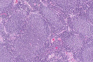
Follicular lymphoma (FL) is a cancer that involves certain types of white blood cells known as lymphocytes. The cancer originates from the uncontrolled division of specific types of B-cells known as centrocytes and centroblasts. These cells normally occupy the follicles in the germinal centers of lymphoid tissues such as lymph nodes. The cancerous cells in FL typically form follicular or follicle-like structures in the tissues they invade. These structures are usually the dominant histological feature of this cancer.

<span class="mw-page-title-main">Primary effusion lymphoma</span> Medical condition

Primary effusion lymphoma (PEL) is classified as a diffuse large B cell lymphoma. It is a rare malignancy of plasmablastic cells that occurs in individuals that are infected with the Kaposi's sarcoma-associated herpesvirus. Plasmablasts are immature plasma cells, i.e. lymphocytes of the B-cell type that have differentiated into plasmablasts but because of their malignant nature do not differentiate into mature plasma cells but rather proliferate excessively and thereby cause life-threatening disease. In PEL, the proliferating plasmablastoid cells commonly accumulate within body cavities to produce effusions, primarily in the pleural, pericardial, or peritoneal cavities, without forming a contiguous tumor mass. In rare cases of these cavitary forms of PEL, the effusions develop in joints, the epidural space surrounding the brain and spinal cord, and underneath the capsule which forms around breast implants. Less frequently, individuals present with extracavitary primary effusion lymphomas, i.e., solid tumor masses not accompanied by effusions. The extracavitary tumors may develop in lymph nodes, bone, bone marrow, the gastrointestinal tract, skin, spleen, liver, lungs, central nervous system, testes, paranasal sinuses, muscle, and, rarely, inside the vasculature and sinuses of lymph nodes. As their disease progresses, however, individuals with the classical effusion-form of PEL may develop extracavitary tumors and individuals with extracavitary PEL may develop cavitary effusions.

<span class="mw-page-title-main">Intravascular lymphomas</span> Medical condition

Intravascular lymphomas (IVL) are rare cancers in which malignant lymphocytes proliferate and accumulate within blood vessels. Almost all other types of lymphoma involve the proliferation and accumulation of malignant lymphocytes in lymph nodes, other parts of the lymphatic system, and various non-lymphatic organs but not in blood vessels.

<span class="mw-page-title-main">Diffuse large B-cell lymphoma</span> Type of blood cancer

Diffuse large B-cell lymphoma (DLBCL) is a cancer of B cells, a type of lymphocyte that is responsible for producing antibodies. It is the most common form of non-Hodgkin lymphoma among adults, with an annual incidence of 7–8 cases per 100,000 people per year in the US and UK. This cancer occurs primarily in older individuals, with a median age of diagnosis at ~70 years, although it can occur in young adults and, in rare cases, children. DLBCL can arise in virtually any part of the body and, depending on various factors, is often a very aggressive malignancy. The first sign of this illness is typically the observation of a rapidly growing mass or tissue infiltration that is sometimes associated with systemic B symptoms, e.g. fever, weight loss, and night sweats.

<span class="mw-page-title-main">Aggressive NK-cell leukemia</span> Medical condition

Aggressive NK-cell leukemia is a disease with an aggressive, systemic proliferation of natural killer cells and a rapidly declining clinical course.

Richter's transformation (RT), also known as Richter's syndrome, is the conversion of chronic lymphocytic leukemia (CLL) or its variant, small lymphocytic lymphoma (SLL), into a new and more aggressively malignant disease. CLL is the circulation of malignant B lymphocytes with or without the infiltration of these cells into lymphatic or other tissues while SLL is the infiltration of these malignant B lymphocytes into lymphatic and/or other tissues with little or no circulation of these cells in the blood. CLL along with its SLL variant are grouped together in the term CLL/SLL.

<span class="mw-page-title-main">Aggressive lymphoma</span> Medical condition

Aggressive lymphoma, also known as high-grade lymphoma, is a group of fast growing non-Hodgkin lymphoma.

<span class="mw-page-title-main">Blastic plasmacytoid dendritic cell neoplasm</span> Medical condition

Blastic plasmacytoid dendritic cell neoplasm (BPDCN) is a rare hematologic malignancy. It was initially regarded as a form of lymphocyte-derived cutaneous lymphoma and alternatively named CD4+CD56+ hematodermic tumor, blastic NK cell lymphoma, and agranular CD4+ NK cell leukemia. Later, however, the disease was determined to be a malignancy of plasmacytoid dendritic cells rather than lymphocytes and therefore termed blastic plasmacytoid dendritic cell neoplasm. In 2016, the World Health Organization designated BPDCN to be in its own separate category within the myeloid class of neoplasms. It is estimated that BPDCN constitutes 0.44% of all hematological malignancies.

<span class="mw-page-title-main">Marginal zone lymphoma</span> Group of lymphomas

Marginal zone lymphomas, also known as marginal zone B-cell lymphomas (MZLs), are a heterogeneous group of lymphomas that derive from the malignant transformation of marginal zone B-cells. Marginal zone B cells are innate lymphoid cells that normally function by rapidly mounting IgM antibody immune responses to antigens such as those presented by infectious agents and damaged tissues. They are lymphocytes of the B-cell line that originate and mature in secondary lymphoid follicles and then move to the marginal zones of mucosa-associated lymphoid tissue, the spleen, or lymph nodes. Mucosa-associated lymphoid tissue is a diffuse system of small concentrations of lymphoid tissue found in various submucosal membrane sites of the body such as the gastrointestinal tract, mouth, nasal cavity, pharynx, thyroid gland, breast, lung, salivary glands, eye, skin and the human spleen.

<span class="mw-page-title-main">Extranodal NK/T-cell lymphoma, nasal type</span> Medical condition

Extranodal NK/T-cell lymphoma, nasal type (ENKTCL-NT) is a rare type of lymphoma that commonly involves midline areas of the nasal cavity, oral cavity, and/or pharynx At these sites, the disease often takes the form of massive, necrotic, and extremely disfiguring lesions. However, ENKTCL-NT can also involve the eye, larynx, lung, gastrointestinal tract, skin, and various other tissues. ENKTCL-NT mainly affects adults; it is relatively common in Asia and to lesser extents Mexico, Central America, and South America but is rare in Europe and North America. In Korea, ENKTCL-NT often involves the skin and is reported to be the most common form of cutaneous lymphoma after mycosis fungoides.

Gene expression profiling has revealed that diffuse large B-cell lymphoma (DLBCL) is composed of at least 3 different sub-groups, each having distinct oncogenic mechanisms that respond to therapies in different ways. Germinal Center B-Cell like (GCB) DLBCLs appear to arise from normal germinal center B cells, while Activated B-cell like (ABC) DLBCLs are thought to arise from postgerminal center B cells that are arrested during plasmacytic differentiation. The differences in gene expression between GCB DLBCL and ABC DLBCL are as vast as the differences between distinct types of leukemia, but these conditions have historically been grouped together and treated as the same disease.

Large B-cell lymphoma arising in HHV8-associated multicentric Castleman's disease is a type of large B-cell lymphoma, recognized in the WHO 2008 classification. It is sometimes called the plasmablastic form of multicentric Castleman disease. It has sometimes been confused with plasmablastic lymphoma in the literature, although that is a dissimilar specific entity. It has variable CD20 expression and unmutated immunoglobulin variable region genes.

Epstein–Barr virus positive diffuse large B-cell lymphoma, not otherwise specified is a form of diffuse large B-cell lymphomas (DLBCL) accounting for around 10-15% of DLBCL cases. DLBCL are lymphomas in which B-cell lymphocytes proliferate excessively, invade multiple tissues, and often causes life-threatening tissue damage. EBV+ DLBCL is distinguished from DLBCL in that virtually all the large B cells in the tissue, infiltrates of the Epstien-Barr virus (EBV) express EBV genes characteristic of the virus's latency III or II phase. EBV is a ubiquitous virus, infecting around 95% of the world population.

Epstein–Barr virus–associated lymphoproliferative diseases are a group of disorders in which one or more types of lymphoid cells, i.e. B cells, T cells, NK cells, and histiocytic-dendritic cells, are infected with the Epstein–Barr virus (EBV). This causes the infected cells to divide excessively, and is associated with the development of various non-cancerous, pre-cancerous, and cancerous lymphoproliferative disorders (LPDs). These LPDs include the well-known disorder occurring during the initial infection with the EBV, infectious mononucleosis, and the large number of subsequent disorders that may occur thereafter. The virus is usually involved in the development and/or progression of these LPDs although in some cases it may be an "innocent" bystander, i.e. present in, but not contributing to, the disease.

Primary cutaneous diffuse large B-cell lymphoma, leg type (PCDLBCL-LT) is a cutaneous lymphoma skin disease that occurs mostly in elderly females. In this disease, B cells become malignant, accumulate in the dermis and subcutaneous tissue below the dermis to form red and violaceous skin nodules and tumors. These lesions typically occur on the lower extremities but in uncommon cases may develop on the skin at virtually any other site. In ~10% of cases, the disease presents with one or more skin lesions none of which are on the lower extremities; the disease in these cases is sometimes regarded as a variant of PCDLBL, LT termed primary cutaneous diffuse large B-cell lymphoma, other (PCDLBC-O). PCDLBCL, LT is a subtype of the diffuse large B-cell lymphomas (DLBCL) and has been thought of as a cutaneous counterpart to them. Like most variants and subtypes of the DLBCL, PCDLBCL, LT is an aggressive malignancy. It has a 5-year overall survival rate of 40–55%, although the PCDLBCL-O variant has a better prognosis than cases in which the legs are involved.

Diffuse large B-cell lymphoma associated with chronic inflammation (DLBCL-CI) is a subtype of the Diffuse large B-cell lymphomas and a rare form of the Epstein–Barr virus-associated lymphoproliferative diseases, i.e. conditions in which lymphocytes infected with the Epstein-Barr virus (EBV) proliferate excessively in one or more tissues. EBV infects ~95% of the world's population to cause no symptoms, minor non-specific symptoms, or infectious mononucleosis. The virus then enters a latency phase in which the infected individual becomes a lifetime asymptomatic carrier of the virus. Some weeks, months, years, or decades thereafter, a very small fraction of these carriers, particularly those with an immunodeficiency, develop any one of various EBV-associated benign or malignant diseases.

Fibrin-associated diffuse large B-cell lymphoma (FA-DLBCL) is an extremely rare form of the diffuse large B-cell lymphomas (DLBCL). DLBCL are lymphomas in which a particular type of lymphocyte, the B-cell, proliferates excessively, invades multiple tissues, and often causes life-threatening tissue damage. DLBCL have various forms as exemplified by one of its subtypes, diffuse large B-cell lymphoma associated with chronic inflammation (DLBCL-CI). DLBCL-CI is an aggressive malignancy that develops in sites of chronic inflammation that are walled off from the immune system. In this protected environment, the B-cells proliferate excessively, acquire malignant gene changes, form tumor masses, and often spread outside of the protected environment. In 2016, the World Health Organization provisionally classified FA-DLBCL as a DLBCL-CI. Similar to DLBCL-CI, FA-DLBCL involves the proliferation of EBV-infected large B-cells in restricted anatomical spaces that afford protection from an individual's immune system. However, FA-DLBCL differs from DLBCL-CI in many other ways, including, most importantly, its comparatively benign nature. Some researchers have suggested that this disease should be regarded as a non-malignant or pre-malignant lymphoproliferative disorder rather than a malignant DLBCL-CI.

<span class="mw-page-title-main">Lymphoid neoplasms with plasmablastic differentiation</span>

Lymphoid neoplasms with plasmablastic differentiation were classified by the World Health Organization, 2017 as a sub-grouping of several distinct but rare lymphomas in which the malignant cells are B-cell lymphocytes that have become plasmablasts, i.e. immature plasma cells. Normally, B-cells take up foreign antigens, move to the germinal centers of secondary lymphoid organs such the spleen and lymph nodes, and at these sites are stimulated by T-cell lymphocytes to differentiate into plasmablasts and thereafter mature plasma cells. Plasmablasts, and to a greater extent, plasma cells make and secrete antibodies that bind the antigens to which their predecessor B-cells were previously exposed. Antibodies function, in part, to neutralize harmful bacteria and viruses by binding antigens that are exposed on their surfaces. Due to their malignant nature, however, the plasmablasts in lymphoid neoplasms with plasmablastic differentiation do not mature into plasma cells or form antibodies but rather uncontrollably proliferate in and damage various tissues and organs. The individual lymphomas in this sub-group of malignancies have heterogeneous clinical, morphological, and gene findings that often overlap with other members of the sub-group. In consequence, correctly diagnosing these lymphomas has been challenging. Nonetheless, it is particularly important to diagnose them correctly because they often have very different prognoses and treatments. The lymphoid neoplasms with plasmacytic differentiation are:

References

  1. 1 2 3 4 5 6 7 8 9 Chen BJ, Chuang SS (March 2020). "Lymphoid Neoplasms With Plasmablastic Differentiation: A Comprehensive Review and Diagnostic Approaches". Advances in Anatomic Pathology. 27 (2): 61–74. doi: 10.1097/PAP.0000000000000253 . PMID   31725418.
  2. 1 2 3 4 5 6 7 8 9 10 11 Lopez A, Abrisqueta P (2018). "Plasmablastic lymphoma: current perspectives". Blood and Lymphatic Cancer: Targets and Therapy. 8: 63–70. doi: 10.2147/BLCTT.S142814 . PMC   6467349 . PMID   31360094.
  3. 1 2 3 4 Korkolopoulou P, Vassilakopoulos T, Milionis V, Ioannou M (July 2016). "Recent Advances in Aggressive Large B-cell Lymphomas: A Comprehensive Review". Advances in Anatomic Pathology. 23 (4): 202–43. doi:10.1097/PAP.0000000000000117. PMID   27271843. S2CID   205915174.
  4. 1 2 Liu F, Asano N, Tatematsu A, Oyama T, Kitamura K, Suzuki K, Yamamoto K, Sakamoto N, Taniwaki M, Kinoshita T, Nakamura S (December 2012). "Plasmablastic lymphoma of the elderly: a clinicopathological comparison with age-related Epstein–Barr virus-associated B cell lymphoproliferative disorder". Histopathology. 61 (6): 1183–97. doi:10.1111/j.1365-2559.2012.04339.x. PMID   22958176. S2CID   205303461.
  5. 1 2 3 4 Dojcinov SD, Fend F, Quintanilla-Martinez L (March 2018). "EBV-Positive Lymphoproliferations of B- T- and NK-Cell Derivation in Non-Immunocompromised Hosts". Pathogens. 7 (1): 28. doi: 10.3390/pathogens7010028 . PMC   5874754 . PMID   29518976.
  6. 1 2 3 Rodrigues-Fernandes CI, de Souza LL, Santos-Costa SFD, Silva AMB, Pontes HAR, Lopes MA, de Almeida OP, Brennan PA, Fonseca FP (November 2018). "Clinicopathological analysis of oral plasmablastic lymphoma: A systematic review". Journal of Oral Pathology & Medicine. 47 (10): 915–922. doi:10.1111/jop.12753. PMID   29917262. S2CID   49298504.
  7. 1 2 3 4 5 6 7 8 Li YJ, Li JW, Chen KL, Li J, Zhong MZ, Liu XL, Yi PY, Zhou H (March 2020). "HIV-negative plasmablastic lymphoma: report of 8 cases and a comprehensive review of 394 published cases". Blood Research. 55 (1): 49–56. doi: 10.5045/br.2020.55.1.49 . PMC   7106118 . PMID   32269975.
  8. 1 2 3 4 5 6 7 8 Bhattacharyya S, Bains APS, Sykes DL, Iverson BR, Sibgatullah R, Kuklani RM (December 2019). "Lymphoid neoplasms of the oral cavity with plasmablastic morphology-a case series and review of the literature". Oral Surgery, Oral Medicine, Oral Pathology and Oral Radiology. 128 (6): 651–659. doi:10.1016/j.oooo.2019.08.001. PMID   31494113. S2CID   201973958.
  9. Zhou J, Nassiri M (April 2022). "Lymphoproliferative Neoplasms With Plasmablastic Morphology". Archives of Pathology & Laboratory Medicine. 146 (4): 407–414. doi: 10.5858/arpa.2021-0117-RA . PMID   34559873. S2CID   237626753.
  10. Tchernonog E, Faurie P, Coppo P, Monjanel H, Bonnet A, Algarte Génin M, Mercier M, Dupuis J, Bijou F, Herbaux C, Delmer A, Fabiani B, Besson C, Le Gouill S, Gyan E, Laurent C, Ghesquieres H, Cartron G (April 2017). "Clinical characteristics and prognostic factors of plasmablastic lymphoma patients: analysis of 135 patients from the LYSA group". Annals of Oncology. 28 (4): 843–848. doi: 10.1093/annonc/mdw684 . PMID   28031174.
  11. Montes-Moreno S, Martinez-Magunacelaya N, Zecchini-Barrese T, Villambrosía SG, Linares E, Ranchal T, Rodriguez-Pinilla M, Batlle A, Cereceda-Company L, Revert-Arce JB, Almaraz C, Piris MA (January 2017). "Plasmablastic lymphoma phenotype is determined by genetic alterations in MYC and PRDM1". Modern Pathology. 30 (1): 85–94. doi: 10.1038/modpathol.2016.162 . PMID   27687004.
  12. 1 2 Rezk SA, Zhao X, Weiss LM (June 2018). "Epstein—Barr virus–associated lymphoid proliferations, a 2018 update". Human Pathology. 79: 18–41. doi:10.1016/j.humpath.2018.05.020. PMID   29885408. S2CID   47010934.
  13. Ott G, Rosenwald A, Campo E (2013). "Understanding MYC-driven aggressive B-cell lymphomas: pathogenesis and classification". Hematology. American Society of Hematology. Education Program. 2013: 575–83. doi: 10.1182/asheducation-2013.1.575 . PMID   24319234.
  14. Bailly J, Jenkins N, Chetty D, Mohamed Z, Verburgh ER, Opie JJ (September 2022). "Plasmablastic lymphoma: An update". International Journal of Laboratory Hematology. 44 (Suppl 1): 54–63. doi:10.1111/ijlh.13863. PMC   9545967 . PMID   36074710.
  15. "A Multicenter, Open-Label Feasibility Study of Daratumumab with Dose-Adjusted EPOCH in Newly Diagnosed Plasmablastic Lymphoma". 18 May 2021.{{cite journal}}: Cite journal requires |journal= (help)
  16. "Safety and Feasibility of Gene Transfer After Frontline Chemotherapy for Non-Hodgkin Lymphoma in AIDS Patients Using Peripheral Blood Stem/Progenitor Cells Treated with a Lentivirus Vector-Encoding Multiple Anti-HIV RNAs". 15 February 2021.{{cite journal}}: Cite journal requires |journal= (help)
  17. Green TL, Eversole LR (April 1989). "Oral lymphomas in HIV-infected patients: association with Epstein–Barr virus DNA". Oral Surgery, Oral Medicine, and Oral Pathology. 67 (4): 437–42. doi:10.1016/0030-4220(89)90388-5. PMID   2542861.
  18. Delecluse HJ, Anagnostopoulos I, Dallenbach F, Hummel M, Marafioti T, Schneider U, Huhn D, Schmidt-Westhausen A, Reichart PA, Gross U, Stein H (February 1997). "Plasmablastic lymphomas of the oral cavity: a new entity associated with the human immunodeficiency virus infection". Blood. 89 (4): 1413–20. doi: 10.1182/blood.V89.4.1413 . PMID   9028965.