
Ophthalmology is a surgical subspecialty within medicine that deals with the diagnosis and treatment of eye disorders.

A cataract is a cloudy area in the lens of the eye that leads to a decrease in vision. Cataracts often develop slowly and can affect one or both eyes. Symptoms may include faded colours, blurry or double vision, halos around light, trouble with bright lights, and difficulty seeing at night. This may result in trouble driving, reading, or recognizing faces. Poor vision caused by cataracts may also result in an increased risk of falling and depression. Cataracts cause 51% of all cases of blindness and 33% of visual impairment worldwide.

Vitrectomy is a surgery to remove some or all of the vitreous humor from the eye.

LASIK or Lasik, commonly referred to as laser eye surgery or laser vision correction, is a type of refractive surgery for the correction of myopia, hyperopia, and an actual cure for astigmatism, since it is in the cornea. LASIK surgery is performed by an ophthalmologist who uses a laser or microkeratome to reshape the eye's cornea in order to improve visual acuity. For most people, LASIK provides a long-lasting alternative to eyeglasses or contact lenses.

Eye surgery, also known as ophthalmic or ocular surgery, is surgery performed on the eye or its adnexa, by an ophthalmologist. Eye surgery is part of ophthalmology. The eye is a fragile organ, and requires due care before, during, and after a surgical procedure to minimize or prevent further damage. An eye surgeon is responsible for selecting the appropriate surgical procedure for the patient, and for taking the necessary safety precautions. Mentions of eye surgery can be found in several ancient texts dating back as early as 1800 BC, with cataract treatment starting in the fifth century BC. It continues to be a widely practiced class of surgery, with various techniques having been developed for treating eye problems.

Refractive eye surgery is optional eye surgery used to improve the refractive state of the eye and decrease or eliminate dependency on glasses or contact lenses. This can include various methods of surgical remodeling of the cornea (keratomileusis), lens implantation or lens replacement. The most common methods today use excimer lasers to reshape the curvature of the cornea. Refractive eye surgeries are used to treat common vision disorders such as myopia, hyperopia, presbyopia and astigmatism.

Phacoemulsification is a cataract surgery method in which the internal lens of the eye which has developed a cataract is emulsified with the tip of an ultrasonic handpiece and aspirated from the eye. Aspirated fluids are replaced with irrigation of balanced salt solution to maintain the volume of the anterior chamber during the procedure. This procedure minimises the incision size and reduces the recovery time and risk of surgery induced astigmatism. It is best suited to relatively soft cataracts, where the ultrasonic energy required is moderate.

An Intraocular lens (IOL) is a lens implanted in the eye usually as part of a treatment for cataracts or for correcting other vision problems such as short sightedness and long sightedness, a form of refractive surgery. If the natural lens is left in the eye, the IOL is known as phakic, otherwise it is a pseudophakic, or false lens. Both kinds of IOLs are designed to provide the same light-focusing function as the natural crystalline lens. This is an alternative to LASIK.

Charles David Kelman was an American ophthalmologist, surgeon, inventor, jazz musician, entertainer, and Broadway producer. Known as the father of phacoemulsification, he developed many of the medical devices, instruments, implant lenses and techniques used in cataract surgery. In the early 1960s, he began the use of cryosurgery to remove cataracts and repair retinal detachments. Cryosurgery for cataracts remained in heavy use until 1978, when phacoemulsification, a procedure Kelman also developed in 1967, became the modern standard treatment. Kelman was given the National Medal of Technology by President George H. W. Bush and recognized as the Ophthalmologist of the Century by the International Congress of Cataract and Refractive Surgery in Montreal, Canada. He was also inducted into the National Inventors Hall of Fame in Akron, Ohio, and received the 2004 Lasker Award.
Cataract surgery, which is also called lens replacement surgery, is the removal of the natural lens of the human eye that has developed a cataract, an opaque or cloudy area. The eye's natural lens is usually replaced with an artificial intraocular lens (IOL).
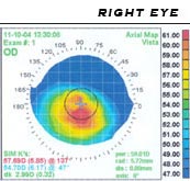
Corneal topography, also known as photokeratoscopy or videokeratography, is a non-invasive medical imaging technique for mapping the anterior curvature of the cornea, the outer structure of the eye. Since the cornea is normally responsible for some 70% of the eye's refractive power, its topography is of critical importance in determining the quality of vision and corneal health.
Capsulorhexis or capsulorrhexis, and the commonly used technique known as continuous curvilinear capsulorhexis (CCC), is a surgical technique used to remove the central anterior part of the capsule of the lens from the eye during cataract surgery by shear and tensile forces. It generally refers to removal of the central part of the anterior lens capsule, but in situations like a developmental cataract a part of the posterior capsule is also removed by a similar technique.
The von Graefe knife was a tool used to make corneal incisions in cataract surgery. Use of the knife demanded a high level of skill and mastery, and was eventually supplanted by modifications of cataract surgery through the Kelman phacoemulsification technique that emphasized a small incision.

An operating microscope or surgical microscope is an optical microscope specifically designed to be used in a surgical setting, typically to perform microsurgery.

Couching is the earliest documented form of cataract surgery. As a cataract is a clouding in the lens of the eye, couching is a technique whereby the lens is dislodged, thus removing the opacity. Although couching is nowadays routinely practiced only in remote areas, it was a precursor to modern cataract surgery and pars plana vitrectomy.
Howard V. Gimbel FRCSC, AOE, FACS, CABES, is a Canadian ophthalmologist, university professor, senior editor, and amateur musician. He is better known for his invention, along with Thomas Neuhann, of the continuous curvilinear capsulorhexis (CCC), a technique employed in modern cataract surgery.

Capsulotomy is a type of eye surgery in which an incision is made into the capsule of the crystalline lens of the eye. In modern cataract operations, the lens capsule is usually not removed. The most common forms of cataract surgery remove nearly all of the crystalline lens but do not remove the crystalline lens capsule. The crystalline lens capsule is retained and used to contain and position the intraocular lens implant (IOL).
Childhood cataract is cataract that occurs at birth or in childhood. It may be congenital or acquired.
Intraocular lens scaffold or IOL scaffold technique is a surgical procedure in ophthalmology. In cases where the posterior lens capsule is ruptured and the cataract has not yet been removed one can insert the intraocular lens (IOL) inside the eye under the cataract. This way the IOL acts as a scaffold and prevents the cataract pieces from falling inside the back of the eye. The cataract can then be removed safely by emulsifying it with ultrasound and aspiration. This technique is called IOL scaffold and was started by Amar Agarwal from Chennai, India, at Dr. Agarwal's Eye Hospital.
Hydrodissection is the use of a directed jet of water to surgically separate tissues. It is generally used to develop tissue planes or divide soft tissues with less trauma than dissection using a cutting instrument. By using an appropriate pressure it will tend to follow the path of least resistance that is close to the direction of the jet.