RNA thermometer

Last updated

The FourU thermometer RNA motif, with the Shine-Dalgarno sequence highlighted. FourU.png
The FourU thermometer RNA motif, with the Shine-Dalgarno sequence highlighted.

An RNA thermometer (or RNA thermosensor) is a temperature-sensitive non-coding RNA molecule which regulates gene expression. RNA thermometers often regulate genes required during either a heat shock or cold shock response, but have been implicated in other regulatory roles such as in pathogenicity and starvation. [1]

Contents

In general, RNA thermometers operate by changing their secondary structure in response to temperature fluctuations. This structural transition can then expose or occlude important regions of RNA such as a ribosome binding site, which then affects the translation rate of a nearby protein-coding gene.

RNA thermometers, along with riboswitches, are used as examples in support of the RNA world hypothesis. This theory proposes that RNA was once the sole nucleic acid present in cells, and was replaced by the current DNA → RNA → protein system. [2]

Examples of RNA thermometers include FourU, [3] the Hsp90 cis-regulatory element, [4] the ROSE element, [5] the Lig RNA thermometer, [6] and the Hsp17 thermometer. [7]

Discovery

The first temperature-sensitive RNA element was reported in 1989. [8] Prior to this research, mutations upstream from the transcription start site in a lambda (λ) phage cIII mRNA were found to affect the level of translation of the cIII protein. [9] This protein is involved in selection of either a lytic or lysogenic life cycle in λ phage, with high concentrations of cIII promoting lysogeny. [9] Further study of this upstream RNA region identified two alternative secondary structures; experimental study found the structures to be interchangeable, and dependent on both magnesium ion concentration and temperature. [8] [10] This RNA thermometer is now thought to encourage entry to a lytic cycle under heat stress in order for the bacteriophage to rapidly replicate and escape the host cell. [1]

The term "RNA thermometer" was not coined until 1999, [11] when it was applied to the rpoH RNA element identified in Escherichia coli . [12] More recently, bioinformatics searches have been employed to uncover several novel candidate RNA thermometers. [13] Traditional sequence-based searches are inefficient, however, as the secondary structure of the element is much more conserved than the nucleic acid sequence. [13]

Distribution

Most known RNA thermometers are located in the 5′ untranslated region (UTR) of messenger RNA encoding heat shock proteins—though it has been suggested this fact may be due, in part, to sampling bias and inherent difficulties of detecting short, unconserved RNA sequences in genomic data. [14] [15]

Though predominantly found in prokaryotes, a potential RNA thermometer has been found in mammals including humans. [16] The candidate thermosensor heat shock RNA-1 (HSR1) activates heat-shock transcription factor 1 (HSF1) and induces protective proteins when cell temperature exceeds 37 °C (body temperature), thus preventing the cells from overheating. [16]

Structure

3D representation of the structure of the ROSE RNA thermometer. PDB 2gio EBI.png
3D representation of the structure of the ROSE RNA thermometer.

RNA thermometers are structurally simple and can be made from short RNA sequences; the smallest is just 44 nucleotides and is found in the mRNA of a heat-shock protein, hsp17, in Synechocystis species PCC 6803. [18] [19] Generally these RNA elements range in length from 60 to 110 nucleotides [20] and they typically contain a hairpin with a small number of mismatched base pairs which reduce the stability of the structure, thereby allowing easier unfolding in response to a temperature increase. [21]

Detailed structural analysis of the ROSE RNA thermometer revealed that the mismatched bases are actually engaged in nonstandard basepairing that preserves the helical structure of the RNA (see figure). The unusual basepairs consist of G-G, U-U, and UC-U pairs. Since these noncanonical base pairs are relatively unstable, increased temperature causes local melting of the RNA structure in this region, exposing the Shine-Dalgarno sequence. [17]

Some RNA thermometers are significantly more complex than a single hairpin, as in the case of a region found in CspA mRNA which is thought to contain a pseudoknot, as well as multiple hairpins. [22] [23]

Synthetic RNA thermometers have been designed with just a simple single-hairpin structure. [24] However, the secondary structure of such short RNA thermometers can be sensitive to mutation, as a single base change can render the hairpin inactive in vivo . [25]

Mechanism

A stable hairpin (left) unwinds at a higher temperature (right). The highlighted Shine-Dalgarno sequence becomes exposed, allowing the binding of the 30S ribosomal subunit. RNA thermometer.svg
A stable hairpin (left) unwinds at a higher temperature (right). The highlighted Shine-Dalgarno sequence becomes exposed, allowing the binding of the 30S ribosomal subunit.

RNA thermometers are found in the 5′ UTR of messenger RNA, upstream of a protein-coding gene. [1] Here they are able to occlude the ribosome binding site (RBS) and prevent translation of the mRNA into protein. [14] As temperature increases, the hairpin structure can 'melt' and expose the RBS or Shine-Dalgarno sequence to permit binding of the small ribosomal subunit (30S), which then assembles other translation machinery. [1] The start codon, typically found 8 nucleotides downstream of the Shine-Dalgarno sequence, [14] signals the beginning of a protein-coding gene which is then translated to a peptide product by the ribosome. In addition to this cis-acting mechanism, a lone example of a trans-acting RNA thermometer has been found in RpoS mRNA where it is thought to be involved in the starvation response. [1]

A specific example of an RNA thermometer motif is the FourU thermometer found in Salmonella enterica . [3] When exposed to temperatures above 45 °C, the stem-loop that base-pairs opposite the Shine-Dalgarno sequence becomes unpaired and allows the mRNA to enter the ribosome for translation to occur. [25] Mg2+ ion concentration has also been shown to affect the stability of FourU. [26] The most well-studied RNA thermometer is found in the rpoH gene in Escherichia coli. [27] This thermosensor upregulates heat shock proteins under high temperatures through σ32, a specialised heat-shock sigma factor. [11]

Though typically associated with heat-induced protein expression, RNA thermometers can also regulate cold-shock proteins. [22] For example, the expression of two 7kDa proteins are regulated by an RNA thermometer in the thermophilic bacterium Thermus thermophilus [28] and a similar mechanism has been identified in Enterobacteriales. [23]

RNA thermometers sensitive to temperatures of 37 °C can be used by pathogens to activate infection-specific genes. [14] For example, the upregulation of prfA, encoding a key transcriptional regulator of virulence genes in Listeria monocytogenes , was demonstrated by fusing the 5′ DNA of prfA to the green fluorescent protein gene; the gene fusion was then transcribed from the T7 promoter in E. coli, and fluorescence was observed at 37 °C but not at 30 °C. [29]

Implications for the RNA world hypothesis

The RNA world hypothesis states that RNA was once both the carrier of hereditary information and enzymatically active, with different sequences acting as biocatalysts, regulators and sensors. [30] The hypothesis then proposes that modern DNA, RNA and protein-based life evolved and selection replaced the majority of RNA's roles with other biomolecules. [2]

RNA thermometers and riboswitches are thought to be evolutionarily ancient due to their wide-scale distribution in distantly-related organisms. [31] It has been proposed that, in the RNA world, RNA thermosensors would have been responsible for temperature-dependent regulation of other RNA molecules. [2] [32] RNA thermometers in modern organisms may be molecular fossils which could hint at a previously more widespread importance in an RNA world. [2]

Other examples

Related Research Articles

<span class="mw-page-title-main">Start codon</span> First codon of a messenger RNA translated by a ribosome

The start codon is the first codon of a messenger RNA (mRNA) transcript translated by a ribosome. The start codon always codes for methionine in eukaryotes and archaea and a N-formylmethionine (fMet) in bacteria, mitochondria and plastids.

Bacterial translation is the process by which messenger RNA is translated into proteins in bacteria.

In genetics, attenuation is a regulatory mechanism for some bacterial operons that results in premature termination of transcription. The canonical example of attenuation used in many introductory genetics textbooks, is ribosome-mediated attenuation of the trp operon. Ribosome-mediated attenuation of the trp operon relies on the fact that, in bacteria, transcription and translation proceed simultaneously. Attenuation involves a provisional stop signal (attenuator), located in the DNA segment that corresponds to the leader sequence of mRNA. During attenuation, the ribosome becomes stalled (delayed) in the attenuator region in the mRNA leader. Depending on the metabolic conditions, the attenuator either stops transcription at that point or allows read-through to the structural gene part of the mRNA and synthesis of the appropriate protein.

<span class="mw-page-title-main">Cobalamin riboswitch</span>

Cobalamin riboswitch is a cis-regulatory element which is widely distributed in 5' untranslated regions of vitamin B12 (Cobalamin) related genes in bacteria.

<span class="mw-page-title-main">PreQ1 riboswitch</span>

The PreQ1-I riboswitch is a cis-acting element identified in bacteria which regulates expression of genes involved in biosynthesis of the nucleoside queuosine (Q) from GTP. PreQ1 (pre-queuosine1) is an intermediate in the queuosine pathway, and preQ1 riboswitch, as a type of riboswitch, is an RNA element that binds preQ1. The preQ1 riboswitch is distinguished by its unusually small aptamer, compared to other riboswitches. Its atomic-resolution three-dimensional structure has been determined, with the PDB ID 2L1V.

<span class="mw-page-title-main">Purine riboswitch</span>

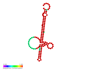
A purine riboswitch is a sequence of ribonucleotides in certain messenger RNA (mRNA) that selectively binds to purine ligands via a natural aptamer domain. This binding causes a conformational change in the mRNA that can affect translation by revealing an expression platform for a downstream gene, or by forming a translation-terminating stem-loop. The ultimate effects of such translational regulation often take action to manage an abundance of the instigating purine, and might produce proteins that facilitate purine metabolism or purine membrane uptake.

<span class="mw-page-title-main">Repression of heat shock gene expression (ROSE) element</span>

The repression of heat shock gene expression (ROSE) element is an RNA element found in the 5' UTR of some heat shock protein's mRNAs. The ROSE element is an RNA thermometer that negatively regulates heat shock gene expression. The secondary structure is thought to be altered by temperature, thus it is an RNA thermometer. This structure blocks access to the ribosome binding site at normal temperatures. During heat shock however, the structure changes freeing the ribosome binding site and allowing expression to occur.

A ribosome binding site, or ribosomal binding site (RBS), is a sequence of nucleotides upstream of the start codon of an mRNA transcript that is responsible for the recruitment of a ribosome during the initiation of translation. Mostly, RBS refers to bacterial sequences, although internal ribosome entry sites (IRES) have been described in mRNAs of eukaryotic cells or viruses that infect eukaryotes. Ribosome recruitment in eukaryotes is generally mediated by the 5' cap present on eukaryotic mRNAs.

<span class="mw-page-title-main">Untranslated region</span> Non-coding regions on either end of mRNA

In molecular genetics, an untranslated region refers to either of two sections, one on each side of a coding sequence on a strand of mRNA. If it is found on the 5' side, it is called the 5' UTR, or if it is found on the 3' side, it is called the 3' UTR. mRNA is RNA that carries information from DNA to the ribosome, the site of protein synthesis (translation) within a cell. The mRNA is initially transcribed from the corresponding DNA sequence and then translated into protein. However, several regions of the mRNA are usually not translated into protein, including the 5' and 3' UTRs.

Cold shock response is a series of neurogenic cardio-respiratory responses caused by sudden immersion in cold water.

<span class="mw-page-title-main">FourU thermometer</span> Class of non-coding RNAs in Salmonella

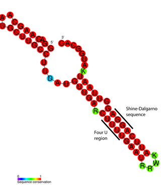
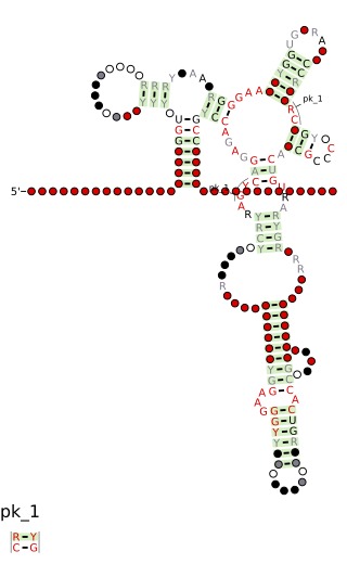
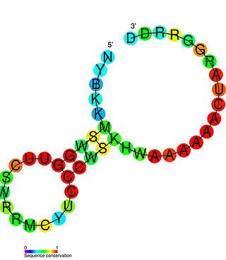
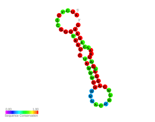
FourU thermometers are a class of non-coding RNA thermometers found in Salmonella. They are named 'FourU' due to the four highly conserved uridine nucleotides found directly opposite the Shine-Dalgarno sequence on hairpin II (pictured). RNA thermometers such as FourU control regulation of temperature via heat shock proteins in many prokaryotes. FourU thermometers are relatively small RNA molecules, only 57 nucleotides in length, and have a simple two-hairpin structure.

<span class="mw-page-title-main">CspA mRNA 5′ UTR</span>

cspA mRNA 5' UTR is the untranslated region of the cspA gene, which is important in the cold shock response in Enterobacteriales such as E. coli. The 5' UTR element acts as an RNA thermometer, regulating the expression of cspA in response to temperature. By regulating temperature, cspA proteins carry out the vital function of homeostasis.

Bacterial small RNAs are small RNAs produced by bacteria; they are 50- to 500-nucleotide non-coding RNA molecules, highly structured and containing several stem-loops. Numerous sRNAs have been identified using both computational analysis and laboratory-based techniques such as Northern blotting, microarrays and RNA-Seq in a number of bacterial species including Escherichia coli, the model pathogen Salmonella, the nitrogen-fixing alphaproteobacterium Sinorhizobium meliloti, marine cyanobacteria, Francisella tularensis, Streptococcus pyogenes, the pathogen Staphylococcus aureus, and the plant pathogen Xanthomonas oryzae pathovar oryzae. Bacterial sRNAs affect how genes are expressed within bacterial cells via interaction with mRNA or protein, and thus can affect a variety of bacterial functions like metabolism, virulence, environmental stress response, and structure.

<i>Escherichia coli</i> sRNA

Escherichia coli contains a number of small RNAs located in intergenic regions of its genome. The presence of at least 55 of these has been verified experimentally. 275 potential sRNA-encoding loci were identified computationally using the QRNA program. These loci will include false positives, so the number of sRNA genes in E. coli is likely to be less than 275. A computational screen based on promoter sequences recognised by the sigma factor sigma 70 and on Rho-independent terminators predicted 24 putative sRNA genes, 14 of these were verified experimentally by northern blotting. The experimentally verified sRNAs included the well characterised sRNAs RprA and RyhB. Many of the sRNAs identified in this screen, including RprA, RyhB, SraB and SraL, are only expressed in the stationary phase of bacterial cell growth. A screen for sRNA genes based on homology to Salmonella and Klebsiella identified 59 candidate sRNA genes. From this set of candidate genes, microarray analysis and northern blotting confirmed the existence of 17 previously undescribed sRNAs, many of which bind to the chaperone protein Hfq and regulate the translation of RpoS. UptR sRNA transcribed from the uptR gene is implicated in suppressing extracytoplasmic toxicity by reducing the amount of membrane-bound toxic hybrid protein.

<span class="mw-page-title-main">IbpB thermometer</span>

The IbpB thermometer is an RNA thermometer element found in the ibpAB operon. The operon contains two heat-shock genes, encoding inclusion body binding proteins A and B (IbpA/B), and is the most drastically upregulated operon under heat-shock in Escherichia coli.

In molecular biology, Toxic Small RNA(tsRNA, not to be confused with tRNA-derived small RNA) is a family of trans-encoded small non-coding RNA found exclusively in intergenic regions of Betaproteobacteria. Several paralogous loci may encode similar tsRNAs in each coding genome. Typically, each species of Burkholderia has 3-5 homologous tsRNAs. Experiments with four species of the Burkholderia lineage showed conserved and constitutive expression of tsRNAs in logarithmic growth phases.

<span class="mw-page-title-main">Hsp17 thermometer</span> RNA element in cynobacteria

In molecular biology, the Hsp17 thermometer is an RNA element found in the 5' UTR of Hsp17 mRNA. Hsp17 is a cyanobacterial heat shock protein belonging to the Hsp20 family.

<span class="mw-page-title-main">Cyanobacterial RNA thermometer</span>

The first cyanobacterial RNA thermometer (RNAT) Hsp17 was found in the 5'UTR of Synechocystis heat shock hsp17 mRNA. Further study demonstrated that cyanobacteria commonly use RNATs to control the translation of their heat shock genes. HspA is a homolog of Hsp17 in thermophilic Thermosynechococcus elongatus. Two more thermometers were found in the 5'UTRs of mesophilic cyanobacteria A. variabilis and Nostocsp. The first RNAT called avashort was shown to regulate translation by masking the AUG translation start site. The second RNAT called avalong, as it has an extended initial hairpin, might involve tertiary interactions and has similarities to the ROSE element.

<span class="mw-page-title-main">Intergenic lcrF RNA thermometer</span>

RNA thermometers regulate gene expression in response to temperature allowing pathogens like Yersinia to switch on silent genes after entering the host organism. Usually, RNA thermometers are located in the 5'UTR, but an intergenic RNA thermometer was found in Yersinia pseudotuberculosis. The LcrFRNA thermometer together with the thermo-labile YmoA protein activates synthesis of the most crucial virulence activator LcrF (VirF). The RNA thermosensor sequence is 100% identical in all human pathogenic Yersinia species.

<span class="mw-page-title-main">Neisseria RNA thermometer</span>

RNA thermometers (RNATs) regulate gene expression in response to temperature, allowing pathogens such as Neisseria meningitidis to switch on silent genes after entering the host organism. However the temperature for expression of Neisseria virulence-associated traits is 42 °C while other bacterial pathogen RNATs require 37 °C. This is probably because N. meningitidis is an obligate commensal of the human nasopharynx and becomes pathogenic during inflammation due to viral infection. Three independent RNA thermosensors were identified in the 5′UTRs of genes needed for: capsule biosynthesis (cssA), the expression of factor H binding protein (fHbp) and sialylation of lipopolysaccharide, which is essential for bacterial resistance against immune killing (lst). The very different nucleotide sequence and predicted inhibitory structures of the three RNATs indicate that they have evolved independently.

References

  1. 1 2 3 4 5 6 Narberhaus F, Waldminghaus T, Chowdhury S (January 2006). "RNA thermometers". FEMS Microbiol. Rev. 30 (1): 3–16. doi:10.1111/j.1574-6976.2005.004.x. PMID   16438677.
  2. 1 2 3 4 Atkins JF, Gesteland RF, Cech T (2006). The RNA world: the nature of modern RNA suggests a prebiotic RNA world. Plainview, N.Y: Cold Spring Harbor Laboratory Press. ISBN   978-0-87969-739-6.
  3. 1 2 Waldminghaus T, Heidrich N, Brantl S, Narberhaus F (July 2007). "FourU: a novel type of RNA thermometer in Salmonella". Mol. Microbiol. 65 (2): 413–424. doi: 10.1111/j.1365-2958.2007.05794.x . PMID   17630972.
  4. 1 2 Ahmed R, Duncan RF (2004). "Translational regulation of Hsp90 mRNA. AUG-proximal 5′-untranslated region elements essential for preferential heat shock translation". J Biol Chem. 279 (48): 49919–49930. doi: 10.1074/jbc.M404681200 . PMID   15347681.
  5. 1 2 Nocker A, Hausherr T, Balsiger S, Krstulovic NP, Hennecke H, Narberhaus F (2001). "A mRNA-based thermosensor controls expression of rhizobial heat shock genes". Nucleic Acids Res. 29 (23): 4800–4807. doi:10.1093/nar/29.23.4800. PMC   96696 . PMID   11726689.
  6. Matsunaga J, Schlax PJ, Haake DA (2013-11-15). "Role for cis-Acting RNA Sequences in the Temperature-Dependent Expression of the Multiadhesive Lig Proteins in Leptospira interrogans". Journal of Bacteriology. 195 (22): 5092–5101. doi:10.1128/jb.00663-13. ISSN   0021-9193. PMC   3811586 . PMID   24013626.
  7. Kortmann J, Sczodrok S, Rinnenthal J, Schwalbe H, Narberhaus F (2011). "Translation on demand by a simple RNA-based thermosensor". Nucleic Acids Res. 39 (7): 2855–2868. doi:10.1093/nar/gkq1252. PMC   3074152 . PMID   21131278.
  8. 1 2 Altuvia S, Kornitzer D, Teff D, Oppenheim AB (1989-11-20). "Alternative mRNA structures of the cIII gene of bacteriophage lambda determine the rate of its translation initiation". Journal of Molecular Biology. 210 (2): 265–280. doi:10.1016/0022-2836(89)90329-X. PMID   2532257.
  9. 1 2 Altuvia S, Oppenheim AB (July 1986). "Translational regulatory signals within the coding region of the bacteriophage lambda cIII gene". Journal of Bacteriology. 167 (1): 415–419. doi:10.1128/jb.167.1.415-419.1986. PMC   212897 . PMID   2941413.
  10. Altuvia S, Kornitzer D, Kobi S, Oppenheim AB (1991-04-20). "Functional and structural elements of the mRNA of the cIII gene of bacteriophage lambda". Journal of Molecular Biology. 218 (4): 723–733. doi:10.1016/0022-2836(91)90261-4. PMID   1827163.
  11. 1 2 Storz G (1999-03-15). "An RNA thermometer". Genes & Development. 13 (6): 633–636. doi: 10.1101/gad.13.6.633 . PMID   10090718.
  12. Morita MT, Tanaka Y, Kodama TS, Kyogoku Y, Yanagi H, Yura T (1999-03-15). "Translational induction of heat shock transcription factor sigma32: evidence for a built-in RNA thermosensor". Genes & Development. 13 (6): 655–665. doi:10.1101/gad.13.6.655. PMC   316556 . PMID   10090722.
  13. 1 2 Waldminghaus T, Gaubig LC, Narberhaus F (November 2007). "Genome-wide bioinformatic prediction and experimental evaluation of potential RNA thermometers". Molecular Genetics and Genomics. 278 (5): 555–564. doi:10.1007/s00438-007-0272-7. PMID   17647020. S2CID   24747327.
  14. 1 2 3 4 Narberhaus F (2010). "Translational control of bacterial heat shock and virulence genes by temperature-sensing mRNAs". RNA Biol. 7 (1): 84–89. doi: 10.4161/rna.7.1.10501 . PMID   20009504 . Retrieved 2011-04-23.
  15. Johansson J (2009). "RNA thermosensors in bacterial pathogens". Bacterial Sensing and Signaling. Contributions to Microbiology. Vol. 16. Basel. pp. 150–160. doi:10.1159/000219378. ISBN   978-3-8055-9132-4. PMID   19494584.{{cite book}}: |journal= ignored (help)CS1 maint: location missing publisher (link)
  16. 1 2 Shamovsky I, Ivannikov M, Kandel ES, Gershon D, Nudler E (March 2006). "RNA-mediated response to heat shock in mammalian cells". Nature. 440 (7083): 556–560. Bibcode:2006Natur.440..556S. doi:10.1038/nature04518. PMID   16554823. S2CID   4311262.
  17. 1 2 Chowdhury S, Maris C, Allain FH, Narberhaus F (2006-06-07). "Molecular basis for temperature sensing by an RNA thermometer". The EMBO Journal. 25 (11): 2487–2497. doi:10.1038/sj.emboj.7601128. PMC   1478195 . PMID   16710302.
  18. Kortmann J, Sczodrok S, Rinnenthal J, Schwalbe H, Narberhaus F (April 2011). "Translation on demand by a simple RNA-based thermosensor". Nucleic Acids Research. 39 (7): 2855–2868. doi:10.1093/nar/gkq1252. PMC   3074152 . PMID   21131278.
  19. Kortmann J, Sczodrok S, Rinnenthal J, Schwalbe H, Narberhaus F (April 2011). "Translation on demand by a simple RNA-based thermosensor". Nucleic Acids Res. 39 (7): 2855–2868. doi:10.1093/nar/gkq1252. PMC   3074152 . PMID   21131278.
  20. Waldminghaus T, Fippinger A, Alfsmann J, Narberhaus F (December 2005). "RNA thermometers are common in alpha- and gamma-proteobacteria". Biol. Chem. 386 (12): 1279–1286. doi:10.1515/BC.2005.145. PMID   16336122. S2CID   84557068.
  21. Narberhaus F (January–February 2010). "Translational control of bacterial heat shock and virulence genes by temperature-sensing mRNAs". RNA Biology. 7 (1): 84–89. doi: 10.4161/rna.7.1.10501 . PMID   20009504.
  22. 1 2 Breaker RR (January 2010). "RNA switches out in the cold". Mol. Cell. 37 (1): 1–2. doi:10.1016/j.molcel.2009.12.032. PMC   5315359 . PMID   20129048.
  23. 1 2 Giuliodori AM, Di Pietro F, Marzi S, Masquida B, Wagner R, Romby P, Gualerzi CO, Pon CL (January 2010). "The cspA mRNA is a thermosensor that modulates translation of the cold-shock protein CspA". Mol. Cell. 37 (1): 21–33. doi: 10.1016/j.molcel.2009.11.033 . PMID   20129052.
  24. Neupert J, Karcher D, Bock R (November 2008). "Design of simple synthetic RNA thermometers for temperature-controlled gene expression in Escherichia coli". Nucleic Acids Research. 36 (19): e124. doi:10.1093/nar/gkn545. PMC   2577334 . PMID   18753148.
  25. 1 2 Nikolova EN, Al-Hashimi HM (September 2010). "Thermodynamics of RNA melting, one base pair at a time". RNA. 16 (9): 1687–1691. doi:10.1261/rna.2235010. PMC   2924531 . PMID   20660079.
  26. Rinnenthal J, Klinkert B, Narberhaus F, Schwalbe H (2011-07-04). "Modulation of the stability of the Salmonella fourU-type RNA thermometer". Nucleic Acids Research. 39 (18): 8258–8270. doi:10.1093/nar/gkr314. PMC   3185406 . PMID   21727085.
  27. Shah P, Gilchrist MA (2010). Spirin AS (ed.). "Is thermosensing property of RNA thermometers unique?". PLOS ONE. 5 (7): e11308. Bibcode:2010PLoSO...511308S. doi: 10.1371/journal.pone.0011308 . PMC   2896394 . PMID   20625392.
  28. Mega R, Manzoku M, Shinkai A, Nakagawa N, Kuramitsu S, Masui R (August 2010). "Very rapid induction of a cold shock protein by temperature downshift in Thermus thermophilus". Biochem. Biophys. Res. Commun. 399 (3): 336–340. doi:10.1016/j.bbrc.2010.07.065. PMID   20655297.
  29. Johansson J, Mandin P, Renzoni A, Chiaruttini C, Springer M, Cossart P (September 2002). "An RNA thermosensor controls expression of virulence genes in Listeria monocytogenes". Cell. 110 (5): 551–561. doi: 10.1016/S0092-8674(02)00905-4 . PMID   12230973.
  30. Gilbert W (February 1986). "The RNA World". Nature . 319 (6055): 618. Bibcode:1986Natur.319..618G. doi: 10.1038/319618a0 .
  31. Serganov A, Patel DJ (October 2007). "Ribozymes, riboswitches and beyond: regulation of gene expression without proteins". Nature Reviews Genetics. 8 (10): 776–790. doi:10.1038/nrg2172. PMC   4689321 . PMID   17846637.
  32. Bocobza SE, Aharoni A (October 2008). "Switching the light on plant riboswitches". Trends in Plant Science. 13 (10): 526–533. doi:10.1016/j.tplants.2008.07.004. PMID   18778966.
  33. Gaubig LC, Waldminghaus T, Narberhaus F (January 2011). "Multiple layers of control govern expression of the Escherichia coli ibpAB heat-shock operon". Microbiology. 157 (Pt 1): 66–76. doi: 10.1099/mic.0.043802-0 . PMID   20864473.
  34. Balsiger S, Ragaz C, Baron C, Narberhaus F (2004). "Replicon-specific regulation of small heat shock genes in Agrobacterium tumefaciens". J Bacteriol. 186 (20): 6824–6829. doi:10.1128/JB.186.20.6824-6829.2004. PMC   522190 . PMID   15466035.