The forearm is the region of the upper limb between the elbow and the wrist. The term forearm is used in anatomy to distinguish it from the arm, a word which is used to describe the entire appendage of the upper limb, but which in anatomy, technically, means only the region of the upper arm, whereas the lower "arm" is called the forearm. It is homologous to the region of the leg that lies between the knee and the ankle joints, the crus.

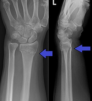
A Colles' fracture is a type of fracture of the distal forearm in which the broken end of the radius is bent backwards. Symptoms may include pain, swelling, deformity, and bruising. Complications may include damage to the median nerve.

A bone fracture is a medical condition in which there is a partial or complete break in the continuity of any bone in the body. In more severe cases, the bone may be broken into several fragments, known as a comminuted fracture. A bone fracture may be the result of high force impact or stress, or a minimal trauma injury as a result of certain medical conditions that weaken the bones, such as osteoporosis, osteopenia, bone cancer, or osteogenesis imperfecta, where the fracture is then properly termed a pathologic fracture.

A distal radius fracture, also known as wrist fracture, is a break of the part of the radius bone which is close to the wrist. Symptoms include pain, bruising, and rapid-onset swelling. The ulna bone may also be broken.

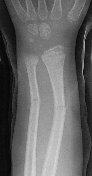
A greenstick fracture is a fracture in a young, soft bone in which the bone bends and breaks. Greenstick fractures occur most often during infancy and childhood when bones are soft. The name is by analogy with green wood which similarly breaks on the outside when bent.

A boxer's fracture is the break of the 5th metacarpal bones of the hand near the knuckle. Occasionally it is used to refer to fractures of the 4th metacarpal as well. Symptoms include pain and a depressed knuckle.

A pulled elbow, also known as nursemaid's elbow or a radial head subluxation, is when the ligament that wraps around the radial head slips off. Often a child will hold their arm against their body with the elbow slightly bent. They will not move the arm as this results in pain. Touching the arm, without moving the elbow, is usually not painful.
A traction splint most commonly refers to a splinting device that uses straps attaching over the pelvis or hip as an anchor, a metal rod(s) to mimic normal bone stability and limb length, and a mechanical device to apply traction to the limb.

A mallet finger, also known as hammer finger or PLF finger or Hannan finger, is an extensor tendon injury at the farthest away finger joint. This results in the inability to extend the finger tip without pushing it. There is generally pain and bruising at the back side of the farthest away finger joint.

Madelung's deformity is usually characterized by malformed wrists and wrist bones and is often associated with Léri-Weill dyschondrosteosis. It can be bilateral or just in the one wrist. It has only been recognized within the past hundred years. Named after Otto Wilhelm Madelung (1846–1926), a German surgeon, who described it in detail, it was noted by others. Guillaume Dupuytren mentioned it in 1834, Auguste Nélaton in 1847, and Joseph-François Malgaigne in 1855.

The styloid process of the ulna is a bony prominence found at distal end of the ulna in the forearm.

The triangular fibrocartilage complex (TFCC) is formed by the triangular fibrocartilage discus (TFC), the radioulnar ligaments (RULs) and the ulnocarpal ligaments (UCLs).

A scaphoid fracture is a break of the scaphoid bone in the wrist. Symptoms generally includes pain at the base of the thumb which is worse with use of the hand. The anatomic snuffbox is generally tender and swelling may occur. Complications may include nonunion of the fracture, avascular necrosis of the proximal part of the bone, and arthritis.

A Barton's fracture is a type of wrist injury where there is a broken bone associated with a dislocated bone in the wrist, typically occurring after falling on top of a bent wrist. It is an intra-articular fracture of the distal radius with dislocation of the radiocarpal joint.
A child bone fracture or a pediatric fracture is a medical condition in which a bone of a child is cracked or broken. About 15% of all injuries in children are fracture injuries. Bone fractures in children are different from adult bone fractures because a child's bones are still growing. Also, more consideration needs to be taken when a child fractures a bone since it will affect the child in his or her growth.
The Essex-Lopresti fracture is a fracture of the radial head of the forearm with concomitant dislocation of the distal radio-ulnar joint along with disruption of the thin interosseous membrane which holds them together. The injury is named after Peter Essex-Lopresti who described it in 1951.

An ulna fracture is a break in the ulna bone, one of the two bones in the forearm. It is often associated with a fracture of the other forearm bone, the radius.

Wrist osteoarthritis is a group of mechanical abnormalities resulting in joint destruction, which can occur in the wrist. These abnormalities include degeneration of cartilage and hypertrophic bone changes, which can lead to pain, swelling and loss of function. Osteoarthritis of the wrist is one of the most common conditions seen by hand surgeons.

Lister's tubercle or dorsal tubercle of radius is a bony prominence located at the distal end of the radius. It is palpable on the dorsum of the wrist.

A medial epicondyle fracture is an avulsion injury to the medial epicondyle of the humerus; the prominence of bone on the inside of the elbow. Medial epicondyle fractures account for 10% elbow fractures in children. 25% of injuries are associated with a dislocation of the elbow.


