Tricho-hepato-enteric syndrome

Last updated
Tricho-hepato-enteric syndrome
Other namesIntractable diarrhea of infancy with facial dysmorphism [1]
THE syndrome (4).jpg
Typical facial abnormalities with prominent forehead and cheeks, broad nasal root and wide-spaced eyes. Abnormal hairs are woolly, easily removed and poorly pigmented.

Tricho-hepato-enteric syndrome (THE), also known as syndromic or phenotypic diarrhea, is an extremely rare congenital bowel disorder which manifests itself as intractable diarrhea in infants with intrauterine growth retardation, hair and facial abnormalities. [2] Many also have liver disease and abnormalities of the immune system. [2] The associated malabsorption leads to malnutrition and failure to thrive. [2]

Contents

It is thought to be a genetic disorder with an autosomal recessive inheritance pattern, although responsible genes have not been found and the exact cause remains unknown. Prognosis is poor; many patients die before the age of 5 (mainly from infections or cirrhosis), although most patients nowadays survive with intravenous feeding (parenteral nutrition).

Symptoms

Tricho-hepato-enteric syndrome is one particular form of intractable diarrhea of infancy, presenting typically in the first month of life. These babies were usually born small for their age and continue to experience failure to thrive, usually with a final short stature. Typical facial features include prominent forehead and cheeks, a broad nasal root and widely spaced eyes (hypertelorism). Their hairs are woolly, easily removed and poorly pigmented. Liver disease is mainly present as cirrhosis or fibrosis, and staining might reveal high iron content of the liver cells (consistent with hemochromatosis). [3] Most evaluated patients had some degree of decrease in intelligence.[ citation needed ]

Genetics

The syndrome appears to be due to mutations in the gene tetratricopeptide repeat domain 37 (TTC37) which encodes the protein Thespin or the SKIV2L gene. [4] [5] This gene is expressed is in the adrenal gland, amniotic fluid, bladder, blood, bone, bone marrow, brain, cervix, connective tissue, ear, epididymis, eye, heart, intestine, kidney, liver, lung, lymph nodes, mammary glands, mouth, muscle, nerve, oesophagus, ovary, pancreas, pharynx, placenta, prostate, pituitary gland, salivary gland, testis, thyroid, tonsil, thymus, trachea, skin, uterus, spleen, spinal cord, stomach and vascular tissue. It is also expressed in ascites and various embryonic tissues. It is expressed at high level in the intestine, lung, lymph nodes, pituitary and vascular tissues. This gene is also known as KIAA0372, MGC32587 and TPR repeat protein 37.[ citation needed ]

This gene is located on the Crick (minus) strand of the long arm of chromosome 5 (5q15). The gene is 91,113 bases in length and encodes a protein of 1564 amino acid residues with twenty tetratricopeptide repeats. It has 43 exons of which exons 1, 2 and 3 are non coding. The predicted molecular weight of the protein is 175.486 kilodaltons and its predicted pI is 7.47. Its function is unknown but it may have adenylate cyclase activity and calcium- and calmodulin-responsive adenylate cyclase activity. A homolog has been identified in the frog ( Xenopus tropicalis ), the mouse ( Mus musculus ) and the rat ( Rattus norvegicus ). In the mouse this gene is located on chromosome 13.[ citation needed ]

Diagnosis

Microscopic analysis of the hair shaft showing breaks located at nodes in the hair (trichorrhexis nodosa) and longitudinal breaks. Tricho-hepato-enteric syndrome (1).jpg
Microscopic analysis of the hair shaft showing breaks located at nodes in the hair (trichorrhexis nodosa) and longitudinal breaks.
Small intestine biopsy of a patient with THE syndrome showing severe villous atrophy with intense mononuclear cell infiltration in the lamina propria. (Courtesy of Prof. Michel Peuchmaur, Hopital Robert Debre, Paris, France) Tricho-hepato-enteric syndrome (3).jpg
Small intestine biopsy of a patient with THE syndrome showing severe villous atrophy with intense mononuclear cell infiltration in the lamina propria. (Courtesy of Prof. Michel Peuchmaur, Hôpital Robert Debré, Paris, France)

Facial features

The typical facial features are low-set ears, prominent eyes with hypertelorism, broad flat nose, prominent forehead and large mouth.[ citation needed ]

Liver

There may be fibrosis with bile duct proliferation, occasional giant cells and regenerative parenchymal nodules. Siderosis is common.[ citation needed ]

Small bowel

Microscopic examination of a biopsy of the small bowel in these patients shows villous atrophy with low or no mononuclear cell infiltration of the lamina propria nor specific abnormalities involving the epithelium. The amount of villous atrophy does not explain the severity of the diarrhea.[ citation needed ]

Studies of enterocyte brush-border ion transporter proteins (sodium-hydrogen exchanger 2, sodium-hydrogen exchanger 3, aquaporin 7, sodium iodide symporter and hydrogen potassium adenosine triphosphatase) showed reduced expression or mislocalization in all patients with different profiles for each. [6]

Hair

Microscopic analysis of the hair shows twisted hairs of unequal size and different shapes (pili torti, aniso- and poikilotrichosis), longitudinal breaks and breaks located at nodes (trichorrhexis nodosa). Scanning electron microscopy might reveal hair budding (trichorrhexis blastysis). Biochemical analysis may reveal sulfur-deficient brittle hair (trichothiodystrophy; note that disulfide bonds determine hair waviness).[ citation needed ]

Platelets

Platelets may be enlarged. The membrane surface connected canalicular system is disrupted with prominent tubules and small membranous vesicles. Alpha granules may be missing from the platelets. Despite these abnormalities there is no increased tendency to bleed in this syndrome.[ citation needed ]

Other

More than 90% of patients present immune defects. Low immunoglobulin level, a defect in antibody production after vaccination, monoclonal hyper IgA, and low lymphocyte count have been reported. In these cases, some patients may need immunoglobulin supplementation. [7]

Treatment

No specific treatment or cure exists. Affected children usually need total parenteral nutrition through a central venous catheter. Further worsening of liver damage should however be avoided if possible. Diarrhea will likely continue even though food stops passing through the gastrointestinal system. [8] They can subsequently be managed with tube feeding, and some may be weaned from nutritional support during adolescence.[ citation needed ]

Epidemiology

Tricho-hepato-enteric syndrome is estimated to affect 1 in 300,000 to 400,000 live births in Western Europe. This syndrome was first reported in 1982 with a report on 2 siblings, [9] and as of 2008 there were around 25 published cases in medical journals. There seem to be no racial differences in its occurrence. It might be more common, as many genetic diseases, in areas with high levels of consanguinity.[ citation needed ]

Footnotes

  1. Fabre A, André N, Breton A, Broué P, Badens C, Roquelaure B (March 2007). "Intractable diarrhea with "phenotypic anomalies" and tricho-hepato-enteric syndrome: two names for the same disorder". Am. J. Med. Genet. A. 143 (6): 584–8. doi:10.1002/ajmg.a.31634. PMID   17318842. S2CID   39567209.
  2. 1 2 3 Goulet O, Vinson C, Roquelaure B, Brousse N, Bodemer C, Cézard JP (2008). "Syndromic (phenotypic) diarrhea in early infancy". Orphanet J Rare Dis. 3: 6. doi: 10.1186/1750-1172-3-6 . PMC   2279108 . PMID   18304370.
  3. Verloes A, Lombet J, Lambert Y, et al. (February 1997). "Tricho-hepato-enteric syndrome: further delineation of a distinct syndrome with neonatal hemochromatosis phenotype, intractable diarrhea, and hair anomalies". Am. J. Med. Genet. 68 (4): 391–5. doi:10.1002/(SICI)1096-8628(19970211)68:4<391::AID-AJMG3>3.0.CO;2-P. PMID   9021008.
  4. Fabre A, Martinez-Vinson C, Roquelaure B, Missirian C, André N, Breton A, Lachaux A, Odul E, Colomb V, Lemale J, Cézard JP, Goulet O, Sarles J, Levy N, Badens C (2011). "Novel mutations in TTC37 associated with tricho-hepato-enteric syndrome" (PDF). Hum Mutat. 32 (3): 277–281. doi:10.1002/humu.21420. PMID   21120949. S2CID   22834121.
  5. Vardi I, Barel O, Sperber M, Schvimer M, Nunberg M, Field M, Ouahed J, Marek-Yagel D, Werner L, Haberman Y, Lahad A, Anikster Y, Rechavi G, Barshack I, McElwee JJ, Maranville J, Somech R, Snapper SB, Weiss B, Shouval DS (May 2018). "Genetic and Structural Analysis of a SKIV2L Mutation Causing Tricho-hepato-enteric Syndrome". Dig Dis Sci. 63 (5): 1192–1199. doi:10.1007/s10620-018-4983-x. PMC   6167312 . PMID   29484573.
  6. Hartley JL, Zachos NC, Dawood B, Donowitz M, Forman J, Pollitt RJ, Morgan NV, Tee L, Gissen P, Kahr WH, Knisely AS, Watson S, Chitayat D, Booth IW, Protheroe S, Murphy S, de Vries E, Kelly DA, Maher ER (2010). "Mutations in TTC37 cause trichohepatoenteric syndrome (phenotypic diarrhea of infancy)". Gastroenterology. 138 (7): 2388–2398. doi:10.1053/j.gastro.2010.02.010. PMC   3166659 . PMID   20176027.
  7. Fabre, A., Martinez-Vinson, C., Goulet, O. et al. Syndromic diarrhea/Tricho-hepato-enteric syndrome. Orphanet J Rare Dis 8, 5 (2013). https://doi.org/10.1186/1750-1172-8-5
  8. Girault D, Goulet O, Le Deist F, et al. (July 1994). "Intractable infant diarrhea associated with phenotypic abnormalities and immunodeficiency". J. Pediatr. 125 (1): 36–42. doi:10.1016/S0022-3476(94)70118-0. PMID   8021782.
  9. Stankler L, Lloyd D, Pollitt RJ, Gray ES, Thom H, Russell G (March 1982). "Unexplained diarrhoea and failure to thrive in 2 siblings with unusual facies and abnormal scalp hair shafts: a new syndrome". Arch. Dis. Child. 57 (3): 212–6. doi:10.1136/adc.57.3.212. PMC   1627586 . PMID   7073301.

Related Research Articles

<span class="mw-page-title-main">Cardiofaciocutaneous syndrome</span> Medical condition

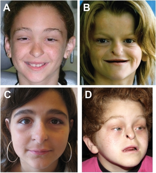
Cardiofaciocutaneous (CFC) syndrome is an extremely rare genetic disorder, and is one of the RASopathies. It was first described in 1986.
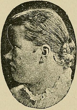
<span class="mw-page-title-main">Alagille syndrome</span> Medical condition

Alagille syndrome (ALGS) is a genetic disorder that affects primarily the liver and the heart. Problems associated with the disorder generally become evident in infancy or early childhood. The disorder is inherited in an autosomal dominant pattern, and the estimated prevalence of Alagille syndrome is 1 in every 30,000 to 1 in every 40,000 live births. It is named after the French pediatrician Daniel Alagille, who first described the condition in 1969.

<span class="mw-page-title-main">McCune–Albright syndrome</span> Mosaic genetic disorder affecting the bone, skin and endocrine systems

McCune–Albright syndrome is a complex genetic disorder affecting the bone, skin and endocrine systems. It is a mosaic disease arising from somatic activating mutations in GNAS, which encodes the alpha-subunit of the Gs heterotrimeric G protein.

<span class="mw-page-title-main">Laron syndrome</span> Medical condition

Laron syndrome (LS), also known as growth hormone insensitivity or growth hormone receptor deficiency (GHRD), is an autosomal recessive disorder characterized by a lack of insulin-like growth factor 1 production in response to growth hormone. It is usually caused by inherited growth hormone receptor (GHR) mutations.

<span class="mw-page-title-main">Muenke syndrome</span> Medical condition

Muenke syndrome, also known as FGFR3-related craniosynostosis, is a human specific condition characterized by the premature closure of certain bones of the skull during development, which affects the shape of the head and face. First described by Maximilian Muenke, the syndrome occurs in about 1 in 30,000 newborns. This condition accounts for an estimated 8 percent of all cases of craniosynostosis.

<span class="mw-page-title-main">Uncombable hair syndrome</span> Rare scalp hair shaft dysplasia

Uncombable hair syndrome (UHS) is a rare structural anomaly of the hair with a variable degree of effect. It is characterized by hair that is silvery, dry, frizzy, wiry, and impossible to comb. It was first reported in the early 20th century. It typically becomes apparent between the ages of 3 months and 12 years. UHS has several names, including pili trianguli et canaliculi (Latin), cheveux incoiffables (French), and "spun-glass hair". This disorder is believed to be autosomal recessive in most instances, but there are a few documented cases where multiple family members display the trait in an autosomal dominant fashion. Based on the current scientific studies related to the disorder, the three genes that have been causally linked to UHS are PADI3, TGM3, and TCHH. These genes encode proteins important for hair shaft formation. Clinical symptoms of the disorder arise between 3 months and 12 years of age. The quantity of hair on the head does not change, but hair starts to grow more slowly and becomes increasingly "uncombable". To be clinically apparent, 50% of all scalp hair shafts must be affected by UHS. This syndrome only affects the hair shaft of the scalp and does not influence hair growth in terms of quantity, textural feel, or appearance on the rest of the body.

Vici syndrome, also called immunodeficiency with cleft lip/palate, cataract, hypopigmentation and absent corpus callosum, is a rare autosomal recessive congenital disorder characterized by albinism, agenesis of the corpus callosum, cataracts, cardiomyopathy, severe psychomotor retardation, seizures, immunodeficiency and recurrent severe infections. To date, about 50 cases have been reported.

<span class="mw-page-title-main">Microvillous inclusion disease</span> Medical condition

Microvillus inclusion disease, previously known as Davidson's disease, congenital microvillus atrophy and, less specifically, microvillus atrophy, is a rare genetic disorder of the small intestine that is inherited in an autosomal recessive pattern.

<span class="mw-page-title-main">SKIV2L</span> Protein-coding gene in the species Homo sapiens

Helicase SKI2W is an enzyme that in humans is encoded by the SKIV2L gene. This enzyme is a human homologue of yeast SKI2, which may be involved in antiviral activity by blocking translation of poly(A) deficient mRNAs. The SKIV2L gene is located in the class III region of the major histocompatibility complex.
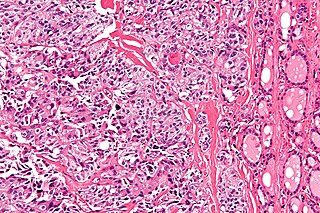
<span class="mw-page-title-main">Keutel syndrome</span> Medical condition

Keutel syndrome (KS) is a rare autosomal recessive genetic disorder characterized by abnormal diffuse cartilage calcification, hypoplasia of the mid-face, peripheral pulmonary stenosis, hearing loss, short distal phalanges (tips) of the fingers and mild mental retardation. Individuals with KS often present with peripheral pulmonary stenosis, brachytelephalangism, sloping forehead, midface hypoplasia, and receding chin. It is associated with abnormalities in the gene coding for matrix gla protein, MGP. Being an autosomal recessive disorder, it may be inherited from two unaffected, abnormal MGP-carrying parents. Thus, people who inherit two affected MGP alleles will likely inherit KS.

<span class="mw-page-title-main">Chylomicron retention disease</span> Medical condition

Chylomicron retention disease is a disorder of fat absorption. It is associated with SAR1B. Mutations in SAR1B prevent the release of chylomicrons in the circulation which leads to nutritional and developmental problems. It is a rare autosomal recessive disorder with around 40 cases reported worldwide. Since the disease allele is recessive, parents usually do not show symptoms.

Congenital hypoplastic anemia is a congenital disorder that occasionally also includes leukopenia and thrombocytopenia and is characterized by deficiencies of red cell precursors.

<span class="mw-page-title-main">Floating–Harbor syndrome</span> Medical condition

Floating–Harbor syndrome, also known as Pelletier–Leisti syndrome, is a rare disease with fewer than 50 cases described in the literature. It is usually diagnosed in early childhood and is characterized by the triad of proportionate short stature with delayed bone age, characteristic facial appearance, and delayed speech development. Although its cause is unknown, it is thought to result from genetic mutation, and diagnosis is established by the presence of a heterozygous SRCAP mutation in those with clinical findings of FHS.

Congenital tufting enteropathy is an inherited disorder of the small intestine that presents with intractable diarrhea in young children.

<span class="mw-page-title-main">Tricho–dento–osseous syndrome</span> Medical condition

Tricho–dento–osseous syndrome (TDO) is a rare, systemic, autosomal dominant genetic disorder that causes defects in hair, teeth, and bones respectively. This disease is present at birth. TDO has been shown to occur in areas of close geographic proximity and within families; most recent documented cases are in Virginia, Tennessee, and North Carolina. The cause of this disease is a mutation in the DLX3 gene, which controls hair follicle differentiation and induction of bone formation. All patients with TDO have two co-existing conditions called enamel hypoplasia and taurodontism in which the abnormal growth patterns of the teeth result in severe external and internal defects. The hair defects are characterized as being rough, course, with profuse shedding. Hair is curly and kinky at infancy but later straightens. Dental defects are characterized by dark-yellow/brownish colored teeth, thin and/or possibly pitted enamel, that is malformed. The teeth can also look normal in color, but also have a physical impression of extreme fragility and thinness in appearance. Additionally, severe underbites where the top and bottom teeth fail to correctly align may be present; it is common for the affected individual to have a larger, more pronounced lower jaw and longer bones. The physical deformities that TDO causes become more noticeable with age, and emotional support for the family as well as the affected individual is frequently recommended. Adequate treatment for TDO is a team based approach, mostly involving physical therapists, dentists, and oromaxillofacial surgeons. Genetic counseling is also recommended.

<span class="mw-page-title-main">Primary pigmented nodular adrenocortical disease</span> Medical condition

Primary pigmented nodular adrenocortical disease (PPNAD) was first coined in 1984 by Carney et al. it often occurs in association with Carney complex (CNC). CNC is a rare syndrome that involves the formation of abnormal tumours that cause endocrine hyperactivity.

<span class="mw-page-title-main">COACH syndrome</span> Medical condition

COACH syndrome, also known as Joubert syndrome with hepatic defect, is a rare autosomal recessive genetic disease. The name is an acronym of the defining signs: cerebellar vermis aplasia, oligophrenia, congenital ataxia, coloboma and hepatic fibrosis. The condition is associated with moderate intellectual disability. It falls under the category of a Joubart Syndrome-related disorder (JSRD).

<span class="mw-page-title-main">Okamoto syndrome</span> Rare genetic condition involving urinary, heart, facial and neurological features

Okamoto syndrome (OS), also known as Au–Kline syndrome (AKS), is a very rare autosomal dominant genetic condition characterised by congenital hydronephrosis, low muscle tone, heart defects, intellectual disability and characteristic facial features. Those affected often have neurological and skeletal abnormalities, as well as frequent urinary tract infections. Language and walking are usually delayed. Facial features include prominent, downturned ears, an open, downturned mouth and drooping eyelids (ptosis).

<span class="mw-page-title-main">HFE H63D gene mutation</span>

The HFE H63D is a single-nucleotide polymorphism in the HFE gene, which results in the substitution of a histidine for an aspartic acid at amino acid position 63 of the HFE protein (p.His63Asp). HFE participates in the regulation of iron absorption.

<span class="mw-page-title-main">TTC37</span> Protein-coding gene in the species Homo sapiens

TTC37 is a protein which in humans is encoded by gene TTC37, located on chromosome 5.

References