Vaginal introital laxity

Last updated
Vaginal introital laxity
DC-116512.jpg
Vaginal introital laxity in a woman, with visible loosened external opening.
Symptoms Sensation of looseness at vaginal introitus
CausesPelvic organ prolapse, vaginal delivery, menopause
Diagnostic method Physical examination (pelvic examination), questionnaires
TreatmentEnergy-based devices, vaginoplasty repairs, dynamic quadripolar satisfaction questionnaires, surgical introital reduction procedures

Vaginal introital laxity is a symptom of pelvic floor dysfunction characterised by a sensation of looseness at vaginal external opening, also known as the vaginal introitus. [1] Possible causes include pelvic organ prolapse (POP), post-pregnancy and vaginal delivery and menopause. [2] Consequences may include experiencing sexual dysfunction, ranging from dyspareunia (i.e. painful intercourse), increased vaginal “wind” to overactive bladder (OAB). These consequences may lead to adverse significant impacts on women’s sexual health, body image and quality of life. [3] Vaginal laxity is often underreported, with approximately 80% of women not seeking treatment or discussing their concerns. [3]

Contents

Diagnosis is based on physical examination, including pelvic examination, as well as validated questionnaires such as vaginal laxity questionnaire (VLQ) and sexual satisfaction questionnaire (SSQ). [4] Possible treatments include nonsurgical treatment with energy-based devices, [2] vaginoplasty repairs [2] and dynamic quadripolar radiofrequency treatment. [4] More severe cases may require surgical introital reduction procedures after the failure of conservative measures. [3] Outcomes following these treatments are generally positive, with reported significant and sustainable long-term effectiveness and improved sexual life quality. [4]

Signs and symptoms

Dyspareunia

Vaginal introital laxity is often associated with a decrease in sensation during sexual intercourse. [5] It may also lead to reduced production of natural lubricating substances in the female body, such as vaginal fluid and cervical mucus, resulting in reduced friction and pressure. [5] Sexual experience may become less enjoyable and may even lead to discomfort. [5] In some serious cases of VIL, the vagina and vaginal introitus may become especially vulnerable during sexual activities when the male penis rubs against the vaginal wall at the external opening. [5] Risks of microtears and vaginal tissue trauma may therefore increase significantly during sexual intercourse, resulting in pain, the feeling of burning and even injuries. [5]

Increased vaginal "wind"

The loosened vaginal walls caused by vaginal introital laxity may not seal the vaginal canal as strongly as a normal one, resulting in the entering of air into the canal during certain activities, such as during vigorous movements, sexual activities or even childbirth. [6] VIL may also lead to weakened pelvic floor muscles and cause improper closure of the vaginal introital, allowing easier entering of air into the vagina. [6] During certain movements and physical activities, the release of this trapped air will result in vaginal “wind” and produce a sound similar to passing a gas or a “popping” sound. [6]

Overactive bladder (OAB)

The mechanism of neural control on contraction and relaxation of smooth muscle within the bladder. Overactivebladder.png
The mechanism of neural control on contraction and relaxation of smooth muscle within the bladder.

Although vaginal introital laxity may not cause OAB directly, there are still some indirect relationships between them that may relate these two conditions. [7] Pelvic floor muscle, being the major muscle supporting the pelvic organs such as the bladder and uterus, is especially vulnerable during vaginal delivery. [7] During childbirth, women are more likely to develop vaginal introital laxity, eventually leading to the weakening of pelvic floor muscles and may develop urinary symptoms such as OAB. [7]

Vaginal introital laxity may also weaken the support structures around the urethra, which is the tube that helps carry urine outside of the body from the bladder. [7] The weakened urethra supporting structures will subsequently cause the urethra to move much more than usual, such as during physical exercise or even coughing. [7] This will contribute to urinary leakage, a symptom of OAB. [7]

Pathophysiology

Pelvic organ prolapse (POP)

An old woman with uterine prolapse, her cervix is protruding through the vulva. Uterine prolapse - standing.jpg
An old woman with uterine prolapse, her cervix is protruding through the vulva.

Vaginal introital laxity is usually suggested to be associated with pelvic organ prolapse, which refers to the descent of one or more of these organs from the normal position. [2] Constant stretching and elongation of the vaginal canal caused by POP may be a cause of vaginal introital laxity. [2] However, the association between this type of prolapse and vaginal introital laxity is still unclear due to the lack of related data. [2]

POP includes (a) the falling out of vagina, bladder and other genito-pelvic structures, (b) vaginal tissue bulging into and through the introitus, or (c) the prolapse of rectal tissues into the vaginal area. [8] It is differentiable between POP and vaginal introital laxity as pelvic organ prolapse involves the descent of one or more pelvic structures, whereas vaginal introital laxity specifically pertains to the looseness of the vaginal introitus. [8]

Vaginal delivery

Stages of childbirth 2920 Stages of Childbirth-ca.svg
Stages of childbirth

Being one of the most dominant causes of vaginal introital laxity, vaginal childbirth in women may cause trauma to the genito-pelvic floor musculature and vagina by stretching the introitus. [8] Along with the hormonal changes that lead to the relaxation of pelvic ligaments and vaginal tissues during pregnancy, these body parts will be further stretched and weakened. [8] During childbirth, a huge pressure may be exerted onto the vaginal tissues during the passing of the baby through the birth canal, especially onto the introitus, causing laxity. [8] In some cases, when the size of the baby is too large, the excessive stretching of the vaginal opening during childbirth may even cause tearing. [8]

Menopause

Another cause of vaginal introital laxity is menopause. Estrogen and progesterone are two of the primary sex hormones, and their levels are directly associated with the thickness and elasticity of the vagina. [7] During menopause, the significant decline of estrogen and progesterone levels may cause reduced production of collagen and elastin, leading to thinner and less rigid vaginal walls. [7] The weakening of pelvic floor muscles due to these sudden hormonal changes is also a contributing factor to potential vaginal introital laxity. [7] Especially for estrogen, its significant decline during menopause may result in a condition known as vaginal atrophy, which refers to the thinning of vaginal walls. [7] This reduces the elasticity of the vaginal introitus. [7] Menopausal women who have given birth may face increased risks compared to younger women due to the combined effects of childbirth-related excessive stretching and menopause-caused hormonal level reduction. [7] These combined factors may eventually weaken the vaginal external opening and cause laxity. [7]

Diagnosis

Diagnosis of vaginal introital laxity involves a comprehensive evaluation of the patient's symptoms and medical history, which may include physical examination and response to several validated questionnaires. [9] Since vaginal introital laxity is usually a patient self-reported condition based on subjective perceptions, there are no objective measurements to quantify its severity. [9]

Physical examination

Healthcare providers will perform pelvic examinations to assess the vaginal tissue. [10] Patients are first asked to empty their bladders to improve access to the pelvic organs, and to alleviate any discomfort or pressure that may arise from a full bladder during the examination. [10] They are then placed in a supine position, usually lying on their back on birthing chairs at 45 degrees with their feet in stirrups, allowing the legs to be comfortably positioned. [11] This position is called the dorsal lithotomy position, which is most commonly used in genital examination. [11] In some cases, alternative positions such as the supine frog leg position or the prone knee chest position may be used. [12]

Application of speculum examination Nulliparous cervix with ectropion.jpg
Application of speculum examination

Next, inspection of external genitalia, including the labia majora and minora, clitoris, perihymenal tissue (vestibule), hymen, posterior fourchette, vagina, and cervix will proceed. [13] Healthcare providers may gently palpate the vaginal introitus and surrounding tissues. [14] Speculum examination is also conducted by inserting a lubricated speculum into the vagina to visualise the vaginal walls and cervix for assessing the vaginal introitus and measuring any laxity or looseness. [14] The speculum is available in different sizes and shapes to accommodate individual anatomy. [14]

Moreover, pelvic floor assessment may be used to evaluate the strength and tone of the muscles. [15] This may involve requesting patients to perform specific movements, such as contracting and relaxing the pelvic floor muscles, coughing, or bearing down. [15] This helps evaluate the muscle function and identify any issues or weaknesses. [15]

Vaginal Laxity Questionnaire (VLQ)

Vaginal Laxity Questionnaire (VLQ) is designed to evaluate the degree of vaginal looseness, which helps guide treatment decisions and monitor the effectiveness of interventions aimed at improving vaginal laxity. [16] The questionnaire covers several aspects: [16]

Sexual Satisfaction Questionnaire (SSQ)

Sexual Satisfaction Questionnaire (SSQ) is used to assess sexual quality of life and sexual function. [17] Since vaginal introital laxity (VIL) may cause decreased genito-pelvic sensation during sexual intercourse, [18] which in turn is associated with decreased sexual desire, arousal difficulties, and orgasmic dysfunction, [17] it can adversely impact the sexual quality of life. Due to this potential cause-and-effect, SSQ can indirectly reveal the occurrence and severity of VIL. [17]

The questionnaire consists of multiple items that individuals can rate on a scale to indicate their level of satisfaction. [19] This method has great reliability due to the high consistency score measured. [19] The questionnaire result can be used to cross-check with physical examinations for inferring the severity extent of VIL. [19]

Treatment

Surgery with energy-based devices

There are two main types of laser modalities – the ablative CO2 and the non-ablative Erbium Yag. [20] The former renders collagen- and elastin-fibre remodelling by denaturing the tissue, while the latter has a deeper secondary thermal effect and controlled heating of the target mucous membrane of the vaginal wall due to a higher affinity of water absorption. [20] Both aim to remodel the subepithelial connective tissue. [20] The safety and efficacy need further investigation. [20]

Vaginoplasty Repairs

In vaginoplasty repairs, a modified anterior and/or high posterior colporrhaphy and/or the excision of lateral vaginal mucosa are used to remove sections of the mucosa from the vaginal fornices. [21] The goal of this procedure is to tighten a rather lax upper vagina. [21]

Dynamic Quadripolar Radiofrequency Treatment

The radiofrequency device emits focused electromagnetic waves that generate 40 °C to 45 °C heat upon tissue impedance. [22] The high temperature stimulates the fibroblasts' production of collagen by activating heat-shock proteins and initiating the inflammatory cascade. [22] With the treatment biophysics, the operator can significantly lower the amount of energy administered by defining the target vulvar area's volume and depth. [23] To provide strict tissue temperature control, it also permits electronic movement and temperature sensor control within the radiofrequency device. [23]

Surgical Introital Reduction Procedures

Comparison between the vaginal condition before and after conducting perineoplasty Perineoplasty Before & After.JPG
Comparison between the vaginal condition before and after conducting perineoplasty

Perineorrhaphy is a common technique that is involved in the procedure, which aims to repair the perineum (i.e. the area between the vagina and anus) surgically. [24] It can be performed with or without levator ani plication to tighten the pelvic floor muscles. [24]

Related Research Articles

<span class="mw-page-title-main">Vagina</span> Part of the female reproductive tract

In mammals and other animals, the vagina is the elastic, muscular reproductive organ of the female genital tract. In humans, it extends from the vulval vestibule to the cervix. The vaginal introitus is normally partly covered by a thin layer of mucosal tissue called the hymen. The vagina allows for copulation and birth. It also channels menstrual flow, which occurs in humans and closely related primates as part of the menstrual cycle.

Dyspareunia is painful sexual intercourse due to medical or psychological causes. The term dyspareunia covers both female dyspareunia and male dyspareunia, but many discussions that use the term without further specification concern the female type, which is more common than the male type. In females, the pain can primarily be on the external surface of the genitalia, or deeper in the pelvis upon deep pressure against the cervix. Medically, dyspareunia is a pelvic floor dysfunction and is frequently underdiagnosed. It can affect a small portion of the vulva or vagina or be felt all over the surface. Understanding the duration, location, and nature of the pain is important in identifying the causes of the pain.

<span class="mw-page-title-main">Kegel exercise</span> Pelvic floor exercise

Kegel exercise, also known as pelvic floor exercise, involves repeatedly contracting and relaxing the muscles that form part of the pelvic floor, now sometimes colloquially referred to as the "Kegel muscles". The exercise can be performed many times a day, for several minutes at a time but takes one to three months to begin to have an effect.

A pessary is a prosthetic device inserted into the vagina for structural and pharmaceutical purposes. It is most commonly used to treat stress urinary incontinence to stop urinary leakage and to treat pelvic organ prolapse to maintain the location of organs in the pelvic region. It can also be used to administer medications locally in the vagina or as a method of contraception.

<span class="mw-page-title-main">Pelvic floor</span> Anatomical structure

The pelvic floor or pelvic diaphragm is an anatomical location in the human body, which has an important role in urinary and anal continence, sexual function and support of the pelvic organs. The pelvic floor includes muscles, both skeletal and smooth, ligaments and fascia. and separates between the pelvic cavity from above, and the perineum from below. It is formed by the levator ani muscle and coccygeus muscle, and associated connective tissue.
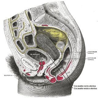
<span class="mw-page-title-main">Rectocele</span> Bulging of the rectum into the vaginal wall

In gynecology, a rectocele or posterior vaginal wall prolapse results when the rectum bulges (herniates) into the vagina. Two common causes of this defect are childbirth and hysterectomy. Rectocele also tends to occur with other forms of pelvic organ prolapse, such as enterocele, sigmoidocele and cystocele.

<span class="mw-page-title-main">Pelvic floor dysfunction</span> Medical condition

Pelvic floor dysfunction is a term used for a variety of disorders that occur when pelvic floor muscles and ligaments are impaired. The condition affects up to 50 percent of women who have given birth. Although this condition predominantly affects women, up to 16 percent of men are affected as well. Symptoms can include pelvic pain, pressure, pain during sex, urinary incontinence (UI), overactive bladder, bowel incontinence, incomplete emptying of feces, constipation, myofascial pelvic pain and pelvic organ prolapse. When pelvic organ prolapse occurs, there may be visible organ protrusion or a lump felt in the vagina or anus. Research carried out in the UK has shown that symptoms can restrict everyday life for women. However, many people found it difficult to talk about it and to seek care, as they experienced embarrassment and stigma.

<span class="mw-page-title-main">Cystocele</span> Protrusion of the bladder into the vagina

The cystocele, also known as a prolapsed bladder, is a medical condition in which a woman's bladder bulges into her vagina. Some may have no symptoms. Others may have trouble starting urination, urinary incontinence, or frequent urination. Complications may include recurrent urinary tract infections and urinary retention. Cystocele and a prolapsed urethra often occur together and is called a cystourethrocele. Cystocele can negatively affect quality of life.

<span class="mw-page-title-main">Pelvic organ prolapse</span> Descent of the pelvic organs from their normal positions

Pelvic organ prolapse (POP) is characterized by descent of pelvic organs from their normal positions into the vagina. In women, the condition usually occurs when the pelvic floor collapses after gynecological cancer treatment, childbirth or heavy lifting. Injury incurred to fascia membranes and other connective structures can result in cystocele, rectocele or both. Treatment can involve dietary and lifestyle changes, physical therapy, or surgery.

<span class="mw-page-title-main">Uterine prolapse</span> Medical condition

Uterine prolapse is a form of pelvic organ prolapse in which the uterus and a portion of the upper vagina protrude into the vaginal canal and, in severe cases, through the opening of the vagina. It is most often caused by injury or damage to structures that hold the uterus in place within the pelvic cavity. Symptoms may include vaginal fullness, pain with sexual intercourse, difficulty urinating, and urinary incontinence. Risk factors include older age, pregnancy, vaginal childbirth, obesity, chronic constipation, and chronic cough. Prevalence, based on physical exam alone, is estimated to be approximately 14%.

A urethrocele is the prolapse of the female urethra into the vagina. Weakening of the tissues that hold the urethra in place may cause it to protrude into the vagina. Urethroceles often occur with cystoceles. In this case, the term used is cystourethrocele.

Urogynecology or urogynaecology is a surgical sub-specialty of urology and gynecology.

Female genital disease is a disorder of the structure or function of the female reproductive system that has a known cause and a distinctive group of symptoms, signs, or anatomical changes. The female reproductive system consists of the ovaries, fallopian tubes, uterus, vagina, and vulva. Female genital diseases can be classified by affected location or by type of disease, such as malformation, inflammation, or infection.

An enterocele is a herniation of a peritoneum-lined sac containing small intestine through the pelvic floor, between the rectum and the vagina. Enterocele is significantly more common in females, especially after hysterectomy.

Urethral hypermobility is a condition of excessive movement of the female urethra due to a weakened urogenital diaphragm. It describes the instability of the urethra in relation to the pelvic floor muscles. A weakened pelvic floor muscle fails to adequately close the urethra and hence can cause stress urinary incontinence. This condition may be diagnosed by primary care providers or urologists. Treatment may include pelvic floor muscle exercises, surgery, or minimally invasive procedures.

The Pelvic Organ Prolapse Quantifications System (POP-Q) is a system for assessing the degree of prolapse of pelvic organs to help standardize diagnosing, comparing, documenting, and sharing of clinical findings. This assessment is the most frequently used among research publications related to pelvic organ prolapse.

A urogenital fistula is an abnormal tract that exists between the urinary tract and bladder, ureters, or urethra. A urogenital fistula can occur between any of the organs and structures of the pelvic region. A fistula allows urine to continually exit through and out the urogenital tract. This can result in significant disability, interference with sexual activity, and other physical health issues, the effects of which may in turn have a negative impact on mental or emotional state, including an increase in social isolation. Urogenital fistulas vary in etiology. Fistulas are usually caused by injury or surgery, but they can also result from malignancy, infection, prolonged and obstructed labor and deliver in childbirth, hysterectomy, radiation therapy or inflammation. Of the fistulas that develop from difficult childbirth, 97 percent occur in developing countries. Congenital urogenital fistulas are rare; only ten cases have been documented. Abnormal passageways can also exist between the vagina and the organs of the gastrointestinal system, and these may also be termed fistulas.

<span class="mw-page-title-main">Vaginal support structures</span> Structures that maintain the position of the vagina within the pelvic cavity

The vaginal support structures are those muscles, bones, ligaments, tendons, membranes and fascia, of the pelvic floor that maintain the position of the vagina within the pelvic cavity and allow the normal functioning of the vagina and other reproductive structures in the female. Defects or injuries to these support structures in the pelvic floor leads to pelvic organ prolapse. Anatomical and congenital variations of vaginal support structures can predispose a woman to further dysfunction and prolapse later in life. The urethra is part of the anterior wall of the vagina and damage to the support structures there can lead to incontinence and urinary retention.

Vaginal rugae are structures of the vagina that are transverse ridges formed out of the supporting tissues and vaginal epithelium in females. Some conditions can cause the disappearance of vaginal rugae and are usually associated with childbirth and prolapse of pelvic structures. The rugae contribute to the resiliency and elasticity of the vagina and its ability to distend and return to its previous state. These structures not only allow expansions and an increase in surface area of the vaginal epithelium, they provide the space necessary for the vaginal microbiota. The shape and structure of the rugae are supported and maintained by the lamina propria of the vagina and the anterior and posterior rugae.

Transvaginal mesh, also known as vaginal mesh implant, is a net-like surgical tool that is used to treat pelvic organ prolapse (POP) and stress urinary incontinence (SUI) among female patients. The surgical mesh is placed transvaginally to reconstruct weakened pelvic muscle walls and to support the urethra or bladder.

References

  1. Dietz, Hans Peter; Stankiewicz, Martyna; Atan, Ixora Kamisan; Ferreira, Caroline Wanderley; Socha, Maciej (2017-07-31). "Vaginal laxity: what does this symptom mean?". International Urogynecology Journal. 29 (5): 723–728. doi:10.1007/s00192-017-3426-0. ISSN   0937-3462. PMID   28762179.
  2. 1 2 3 4 5 6 Polland, Allison; Duong, Vi; Furuya, Rachel; Fitzgerald, Jocelyn J.; Wang, Haijun; Iwamoto, Aya; Bradley, Sarah; Iglesia, Cheryl B. (2021-10-08). "Description of Vaginal Laxity and Prolapse and Correlation With Sexual Function (DeVeLoPS)". Sexual Medicine. 9 (6): 100443. doi:10.1016/j.esxm.2021.100443. ISSN   2050-1161. PMC   8766263 . PMID   34629323.
  3. 1 2 3 Al Salehi, Asma; Zemmache, Mohammed Zakarya; Allegre, Lucie; Fatton, Brigitte; de Tayrac, Renaud (2023-07-01). "Functional and sexual outcomes following surgical vaginal introital reduction". Progrès en Urologie. 33 (7): 370–376. doi:10.1016/j.purol.2023.05.003. ISSN   1166-7087. PMID   37263901.
  4. 1 2 3 Vicariotto, Franco; DE Seta, Francesco; Faoro, Valentina; Raichi, Mauro (2017). "Dynamic quadripolar radiofrequency treatment of vaginal laxity/menopausal vulvo-vaginal atrophy: 12-month efficacy and safety". Minerva Ginecologica. 69 (4): 342–349. doi:10.23736/S0026-4784.17.04072-2. ISSN   1827-1650. PMID   28608667.
  5. 1 2 3 4 5 Pauls, Rachel N.; Fellner, Angela N.; Davila, G. Willy (2012-10-01). "Vaginal laxity: a poorly understood quality of life problem; a survey of physician members of the International Urogynecological Association (IUGA)". International Urogynecology Journal. 23 (10): 1435–1448. doi:10.1007/s00192-012-1757-4. ISSN   1433-3023. PMID   22669419.
  6. 1 2 3 Talab, Sali; Al-Badr, Ahmed; AlKusayer, Ghadeer M.; Dawood, Ashraf; Bazi, Tony (2019). "Correlates of vaginal laxity symptoms in women attending a urogynecology clinic in Saudi Arabia". International Journal of Gynecology & Obstetrics. 145 (3): 278–282. doi:10.1002/ijgo.12810. ISSN   0020-7292. PMC   7027493 . PMID   30924525.
  7. 1 2 3 4 5 6 7 8 9 10 11 12 13 https://www.researchgate.net/profile/Ahmed-Al-Badr/publication/367150313_Vaginal_Laxity_Prevalence_Risk_Factors_Diagnostic_and_Therapeutic_Approaches/links/63c2f7526fe15d6a571bc894/Vaginal-Laxity-Prevalence-Risk-Factors-Diagnostic-and-Therapeutic-Approaches.pdf
  8. 1 2 3 4 5 6 Sekiguchi, Yuki; Utsugisawa, Yukari; Azekosi, Yoko; Kinjo, Manami; Song, Mihyon; Kubota, Yodhinobu; Kingsberg, Sheryl A.; Krychman, Michael L. (2013). "Laxity of the Vaginal Introitus After Childbirth: Nonsurgical Outpatient Procedure for Vaginal Tissue Restoration and Improved Sexual Satisfaction Using Low-Energy Radiofrequency Thermal Therapy". Journal of Women's Health. 22 (9): 775–781. doi:10.1089/jwh.2012.4123. ISSN   1540-9996. PMID   23952177.
  9. 1 2 "Vaginal Laxity Issues, Answers and Implications for Female Sexual Function". academic.oup.com. Retrieved 2024-03-27.
  10. 1 2 Haylen, Bernard T. (2007). "The empty bladder". International Urogynecology Journal and Pelvic Floor Dysfunction. 18 (3): 237–239. doi:10.1007/s00192-006-0111-0. PMID   16791705.
  11. 1 2 Barber, Matthew D.; Lambers, Anouk R.; Visco, Anthony G.; Bump, Richard C. (2000). "Effect of Patient Position on Clinical Evaluation of Pelvic Organ Prolapse". Obstetrics & Gynecology. 96 (1): 18–22. doi:10.1016/s0029-7844(00)00859-0. ISSN   0029-7844. PMID   10862835.
  12. Goldberg, Murray G.; Surya, Babu V.; Catanese, Anthony; Johanson, Karl-Eric; Brown, Jordan (1991-11-01). "Effect of Patient Positioning on Urethral Mobility: Implications for Radical Pelvic Surgery". The Journal of Urology. 146 (5): 1252–1254. doi:10.1016/S0022-5347(17)38061-8. ISSN   0022-5347. PMID   1942273.
  13. Polland, Allison; Duong, Vi; Furuya, Rachel; Fitzgerald, Jocelyn J.; Wang, Haijun; Iwamoto, Aya; Bradley, Sarah; Iglesia, Cheryl B. (2021). "Description of Vaginal Laxity and Prolapse and Correlation With Sexual Function (DeVeLoPS)". Sexual Medicine. 9 (6): 100443. doi:10.1016/j.esxm.2021.100443. ISSN   2050-1161. PMC   8766263 . PMID   34629323.
  14. 1 2 3 Evans, Devon; Goldstein, Susan; Loewy, Amanda; Altman, Alon D. (2019-08-01). "No. 385-Indications for Pelvic Examination". Journal of Obstetrics and Gynaecology Canada. 41 (8): 1221–1234. doi:10.1016/j.jogc.2018.12.007. ISSN   1701-2163. PMID   31331610.
  15. 1 2 3 Dietz, Hans Peter (2009). "Pelvic Floor Assessment". Fetal and Maternal Medicine Review. 20 (1): 49–66. doi:10.1017/S096553950900237X. ISSN   1469-5065.
  16. 1 2 "Treatment of women with vaginal laxity: systematic review with meta-analysis". academic.oup.com. doi:10.1093/jsxmed/qdae028/7632419 (inactive 1 November 2024). Retrieved 2024-03-27.{{cite web}}: CS1 maint: DOI inactive as of November 2024 (link)
  17. 1 2 3 FU, Lixia; Long, Senyang; LI, Qin; XU, Hainan; Guo, Ling; Wang, Huarong; Zheng, Zhongyan; Zhang, Jing (2023-03-23). "The efficacy and safety of temperature controlled dual-mode radiofrequency in women with vaginal laxity". BMC Women's Health. 23 (1): 121. doi: 10.1186/s12905-023-02261-y . ISSN   1472-6874. PMC   10035145 . PMID   36959573.
  18. "Description of Vaginal Laxity and Prolapse and Correlation With Sexual Function (DeVeLoPS)". academic.oup.com. Retrieved 2024-03-27.
  19. 1 2 3 http://archive.sciendo.com/PJAP/pjap.2014.12.issue-3/pjap-2015-0017/pjap-2015-0017.pdf
  20. 1 2 3 4 Shobeiri, S. Abbas; Kerkhof, M. H.; Minassian, Vatche A.; Bazi, Tony; on behalf of the IUGA Research and Development Committee (2019-03-01). "IUGA committee opinion: laser-based vaginal devices for treatment of stress urinary incontinence, genitourinary syndrome of menopause, and vaginal laxity". International Urogynecology Journal. 30 (3): 371–376. doi:10.1007/s00192-018-3830-0. ISSN   1433-3023. PMID   30523374.
  21. 1 2 "A Large Multicenter Outcome Study of Female Genital Plastic Surgery". academic.oup.com. Retrieved 2024-03-27.
  22. 1 2 Robinson, Dudley; Flint, Richard; Veit-Rubin, Nikolaus; Araklitis, George; Cardozo, Linda (2020). "Is there enough evidence to justify the use of laser and other thermal therapies in female lower urinary tract dysfunction? Report from the ICI-RS 2019". Neurourology and Urodynamics. 39 (S3): S140–S147. doi:10.1002/nau.24298. ISSN   0733-2467. PMID   32040871.
  23. 1 2 Vicariotto, Franco; Raichi, Mauro (2016-06-01). "Technological evolution in the radiofrequency treatment of vaginal laxity and menopausal vulvo-vaginal atrophy and other genitourinary symptoms: first experiences with a novel dynamic quadripolar device". Minerva Ginecologica. 68 (3): 225–236. ISSN   1827-1650. PMID   27206062.
  24. 1 2 Al Salehi, Asma; Zemmache, Mohammed Zakarya; Allegre, Lucie; Fatton, Brigitte; de Tayrac, Renaud (2023). "Functional and sexual outcomes following surgical vaginal introital reduction". Progrès en Urologie. 33 (7): 370–376. doi:10.1016/j.purol.2023.05.003. ISSN   1166-7087. PMID   37263901.