Visible Human Project

Last updated
Cryosection through the head of a human male NLM Visible Human Project - Section through the head of a human male.jpg
Cryosection through the head of a human male

The Visible Human Project is an effort to create a detailed data set of cross-sectional photographs of the human body, in order to facilitate anatomy visualization applications. It is used as a tool for the progression of medical findings, in which these findings link anatomy to its audiences. [1] A male and a female cadaver were cut into thin slices, which were then photographed and digitized. The project is run by the U.S. National Library of Medicine (NLM) under the direction of Michael J. Ackerman. Planning began in 1986; [2] the data set of the male was completed in November 1994 and the one of the female in November 1995. The project can be viewed today at the NLM in Bethesda, Maryland. [3] There are currently efforts to repeat this project with higher resolution images but only with parts of the body instead of a cadaver.

Contents

Data

Cryosection through the abdomen of a human male, including the upper extremities NLM Visible Human Project - section through the abdomen of a human male.jpg
Cryosection through the abdomen of a human male, including the upper extremities

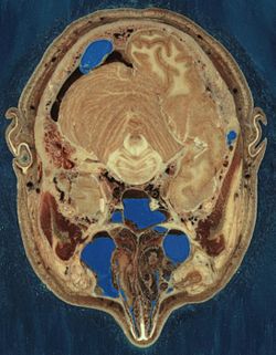
The male cadaver was encased and frozen in a gelatin and water mixture in order to stabilize the specimen for cutting. The specimen was then "cut" in the axial plane at 1-millimeter intervals. Each of the resulting 1,871 "slices" was photographed in both analog and digital, yielding 15 gigabytes of data. In 2000, the photos were rescanned at a higher resolution, yielding more than 65 gigabytes. The female cadaver was cut into slices at 0.33-millimeter intervals, resulting in some 40 gigabytes of data.

The term "cut" is a bit of a misnomer, yet it is used to describe the process of grinding away the top surface of a specimen at regular intervals. The term "slice", also a misnomer, refers to the revealed surface of the specimen to be photographed; the process of grinding the surface away is entirely destructive to the specimen and leaves no usable or preservable "slice" of the cadaver.

The data are supplemented by axial sections of the whole body obtained by computed tomography, axial sections of the head and neck obtained by magnetic resonance imaging (MRI), and coronal sections of the rest of the body also obtained by MRI.

The scanning, slicing, and photographing took place at the University of Colorado Anschutz Medical Campus, where additional cutting of anatomical specimens continues to take place.

Donors

The male cadaver is from Joseph Paul Jernigan, a 39-year-old Texas murderer who was executed by lethal injection on August 5, 1993. At the prompting of a prison chaplain he had agreed to donate his body for scientific research or medical use, without knowing about the Visible Human Project. Some people have voiced ethical concerns over this. One of the most notable statements came from the University of Vienna, which demanded that the images be withdrawn with reference to the point that the medical profession should have no association with executions, and that the donor's informed consent could be scrutinised. [4]

The 59-year-old female donor remains anonymous. In the press she has been described as a Maryland housewife who died from a heart attack and whose husband requested that she be part of the project.

In 2000, Susan Potter—a cancer patient and a disability rights activist—became the third body donor to the project, spending the 15 following years until her death by pneumonia in 2015 as an outspoken advocate for medical education and a mentor of medical students at the University of Colorado. [5] For nearly two decades, [6] National Geographic documented the story of Susan Potter and Dr. Victor M. Spitzer, the director of the Center for Human Simulation at the University of Colorado Anschutz Medical Campus who led the NIH-funded project, releasing a video documentary in 2018. [7] By the time Potter met Spitzer in 2000, she had gone through 26 surgeries and had been diagnosed with melanoma, breast cancer and diabetes: [8] her participation in the Visible Human Project marked a significant departure from the original goals of the project, which up until then had only focused on the dissection and imaging of healthy bodies. [5]

Problems with the data sets

Freezing caused the brain of the man to be slightly swollen, and his middle ear ossicles were lost during preparation of the slices. Nerves are hard to make out since they have almost the same color as fat, but many have nevertheless been identified. Small blood vessels were collapsed by the freezing process. Tendons are difficult to cut cleanly, and they occasionally smear across the slice surfaces.

The male has only one testicle, is missing his appendix, and has tissue deterioration at the site of lethal injection. Also visible are tissue damage to the dorsum of each forearm by formalin injection and damage to the right sartorius from opening the right femoral vein for drainage. The male was also not "cut" while in standard anatomical position, so the cuts through his arms are oblique. The female was missing 14 body parts, including nose cartilage. [9]

The reproductive organs of the woman are not representative of those of a young woman. The specimen contains several pathologies, including cardiovascular disease and diverticulitis.

Discoveries

By studying the data set, researchers at Columbia University found several errors in anatomy textbooks related to males, regarding the shape of a muscle in the pelvic region and the location of the urinary bladder and prostate. [10]

License

The data may be bought on tape or downloaded free of charge; Currently no license agreement is required to access or download the dataset, however general terms and conditions [11] apply requiring acknowledgement of the National Library of Medicine with any use. Prior to 2019, one had to specify the intended use and sign a license agreement that allows NLM to use and modify the resulting application. NLM can cancel the agreement at any time, at which point the user has to erase the data files.


Applications using the data

Various projects to make the raw data more useful for educational purposes are under way. It is necessary to build a three-dimensional virtual model of the body where the organs are labeled, may be removed selectively and viewed from all sides, and ideally are even animated. Two commercial software products accomplish the majority of these goals, the VH Dissector from Touch of Life Technologies and "Voxel-Man 3D-Navigator" from the University of Hamburg [12] NLM itself has started an open source project, the Insight Toolkit, whose aim is to automatically deduce organ boundaries from the data.

The data were used for Alexander Tsiaras's book and CD-ROM Body Voyage, which features a three-dimensional tour through the body. [13]

A "Virtual Radiography" application creates Digitally Reconstructed Radiographs and "virtual surgery", where endoscopic procedures or balloon angioplasty are simulated: the surgeon can view the progress of the instrument on a screen and receives realistic tactile feedback according to what kind of tissue the instrument would currently be touching.

Several other educational applications utilized form the visible human project include: multiple interactive anatomy computer software programs (Primal Pictures/Anatomy.tv, Anatomage), multimodality image restoration for hospital patients, body system relationships, and volumetric data. [14] [15]

The male data set was used in "Project 12:31", a series of photographic light paintings by Croix Gagnon and Frank Schott, and is the male Caucasian cadaver on the Anatomage Table 6.0 application.

See also

Related Research Articles

<span class="mw-page-title-main">History of anatomy</span> Aspect of history

The history of anatomy extends from the earliest examinations of sacrificial victims to the sophisticated analyses of the body performed by modern anatomists and scientists. Written descriptions of human organs and parts can be traced back thousands of years to ancient Egyptian papyri, where attention to the body was necessitated by their highly elaborate burial practices.

<span class="mw-page-title-main">Autopsy</span> Medical examination of a corpse

An autopsy is a surgical procedure that consists of a thorough examination of a corpse by dissection to determine the cause, mode, and manner of death; or the exam may be performed to evaluate any disease or injury that may be present for research or educational purposes. For animals, the term necropsy is generally reserved.

<span class="mw-page-title-main">Dissection</span> Cutting procedure used in anatomy

Dissection is the dismembering of the body of a deceased animal or plant to study its anatomical structure. Autopsy is used in pathology and forensic medicine to determine the cause of death in humans. Less extensive dissection of plants and smaller animals preserved in a formaldehyde solution is typically carried out or demonstrated in biology and natural science classes in middle school and high school, while extensive dissections of cadavers of adults and children, both fresh and preserved are carried out by medical students in medical schools as a part of the teaching in subjects such as anatomy, pathology and forensic medicine. Consequently, dissection is typically conducted in a morgue or in an anatomy lab.

<span class="mw-page-title-main">3D scanning</span> Scanning of an object or environment to collect data on its shape

3D scanning is the process of analyzing a real-world object or environment to collect three dimensional data of its shape and possibly its appearance. The collected data can then be used to construct digital 3D models.

<span class="mw-page-title-main">Mimics</span>

Materialise Mimics is an image processing software for 3D design and modeling, developed by Materialise NV, a Belgian company specialized in additive manufacturing software and technology for medical, dental and additive manufacturing industries. Materialise Mimics is used to create 3D surface models from stacks of 2D image data. These 3D models can then be used for a variety of engineering applications. Mimics is an acronym for Materialise Interactive Medical Image Control System. It is developed in an ISO environment with CE and FDA 510k premarket clearance. Materialise Mimics is commercially available as part of the Materialise Mimics Innovation Suite, which also contains Materialise3-matic, a design and meshing software for anatomical data. The current version is 24.0(released in 2021), and it supports Windows 10, Windows 7, Vista and XP in x64.

A tissue bank is an establishment that collects and recovers human cadaver tissue for the purposes of medical research, education and allograft transplantation. A tissue bank may also refer to a location where biomedical tissue is stored under cryogenic conditions and is generally used in a more clinical sense.

<span class="mw-page-title-main">Cadaver</span> Dead body used for study or instruction

A cadaver or corpse is a dead human body. Cadavers are used by medical students, physicians and other scientists to study anatomy, identify disease sites, determine causes of death, and provide tissue to repair a defect in a living human being. Students in medical school study and dissect cadavers as a part of their education. Others who study cadavers include archaeologists and arts students. In addition, a cadaver may be used in the development and evaluation of surgical instruments.

Virtopsy is a virtual alternative to a traditional autopsy, conducted with scanning and imaging technology. The name is a portmanteau of "virtual" and "autopsy" and is a trademark registered to Richard Dirnhofer (de), the former head of the Institute of Forensic Medicine of the University of Bern, Switzerland.

Professor Paul Gerard McMenamin is an Australian academic and researcher specialising in the structure and immunology of the eye.

3D Indiana is a commercial Educational software for teaching and research on the human anatomy. The name is an acronym for Three-Dimensional Interactive Digital Anatomy. This software is based on the principles of volumetric anatomy which uses three intersecting coordinate planes to locate the organs of the human body based on mathematical calculations. This is in contrast to the traditional method of describing the location of the organs in relation to one another. The Gall bladder is traditionally described thus: 'It is situated on the right side of the body closely in contact with the inferior surface of the liver, along the right edge of the quadrate lobe in a shallow fossa extending from the right edge of the porta hepatis to the inferior lobe of the liver'.

<span class="mw-page-title-main">Voxel-Man</span>

VOXEL-MAN is the name of a set of a computer programs for creation and visualization of three-dimensional digital models of the human body derived from cross-sectional images of computer tomography, magnetic resonance tomography or photography. It was developed at the University Medical Center Hamburg-Eppendorf. Applications include diagnostic imaging, digital anatomical atlases and surgery simulators. The 3D interactive atlases of anatomy and radiology for brain/skull and inner organs are available for free download. The name Voxel-Man is derived from the term voxel, the elementary cuboid component of a digital representation of a three-dimensional object. Occasionally the name Voxel-Man is also used as a general term for a digital representation of the human body.

<span class="mw-page-title-main">GIMIAS</span>

GIMIAS is a workflow-oriented environment focused on biomedical image computing and simulation. The open-source framework is extensible through plug-ins and is focused on building research and clinical software prototypes. Gimias has been used to develop clinical prototypes in the fields of cardiac imaging and simulation, angiography imaging and simulation, and neurology

<span class="mw-page-title-main">3D modeling</span> Form of computer-aided engineering

In 3D computer graphics, 3D modeling is the process of developing a mathematical coordinate-based representation of any surface of an object in three dimensions via specialized software by manipulating edges, vertices, and polygons in a simulated 3D space.

<span class="mw-page-title-main">Computer-generated imagery</span> Application of computer graphics to create or contribute to images

Computer-generated imagery (CGI) is a specific-technology or application of computer graphics for creating or improving images in art, printed media, simulators, videos and video games. These images are either static or dynamic. CGI both refers to 2D computer graphics and 3D computer graphics with the purpose of designing characters, virtual worlds, or scenes and special effects. The application of CGI for creating/improving animations is called computer animation, or CGI animation.

Computational human phantoms are models of the human body used in computerized analysis. Since the 1960s, the radiological science community has developed and applied these models for ionizing radiation dosimetry studies. These models have become increasingly accurate with respect to the internal structure of the human body.

A medical animation is a short educational film, usually based around a physiological or surgical topic, that is rendered using 3D computer graphics. While it may be intended for an array of audiences, the medical animation is most commonly utilized as an instructional tool for medical professionals or their patients.

Alexander Tsiaras is an American photographer, entrepreneur, technology innovator, and journalist whose work has appeared on the cover of over 150 magazines. He is the founder, CEO, and editor-in-chief of TheVisualMD and StoryMD.

Cultural property imaging is a necessary part of long term preservation of cultural heritage. While the physical conditions of objects will change over time, imaging serves as a way to document and represent heritage in a moment in time of the life of the item. Different methods of imaging produce results that are applicable in various circumstances. Not every method is appropriate for every object, and not every object needs to be imaged by multiple methods. In addition to preservation and conservation-related concerns, imaging can also serve to enhance research and study of cultural heritage.

<span class="mw-page-title-main">Susan Potter</span> German cancer survivor, disability rights activist and body donor (1927–2015)

Susan Christina Potter was a cancer survivor, a disability rights activist and a body donor for the Visible Human Project. During the 15 years between signing on to the project in 2000 and her death by pneumonia in 2015 at the age of 87, Potter became a public figure and an outspoken advocate for medical education, mentoring medical students at the University of Colorado.

The Visible Embryo Project (VEP) is a multi-institutional, multidisciplinary research project originally created in the early 1990s as a collaboration between the Developmental Anatomy Center at the National Museum of Health and Medicine and the Biomedical Visualization Laboratory (BVL) at the University of Illinois at Chicago, "to develop software strategies for the development of distributed biostructural databases using cutting-edge technologies for high-performance computing and communications (HPCC), and to implement these tools in the creation of a large-scale digital archive of multidimensional data on normal and abnormal human development." This project related to BVL's other research in the areas of health informatics, educational multimedia, and biomedical imaging science. Over the following decades, the list of VEP collaborators grew to include over a dozen universities, national laboratories, and companies around the world.

References

  1. Waldby, Catherine (September 2003). The Visible Human Project: Informatic Bodies and Posthuman Medicine. Routledge. p. 4. ISBN   978-0-203-36063-7 . Retrieved 2 February 2017.
  2. Burke, L., & Weill, B. (2009). Chapter 10. Information technology for the health professions (3rd ed., p. 212). Upper Saddle River, N.J.: Pearson Prentice Hall.
  3. "The Visible Humans | NIH MedlinePlus the Magazine". medlineplus.gov. Retrieved 2019-01-18.
  4. Roeggla G., U. Landesmann and M. Roeggla: Ethics of executed person on Internet. [Letter]. Lancet. 28. January 1995; 345(0):260.
  5. 1 2 Newman, Cathy (December 13, 2018). "She gave her body to science. Her corpse became immortal". National Geographic. Retrieved 2018-12-15.
  6. Pescovitz, David (December 14, 2018). "The incredible story of Susan Potter, the "immortal corpse"". Boing Boing. Retrieved 2018-12-15.
  7. "She donated her body to science, and now she'll live forever", National Geographic, 2018, retrieved 2018-12-15
  8. Rivas, Anthony (2018-12-14). "Why one woman agreed to become an 'Immortal Corpse' for science". ABC News. Retrieved 2018-12-15.
  9. Hamzelou, Jessica. "Virtual human built from more than 5000 slices of a real woman" . Retrieved 2016-09-26.
  10. Venuti, J.; Imielinska, C.; Molholt, P. (2004). "New Views of Male Pelvic Anatomy: Role of Computer Generated 3D Images". Clinical Anatomy. 17: 261–271.
  11. https://www.nlm.nih.gov/databases/download/terms_and_conditions.html
  12. Hoehne K. H. et al.: VOXEL-MAN 3D-Navigator: Inner Organs. Regional, Systemic and Radiological Anatomy. Springer Electronic Media, New York, 2003 (DVD-ROM, ISBN   978-3-540-40069-1).
  13. Tsiaras A.: Body Voyage: A Three-Dimensional Tour of a Real Human Body. Warner Books, New York, 1997 ( ISBN   0-446-52009-8).
  14. "The Visible Human Project Projects Based on the Visible Human Data Set Applications for viewing images". U.S. National Library of Medicine. Retrieved 26 September 2016.
  15. "Projects Based on the Visible Human Data Set: Products". U.S. National Library of Medicine. Retrieved 30 September 2022.