Submasseteric space

Last updated
Submasseteric space
Masseter muscle animation small.gif
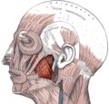
Location of the masseter muscles. The submasseteric space is between the masseter and the mandible.
Masticator space.png
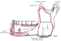
The four compartents of the right masticator space. A Temporalis muscle, B Masseter muscle, C Lateral pterygoid muscle, D Medial ptaerygoid muscle, E Superficial temporal space, F Deep temporal space, G Submasseteric space, H Pterygomandibular space, I Approximate location of infratemporal space
Anatomical terminology

The submasseterric space (also termed the masseteric space) is a fascial space of the head and neck (sometimes also termed fascial spaces or tissue spaces). It is a potential space in the face over the angle of the jaw, and is paired on each side. It is located between the lateral aspect of the mandible and the medial aspect of the masseter muscle and its investing fascia. The term is derived from sub- meaning "under" in Latin and masseteric which refers to the masseter muscle. The submasseteric space is one of the four compartments of the masticator space. [1] Sometimes the submasseteric space is described as a series of spaces, created because the masseter muscle has multiple insertions that cover most of the lateral surface of the ramus of the mandible.

Contents

Structure

Anatomic boundaries

The boundaries of each submasseteric space are:

Communications

The communications of each submasseteric space are:

Contents

In health, the space contains:

Clinical significance

Submasseteric abscesses are relatively rare, and may be confused with a parotid abscess or parotitis. [2] They tend to be chronic. [3] The submasseteric space may be involved by infections that spread from the buccal space. [1] Sometimes mandibular fractures in the region of the angle of the mandible may cause an infection of the submasseteric space. [1] The signs and symptoms of a submasseteric abscess may include marked trismus (i.e. difficulty opening the mouth, since the masseter elevates the mandible and it becomes restricted) and swelling in the region of the masseter muscle. [1] The treatment of a submasseteric space infection is usually by surgical incision and drainage, and the incision is placed intra-orally (inside the mouth) or both intra and extra-orally if other parts of the masticator space are involved.

Odontogenic infections

The submasseteric space is sometimes involved by the spread of odontogenic infections, such as a pericoronal abscess associated with an impacted mandibular third molar (lower wisdom tooth) when the apices of the tooth lie very close to or within the space. [4]

Additional images

Related Research Articles

<span class="mw-page-title-main">Temporomandibular joint</span> Joints connecting the jawbone to the skull

In anatomy, the temporomandibular joints (TMJ) are the two joints connecting the jawbone to the skull. It is a bilateral synovial articulation between the temporal bone of the skull above and the mandible below; it is from these bones that its name is derived. This joint is unique in that it is a bilateral joint that functions as one unit. Since the TMJ is connected to the mandible, the right and left joints must function together and therefore are not independent of each other.

<span class="mw-page-title-main">Mandibular nerve</span> Branch of the trigeminal nerve responsible for the lower face and jaw

In neuroanatomy, the mandibular nerve (V3) is the largest of the three divisions of the trigeminal nerve, the fifth cranial nerve (CN V). Unlike the other divisions of the trigeminal nerve (ophthalmic nerve, maxillary nerve) which contain only afferent fibers, the mandibular nerve contains both afferent and efferent fibers. These nerve fibers innervate structures of the lower jaw and face, such as the tongue, lower lip, and chin. The mandibular nerve also innervates the muscles of mastication.

<span class="mw-page-title-main">Parotid gland</span> Major salivary gland in many animals

The parotid gland is a major salivary gland in many animals. In humans, the two parotid glands are present on either side of the mouth and in front of both ears. They are the largest of the salivary glands. Each parotid is wrapped around the mandibular ramus, and secretes serous saliva through the parotid duct into the mouth, to facilitate mastication and swallowing and to begin the digestion of starches. There are also two other types of salivary glands; they are submandibular and sublingual glands. Sometimes accessory parotid glands are found close to the main parotid glands.

<span class="mw-page-title-main">Masseter muscle</span> One of the masticatory muscles in mammals

In human anatomy, the masseter is one of the muscles of mastication. Found only in mammals, it is particularly powerful in herbivores to facilitate chewing of plant matter. The most obvious muscle of mastication is the masseter muscle, since it is the most superficial and one of the strongest.

<span class="mw-page-title-main">Medial pterygoid muscle</span> Muscle involved in chewing

The medial pterygoid muscle, is a thick, quadrilateral muscle of the face. It is supplied by the mandibular branch of the trigeminal nerve (V). It is important in mastication (chewing).

<span class="mw-page-title-main">Buccal nerve</span> Nerve in the human face

The buccal nerve is a nerve in the face. It is a branch of the mandibular nerve and transmits sensory information from skin over the buccal membrane and from the second and third molar teeth. Not to be confused with the buccal branch of the facial nerve which transmits motor information to the buccinator muscle.

<span class="mw-page-title-main">Maxillary artery</span>

The maxillary artery supplies deep structures of the face. It branches from the external carotid artery just deep to the neck of the mandible.

<span class="mw-page-title-main">Infratemporal fossa</span> Cavity that is part of the skull

The infratemporal fossa is an irregularly shaped cavity that is a part of the skull. It is situated below and medial to the zygomatic arch. It is not fully enclosed by bone in all directions. It contains superficial muscles, including the lower part of the temporalis muscle, the lateral pterygoid muscle, and the medial pterygoid muscle. It also contains important blood vessels such as the middle meningeal artery, the pterygoid plexus, and the retromandibular vein, and nerves such as the mandibular nerve (CN V3) and its branches.

<span class="mw-page-title-main">Deep cervical fascia</span>

The deep cervical fascia lies under cover of the platysma, and invests the muscles of the neck; it also forms sheaths for the carotid vessels, and for the structures situated in front of the vertebral column. Its attachment to the hyoid bone prevents the formation of a dewlap.

<span class="mw-page-title-main">Buccal space</span>

The buccal space is a fascial space of the head and neck. It is a potential space in the cheek, and is paired on each side. The buccal space is superficial to the buccinator muscle and deep to the platysma muscle and the skin. The buccal space is part of the subcutaneous space, which is continuous from head to toe.

<span class="mw-page-title-main">Buccal fat pad</span>

The buccal fat pad is one of several encapsulated fat masses in the cheek. It is a deep fat pad located on either side of the face between the buccinator muscle and several more superficial muscles. The inferior portion of the buccal fat pad is contained within the buccal space. It should not be confused with the malar fat pad, which is directly below the skin of the cheek. It should also not be confused with jowl fat pads. It is implicated in the formation of hollow cheeks and the nasolabial fold, but not in the formation of jowls.

Inferior alveolar nerve block is a nerve block technique which induces anesthesia (numbness) in the areas of the mouth and face innervated by one of the inferior alveolar nerves which are paired on the left and right side. These areas are the skin and mucous membranes of the lower lip, the skin of the chin, the lower teeth and the labial gingiva of the anterior teeth, all unilaterally to the midline of the side on which the block is administered. However, depending on technique, the long buccal nerve may not be anesthetized by an IANB and therefore an area of buccal gingiva adjacent to the lower posterior teeth will retain normal sensation unless that nerve is anesthetized separately, via a (long) buccal nerve block. The inferior alveolar nerve is a branch of the mandibular nerve, the third division of the trigeminal nerve. This procedure attempts to anaesthetise the inferior alveolar nerve prior to it entering the mandibular foramen on the medial surface of the mandibular ramus.

<span class="mw-page-title-main">Parapharyngeal space</span>

The parapharyngeal space, is a potential space in the head and the neck. It has clinical importance in otolaryngology due to parapharyngeal space tumours and parapharyngeal abscess developing in this area. It is also a key anatomic landmark for localizing disease processes in the surrounding spaces of the neck; the direction of its displacement indirectly reflects the site of origin for masses or infection in adjacent areas, and consequently their appropriate differential diagnosis.

<span class="mw-page-title-main">Sublingual space</span>

The sublingual space is a fascial space of the head and neck. It is a potential space located below the mouth and above the mylohyoid muscle, and is part of the suprahyoid group of fascial spaces.

<span class="mw-page-title-main">Submandibular space</span>

The submandibular space is a fascial space of the head and neck. It is a potential space, and is paired on either side, located on the superficial surface of the mylohyoid muscle between the anterior and posterior bellies of the digastric muscle. The space corresponds to the anatomic region termed the submandibular triangle, part of the anterior triangle of the neck.

<span class="mw-page-title-main">Submental space</span>

The submental space is a fascial space of the head and neck. It is a potential space located between the mylohyoid muscle superiorly, the platysma muscle inferiorly, under the chin in the midline. The space coincides with the anatomic region termed the submental triangle, part of the anterior triangle of the neck.

Fascial spaces are potential spaces that exist between the fasciae and underlying organs and other tissues. In health, these spaces do not exist; they are only created by pathology, e.g. the spread of pus or cellulitis in an infection. The fascial spaces can also be opened during the dissection of a cadaver. The fascial spaces are different from the fasciae themselves, which are bands of connective tissue that surround structures, e.g. muscles. The opening of fascial spaces may be facilitated by pathogenic bacterial release of enzymes which cause tissue lysis. The spaces filled with loose areolar connective tissue may also be termed clefts. Other contents such as salivary glands, blood vessels, nerves and lymph nodes are dependent upon the location of the space. Those containing neurovascular tissue may also be termed compartments.

<span class="mw-page-title-main">Pterygomandibular space</span>

The pterygomandibular space is a fascial space of the head and neck. It is a potential space in the head and is paired on each side. It is located between the lateral pterygoid muscle and the medial surface of the ramus of the mandible. The pterygomandibular space is one of the four compartments of the masticator space.

<span class="mw-page-title-main">Canine space</span>

The canine space, is a fascial space of the head and neck. It is a thin potential space on the face, and is paired on either side. It is located between the levator anguli oris muscle inferiorly and the levator labii superioris muscle superiorly. The term is derived from the fact that the space is in the region of the canine fossa, and that infections originating from the maxillary canine tooth may spread to involve the space. Infra-orbital is derived from infra- meaning below and orbit which refers to the eye socket.

<span class="mw-page-title-main">Infratemporal space</span>

The Infratemporal space is a fascial space of the head and neck. It is a potential space in the side of the head, and is paired on either side. It is located posterior to the maxilla, between the lateral pterygoid plate of the sphenoid bone medially and by the base of skull superiorly. The term is derived from infra- meaning below and temporal which refers to the temporalis muscle.

References

  1. 1 2 3 4 5 6 7 8 9 10 Hupp JR, Ellis E, Tucker MR (2008). Contemporary oral and maxillofacial surgery (5th ed.). St. Louis, Mo.: Mosby Elsevier. pp. 317–333. ISBN   9780323049030.
  2. Rai, Ashutosh; Rajput, Ruchi; Khatua, RabindraK; Singh, Mangal (1 January 2011). "Submasseteric abscess: A rare head and neck abscess". Indian Journal of Dental Research. 22 (1): 166–8. doi: 10.4103/0970-9290.79990 . PMID   21525699.
  3. Jones, KC; Silver, J; Millar, WS; Mandel, L (Jun–Jul 2003). "Chronic submasseteric abscess: anatomic, radiologic, and pathologic features". American Journal of Neuroradiology. 24 (6): 1159–63. PMC   8149006 . PMID   12812946.
  4. Hargreaves KM, Cohen S (editors), Berman LH (web editor) (2010). Cohen's pathways of the pulp (10th ed.). St. Louis, Mo.: Mosby Elsevier. pp. 590–594. ISBN   978-0-323-06489-7.{{cite book}}: |last= has generic name (help)CS1 maint: multiple names: authors list (link)