Viral protein

Last updated
Diagram of how a virus capsid can be constructed using multiple copies of just two protein molecules Hexon.svg
Diagram of how a virus capsid can be constructed using multiple copies of just two protein molecules

The term viral protein refers to both the products of the genome of a virus and any host proteins incorporated into the viral particle. Viral proteins are grouped according to their functions, and groups of viral proteins include structural proteins, nonstructural proteins, regulatory proteins, and accessory proteins. [1] Viruses are non-living and do not have the means to reproduce on their own, instead depending on their host cell's machinery to do this. Thus, viruses do not code for most of the proteins required for their replication and the translation of their mRNA into viral proteins, but use proteins encoded by the host cell for this purpose. [2]

Contents

Viral structural proteins

Most viral structural proteins are components for the capsid and the envelope of the virus.

Capsid

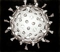
The genetic material of a virus is stored within a viral protein structure called the capsid. The capsid is a "shield" that protects the viral nucleic acids from getting degraded by host enzymes or other types of pesticides or pestilences. It also functions to attach the virion to its host, and enable the virion to penetrate the host cell membrane. Many copies of a single viral protein or a number of different viral proteins make up the capsid, and each of these viral proteins are coded for by one gene from the viral genome. The structure of the capsid allows the virus to use a small number of viral genes to make a large capsid. [3]

Several protomers, oligomeric (viral) protein subunits, combine to form capsomeres, and capsomeres come together to form the capsid. [1] Capsomeres can arrange into an icosahedral, helical, or complex capsid, but in many viruses, such as the herpes simplex virus, an icosahedral capsid is assembled. [2] Three asymmetric and nonidentical viral protein units make up each of the twenty identical triangular faces in the icosahedral capsid. [2]

Viral envelope

The capsid of some viruses are enclosed in a membrane called the viral envelope. In most cases, the viral envelope is obtained by the capsid from the host cell's plasma membrane when a virus leaves its host cell through a process called budding. [4] The viral envelope is made up of a lipid bilayer embedded with viral proteins, including viral glycoproteins. [1] These viral glycoproteins bind to specific receptors and coreceptors on the membrane of host cells, and they allow viruses to attach onto their target host cells. [1] Some of these glycoproteins include:

Viral glycoproteins play a critical role in virus-to-cell fusion. Virus-to-cell fusion is initiated when viral glycoproteins bind to cellular receptors. [5]

Viral membrane fusion proteins

The fusion of the viral envelope with the cellular membrane requires high energy to occur. Viral membrane fusion proteins act as catalysts to overcome this high energy barrier. [6] Following viral glycoprotein binding to cellular receptors, viral membrane fusion proteins undergo a change in structure conformation. [6] This change in conformation then facilitates the destabilization and fusion of the viral envelope with the cellular membrane by allowing fusion loops (FLs) or hydrophobic fusion peptides (FPs) on the viral envelope to interact with the cell membrane. [6] Most viral membrane fusion proteins would end up in a hairpin-like conformation after fusion, in which FLs/FPs and the transmembrane domain are all on the same side of the protein. [6]

Viral glycoproteins and their three-dimensional structures, before and after fusion, have allowed a wide range of structural conformations to be discovered. [6] Viral membrane fusion proteins have been grouped into four different classes, and each class is identified by characteristic structural conformations:

  • Class I: Post-fusion conformation has a distinct central coiled-coil structure composed of signature trimer of α-helical hairpins. An example of a Class I viral fusion protein is the HIV glycoprotein, gp41. [6]
  • Class II: Protein lacks the central coiled-coil structure. Contains a characteristic elongated β- sheet ectodomain structure that refolds to give a trimer of hairpins. Examples of class II viral fusion proteins include the dengue virus E protein, and the west nile virus E protein. [5] [6]
  • Class III: Structural conformation is a combination of features from Class I and Class II viral membrane fusion proteins. An example of a Class III viral fusion protein is the rabies virus glycoprotein, G. [6]
  • Class IV: Class IV viral fusion proteins are fusion-associated small transmembrane (FAST) proteins. They do not form trimers of hairpins or hairpin structures themselves, and they are the smallest known viral fusion proteins. FAST proteins are coded for by members of the nonenveloped reoviridae family of viruses. [6]

Viral nonstructural proteins

Viral nonstructural proteins are proteins coded for by the genome of the virus and are expressed in infected cells. [1] However, these proteins are not assembled in the virion. [1] During the replication of viruses, some viral nonstructural proteins carry out important functions that affect the replication process itself. [1] Similarly, during the assembly of viruses, some of these proteins also carry out important functions that affect the assembly process. [1] Some of these viral nonstructural protein functions are replicon formation, immunomodulation, and transactivation of viral structural protein encoding genes. [1]

Replicon formation

Viral nonstructural proteins interact with host cell proteins to form the replicon, otherwise known as the replication complex. [1] In the hepatitis C virus, viral nonstructural proteins interact with cellular vesicle membrane transport protein, hVAP-33, to assemble the replicon. [1] Viral nonstructural 4b (NS4B) protein alters the host cell's membrane and starts the formation process of the replication complex. [1] [7] Other viral nonstructural proteins such as NS5A, NS5B, and NS3, are also recruited to the complex, and NS4B interacts with them and binds to viral RNA. [1] [7]

Immunomodulation

The immune response of a host to an infected cell can be adjusted through the immunomodulatory properties of viral nonstructural proteins. [1] Many species of large DNA viruses encode proteins which subvert host immune response, allowing proliferation of the virus. [8] Such proteins hold potential in developing new bio-pharmaceutical treatments for inflammatory disease in humans, as the proteins have been proven to subvert inflammatory immune mediators. [9] Viral nonstructural protein NS1 in the West Nile virus prevents complement activation through its binding to a complement control protein, factor H. [1] As a result, complement recognition of infected cells is reduced, and infected cells remain unharmed by the host's immune system. [1] [10]

Viral regulatory and accessory proteins

Viral regulatory and accessory proteins have many functions. These viral proteins control and influence viral gene expressions in the viral genome, including viral structural gene transcription rates. [1] Viral regulatory and accessory proteins also influence and adjust cellular functions of the host cell, such as the regulation of genes, and apoptosis. [1]

In DNA viruses and retroviruses, viral regulatory proteins can enhance viral gene transcription, likewise, these proteins can also enhance host cellular gene transcription too. [11]

Viral accessory proteins, also known as auxiliary proteins, are coded for by the genome of retroviruses. [12] Most viral accessory proteins only carry out their functions in specific types of cells. [12] Also, they do not have much influence on the replication of the virus. [12] However, in some instances, maintaining the replication of viruses would require the help (and function) of viral accessory proteins. [12]

Endogenous retroviral proteins

Syncytin is an endogenous retrovirus protein that has been captured in the mammalian genome to allow membrane fusion in placental morphogenesis. [13]

Related Research Articles

<span class="mw-page-title-main">Capsid</span> Protein shell of a virus

A capsid is the protein shell of a virus, enclosing its genetic material. It consists of several oligomeric (repeating) structural subunits made of protein called protomers. The observable 3-dimensional morphological subunits, which may or may not correspond to individual proteins, are called capsomeres. The proteins making up the capsid are called capsid proteins or viral coat proteins (VCP). The capsid and inner genome is called the nucleocapsid.

<span class="mw-page-title-main">Retrovirus</span> Family of viruses

A retrovirus is a type of virus that inserts a DNA copy of its RNA genome into the DNA of a host cell that it invades, thus changing the genome of that cell. After invading a host cell's cytoplasm, the virus uses its own reverse transcriptase enzyme to produce DNA from its RNA genome, the reverse of the usual pattern, thus retro (backwards). The new DNA is then incorporated into the host cell genome by an integrase enzyme, at which point the retroviral DNA is referred to as a provirus. The host cell then treats the viral DNA as part of its own genome, transcribing and translating the viral genes along with the cell's own genes, producing the proteins required to assemble new copies of the virus. Many retroviruses cause serious diseases in humans, other mammals, and birds.

<span class="mw-page-title-main">Rubella virus</span> Species of virus

Rubella virus (RuV) is the pathogenic agent of the disease rubella, transmitted only between humans via the respiratory route, and is the main cause of congenital rubella syndrome when infection occurs during the first weeks of pregnancy.

<i>Alphavirus</i> Genus of viruses

Alphavirus is a genus of RNA viruses, the sole genus in the Togaviridae family. Alphaviruses belong to group IV of the Baltimore classification of viruses, with a positive-sense, single-stranded RNA genome. There are 32 alphaviruses, which infect various vertebrates such as humans, rodents, fish, birds, and larger mammals such as horses, as well as invertebrates. Alphaviruses that could infect both vertebrates and arthropods are referred dual-host alphaviruses, while insect-specific alphaviruses such as Eilat virus and Yada yada virus are restricted to their competent arthropod vector. Transmission between species and individuals occurs mainly via mosquitoes, making the alphaviruses a member of the collection of arboviruses – or arthropod-borne viruses. Alphavirus particles are enveloped, have a 70 nm diameter, tend to be spherical, and have a 40 nm isometric nucleocapsid.

The genome and proteins of HIV (human immunodeficiency virus) have been the subject of extensive research since the discovery of the virus in 1983. "In the search for the causative agent, it was initially believed that the virus was a form of the Human T-cell leukemia virus (HTLV), which was known at the time to affect the human immune system and cause certain leukemias. However, researchers at the Pasteur Institute in Paris isolated a previously unknown and genetically distinct retrovirus in patients with AIDS which was later named HIV." Each virion comprises a viral envelope and associated matrix enclosing a capsid, which itself encloses two copies of the single-stranded RNA genome and several enzymes. The discovery of the virus itself occurred two years following the report of the first major cases of AIDS-associated illnesses.

Rous sarcoma virus (RSV) is a retrovirus and is the first oncovirus to have been described. It causes sarcoma in chickens.

The murine leukemia viruses are retroviruses named for their ability to cause cancer in murine (mouse) hosts. Some MLVs may infect other vertebrates. MLVs include both exogenous and endogenous viruses. Replicating MLVs have a positive sense, single-stranded RNA (ssRNA) genome that replicates through a DNA intermediate via the process of reverse transcription.

<span class="mw-page-title-main">Viral envelope</span> Outermost layer of many types of the infectious agent

A viral envelope is the outermost layer of many types of viruses. It protects the genetic material in their life cycle when traveling between host cells. Not all viruses have envelopes. A viral envelope protein or E protein is a protein in the envelope, which may be acquired by the capsid from an infected host cell.

<span class="mw-page-title-main">Herpes simplex virus</span> Species of virus

Herpes simplex virus1 and 2, also known by their taxonomic names Human alphaherpesvirus 1 and Human alphaherpesvirus 2, are two members of the human Herpesviridae family, a set of viruses that produce viral infections in the majority of humans. Both HSV-1 and HSV-2 are very common and contagious. They can be spread when an infected person begins shedding the virus.

Simian foamy virus (SFV) is a species of the genus Spumavirus that belongs to the family of Retroviridae. It has been identified in a wide variety of primates, including prosimians, New World and Old World monkeys, as well as apes, and each species has been shown to harbor a unique (species-specific) strain of SFV, including African green monkeys, baboons, macaques, and chimpanzees. As it is related to the more well-known retrovirus human immunodeficiency virus (HIV), its discovery in primates has led to some speculation that HIV may have been spread to the human species in Africa through contact with blood from apes, monkeys, and other primates, most likely through bushmeat-hunting practices.

<span class="mw-page-title-main">Viral entry</span> Earliest stage of infection in the viral life cycle

Viral entry is the earliest stage of infection in the viral life cycle, as the virus comes into contact with the host cell and introduces viral material into the cell. The major steps involved in viral entry are shown below. Despite the variation among viruses, there are several shared generalities concerning viral entry.

Env is a viral gene that encodes the protein forming the viral envelope. The expression of the env gene enables retroviruses to target and attach to specific cell types, and to infiltrate the target cell membrane.

<span class="mw-page-title-main">Herpesvirus glycoprotein B</span> Viral glycoprotein

Herpesvirus glycoprotein B is a viral glycoprotein that is involved in the viral cell entry of Herpes simplex virus (HSV). Herpesviruses have a lipid bilayer, called the envelope, which contains twelve surface glycoproteins. For infectivity to be attained, the double stranded DNA genome of HSV must enter the host cell through means of fusion of its envelope with the cellular membrane or via endocytosis. Other viral glycoproteins involved in the process of viral cell entry include gC, gB, gD, gH, and gL, but only gC, gB, gD, and gH are required for the fusion of the HSV's envelope with the cellular membrane. It can be noted that all herpesviruses have glycoproteins gB, gH, and gL.

Adenovirus genomes are linear, non-segmented double-stranded (ds) DNA molecules that are typically 26-46 Kbp long, containing 23-46 protein-coding genes. The example used for the following description is Human adenovirus E, a mastadenovirus with a 36 Kbp genome containing 38 protein-coding genes. While the precise number and identity of genes varies among adenoviruses, the basic principles of genome organization and the functions of most of the genes described in this article are shared among all adenoviruses.

<i>Pneumoviridae</i> Family of viruses

Pneumoviridae is a family of negative-strand RNA viruses in the order Mononegavirales. Humans, cattle, and rodents serve as natural hosts. Respiratory tract infections are associated with member viruses such as human respiratory syncytial virus. There are five species in the family which are divided between the genera Metapneumovirus and Orthopneumovirus. The family used to be considered as a sub-family of Paramyxoviridae, but has been reclassified as of 2016.

<i>Middelburg virus</i> Species of virus

Middelburg virus (MIDV) is an alphavirus of the Old World Group that has likely endemic and zoonotic potential. It is of the viral family Togaviridae. It was isolated from mosquitos in 1957 in South Africa, MDIV antigens have now been found in livestock, horses, and humans.

Batravirus ranidallo1, also known as Ranid herpesvirus 1 (RaHV-1), is a double-stranded DNA virus within the order Herpesvirales. The virus was initially observed within renal tumors in 1934 by Baldwin Lucké, and more recently has become identifiable through the use of PCR in samples isolated from frog tumors. RaHV-1 causes renal tumors within the northern leopard frog, Rana pipiens. The virus has not yet been isolated in vitro within cell lines, meaning that while its existence and symptoms are fairly evident, its methods of transmission, cell infection, and reproduction are largely unknown.

<i>Sepik virus</i> Mosquito transmitted virus endemic to Papua New Guinea

Sepik virus (SEPV) is an arthropod-borne virus (arbovirus) of the genus Flavivirus and family Flaviviridae. Flaviviridae is one of the most well characterized viral families, as it contains many well-known viruses that cause diseases that have become very prevalent in the world, like Dengue virus. The genus Flavivirus is one of the largest viral genera and encompasses over 50 viral species, including tick and mosquito borne viruses like Yellow fever virus and West Nile virus. Sepik virus is much less well known and has not been as well-classified as other viruses because it has not been known of for very long. Sepik virus was first isolated in 1966 from the mosquito Mansoniaseptempunctata, and it derives its name from the Sepik River area in Papua New Guinea, where it was first found. The geographic range of Sepik virus is limited to Papua New Guinea, due to its isolation.

<i>Woolly monkey hepatitis B virus</i> Species of virus

The woolly monkey hepatitis B virus (WMHBV) is a viral species of the Orthohepadnavirus genus of the Hepadnaviridae family. Its natural host is the woolly monkey (Lagothrix), an inhabitant of South America categorized as a New World primate. WMHBV, like other hepatitis viruses, infects the hepatocytes, or liver cells, of its host organism. It can cause hepatitis, liver necrosis, cirrhosis, and hepatocellular carcinoma. Because nearly all species of Lagothrix are threatened or endangered, researching and developing a vaccine and/or treatment for WMHBV is important for the protection of the whole woolly monkey genus.

Rio Negro virus is an alphavirus that was first isolated in Argentina in 1980. The virus was first called Ag80-663 but was renamed to Rio Negro virus in 2005. It is a former member of the Venezuelan equine encephalitis complex (VEEC), which are a group of alphaviruses in the Americas that have the potential to emerge and cause disease. Río Negro virus was recently reclassified as a distinct species. Closely related viruses include Mucambo virus and Everglades virus.

References

  1. 1 2 3 4 5 6 7 8 9 10 11 12 13 14 15 16 17 18 19 Vladimir N. Uversky; Sonia Longhi (2011). Flexible Viruses. Wiley. p. 4. ISBN   978-1-118-13554-9.
  2. 1 2 3 Joan L. Slonczewski; ‎John W. Foster (2013). Microbiology: An Evolving Science. Third Edition. W. W. Norton & Company. pp. 192–195. ISBN   978-0-393-12367-8.
  3. Lodish, Harvey; Berk, Arnold; Zipursky, S. Lawrence; Matsudaira, Paul; Baltimore, David; Darnell, James (2000-01-01). Molecular Cell Biology: Viruses: Structure, Function, and Uses . Retrieved 9 April 2016.
  4. Pornillos, Owen; Garrus, Jennifer E; Sundquist, Wesley I (2002-12-01). "Mechanisms of enveloped RNA virus budding". Trends in Cell Biology. 12 (12): 569–579. doi:10.1016/S0962-8924(02)02402-9. PMID   12495845.
  5. 1 2 White, Judith M.; Delos, Sue E.; Brecher, Matthew; Schornberg, Kathryn (2008-01-01). "Structures and Mechanisms of Viral Membrane Fusion Proteins". Critical Reviews in Biochemistry and Molecular Biology. 43 (3): 189–219. doi:10.1080/10409230802058320. ISSN   1040-9238. PMC   2649671 . PMID   18568847.
  6. 1 2 3 4 5 6 7 8 9 Podbilewicz, Benjamin (2014). "Virus and Cell Fusion Mechanisms". Annual Review of Cell and Developmental Biology. 30 (1): 111–139. doi:10.1146/annurev-cellbio-101512-122422. PMID   25000995.
  7. 1 2 Gouttenoire, Jérôme; Penin, François; Moradpour, Darius (2010-03-01). "Hepatitis C virus nonstructural protein 4B: a journey into unexplored territory". Reviews in Medical Virology. 20 (2): 117–129. doi:10.1002/rmv.640. ISSN   1099-1654. PMID   20069613. S2CID   38778113.
  8. Engel, P; Angulo, A (2012). "Viral Immunomodulatory Proteins: Usurping Host Genes as a Survival Strategy". Self and Nonself. Advances in Experimental Medicine and Biology. Vol. 738. pp. 256–278. doi:10.1007/978-1-4614-1680-7_15. ISBN   978-1-4614-1679-1. PMID   22399384.
  9. Lucas, A; McFadden, G (2004). "Secreted immunomodulatory viral proteins as novel biotherapeutics". J Immunol. 173 (8): 4765–74. doi: 10.4049/jimmunol.173.8.4765 . PMID   15470015.
  10. Chung, Kyung Min; Liszewski, M. Kathryn; Nybakken, Grant; Davis, Alan E.; Townsend, R. Reid; Fremont, Daved H.; Atkinson, John P.; Diamond, Michael S. (2006-12-12). "West Nile virus nonstructural protein NS1 inhibits complement activation by binding the regulatory protein factor H". Proceedings of the National Academy of Sciences of the United States of America. 103 (50): 19111–19116. doi: 10.1073/pnas.0605668103 . ISSN   0027-8424. PMC   1664712 . PMID   17132743.
  11. Flint, Jane; Shenk, Thomas (1997). "Viral Transactivating Proteins". Annual Review of Genetics. 31 (1): 177–212. doi:10.1146/annurev.genet.31.1.177. PMID   9442894.
  12. 1 2 3 4 Coffin, John M.; Hughes, Stephen H.; Varmus, Harold E. (1997-01-01). Retroviruses: Accessory Proteins and Assembly. Cold Spring Harbor Laboratory Press. Retrieved 13 April 2016.
  13. Mi S, Lee X, Li X, Veldman GM, Finnerty H, Racie L, LaVallie E, Tang XY, Edouard P, Howes S, Keith JC Jr, McCoy JM (2000). "Syncytin is a captive retroviral envelope protein involved in human placental morphogenesis". Nature. 403 (6771): 785–9. Bibcode:2000Natur.403..785M. doi:10.1038/35001608. PMID   10693809. S2CID   4367889.