Vpu protein

Last updated
Vpu
PDB 1pje EBI.jpg
structure of the channel-forming trans-membrane domain of virus protein "u" (vpu) from HIV-1
Identifiers
SymbolVpu
Pfam PF00558
InterPro IPR008187
SCOP2 1vpu / SCOPe / SUPFAM
TCDB 1.A.40
OPM superfamily 262
OPM protein 2k7y
Available protein structures:
Pfam   structures / ECOD  
PDB RCSB PDB; PDBe; PDBj
PDBsum structure summary
Viral Protein Unique
Identifiers
Organism HIV-1
Symbolvpu
Entrez 155945
RefSeq (Prot) NP_057855.1
UniProt P05919
Other data
Chromosome viral genome: 0.01 - 0.01 Mb
Search for
Structures Swiss-model
Domains InterPro

Vpu is an accessory protein that in HIV is encoded by the vpu gene. Vpu stands for "Viral Protein U". The Vpu protein acts in the degradation of CD4 in the endoplasmic reticulum and in the enhancement of virion release from the plasma membrane of infected cells. [1] Vpu induces the degradation of the CD4 viral receptor and therefore participates in the general downregulation of CD4 expression during the course of HIV infection. Vpu-mediated CD4 degradation is thought to prevent CD4-Env binding in the endoplasmic reticulum to facilitate proper Env assembly into virions. [2] It is found in the membranes of infected cells, but not the virus particles themselves.

Contents

The Vpu gene is found exclusively in HIV-1 and some HIV-1-related simian immunodeficiency virus (SIV) isolates, such as SIVcpz, SIVgsn, and SIVmon, but not in HIV-2 or the majority of SIV isolates. [3] Structural similarities between Vpu and another small viral protein, M2, encoded by influenza A virus were first noted soon after the discovery of Vpu. Since then, Vpu has been shown to form cation-selective ion channels when expressed in Xenopus oocytes or mammalian cells and also when purified and reconstituted into planar lipid bilayers. [4] Vpu also permeabilizes membranes of bacteria and mammalian cells to small molecules. [5] Therefore, it is considered a member of the Viroporins family. [6]

Expression

Vpu and Env are expressed from the same bicistronic mRNA in a Rev-dependent manner, presumably by leaky scanning of ribosomes through the vpu initiation codon. [7] In fact Vpu gene overlaps at its 3′-end with the env gene. Several HIV-1 isolates were found to carry point mutations in the Vpu translation initiation codon but have otherwise intact vpu genes. Since removal of the Vpu initiation codon results in increased expression of the downstream env gene, it is possible that HIV-1 actually uses this mechanism as a molecular switch to regulate the relative expression of Vpu or Env in infected cells. The possible benefits of such a regulation are unclear. [8]

Function

Two main functions have been assigned to the Vpu protein. The first function is known to induce degradation of the viral receptor molecule CD4, and the second function is to enhance the release of newly formed virions from the cell surface. Vpu accomplishes these two functions through two distinct mechanisms. In the case of CD4, Vpu acts as a molecular adaptor to connect CD4 to an E3 ubiquitin ligase complex resulting in CD4 degradation by cellular proteasomes. This requires signals located in Vpu’s cytoplasmic domain. Enhancement of virus release on the other hand involves the neutralization of a cellular host factor, BST-2 (also known as CD317, HM1.24, or tetherin) and requires Vpu’s TM domain. [9] however, the exact mechanism of how Vpu counteracts BST-2 is still unclear. [8] In the absence of Vpu, tetherin binds to the viral envelope and ties it to the cell membrane and other viral particles, impeding release of the viral particles. Recent data suggest that the BST-2 transmembrane domain is crucial for interference by Vpu. The interaction of Vpu and BST-2 results in the downregulation of BST-2 from the cell surface. [10]

BST-2, which is an interferon (IFN)-inducible cell surface protein, appears to “tether” HIV to the cell in the absence of Vpu. BST-2 is a heavily glycosylated 29- to 33-kDa integral membrane protein with both a transmembrane domain and a putative glycosylphosphatidylinositol anchor (GPI). [11] At the cell surface, BST2 resides in lipid rafts through the GPI anchor, whereas its TM domain lies outside them, indirectly interacting with the actin cytoskeleton. Vpu primary site of action is the plasma membrane, where this protein targets cell-surface BST-2 through their mutual TM-to-TM binding, leading to lysosomes, partially dependent on βTrCP. [12]

Structure

Viral protein "u" (Vpu) is an oligomeric, 81-amino acid type I membrane protein (16 kDa) that is translated from vpu-env bicistronic mRNA. The N-terminus of Vpu encoding the transmembrane (TM) anchor represents an active domain important for the regulation of virus release but not CD4 degradation. The C-terminal cytoplasmic domain (54 residues) that contains a pair of serine residues (at positions 52 and 56) constitutively phosphorylated by casein kinase 2. The phosphorylation of two serine residues in the cytoplasmic domain is critical for CD4 degradation in the ER. [13] Based on 2D 1H NMR spectroscopy of a peptide corresponding to the cytoplasmic domain of Vpu, it was proposed that the cytoplasmic domain of Vpu contains two α-helical domains, helix-1 and helix-2, which are connected by an unstructured region containing the two conserved phosphoseryl residues. In addition, computer models predict a third α-helical domain in the transmembrane domain of Vpu, which could play an important role in the formation of ion channels. [14]

See also

Related Research Articles

The genome and proteins of HIV (human immunodeficiency virus) have been the subject of extensive research since the discovery of the virus in 1983. "In the search for the causative agent, it was initially believed that the virus was a form of the Human T-cell leukemia virus (HTLV), which was known at the time to affect the human immune system and cause certain leukemias. However, researchers at the Pasteur Institute in Paris isolated a previously unknown and genetically distinct retrovirus in patients with AIDS which was later named HIV." Each virion comprises a viral envelope and associated matrix enclosing a capsid, which itself encloses two copies of the single-stranded RNA genome and several enzymes. The discovery of the virus itself occurred two years following the report of the first major cases of AIDS-associated illnesses.

<span class="mw-page-title-main">Envelope glycoprotein GP120</span> Glycoprotein exposed on the surface of the HIV virus

Envelope glycoprotein GP120 is a glycoprotein exposed on the surface of the HIV envelope. It was discovered by Professors Tun-Hou Lee and Myron "Max" Essex of the Harvard School of Public Health in 1988. The 120 in its name comes from its molecular weight of 120 kDa. Gp120 is essential for virus entry into cells as it plays a vital role in attachment to specific cell surface receptors. These receptors are DC-SIGN, Heparan Sulfate Proteoglycan and a specific interaction with the CD4 receptor, particularly on helper T-cells. Binding to CD4 induces the start of a cascade of conformational changes in gp120 and gp41 that lead to the fusion of the viral membrane with the host cell membrane. Binding to CD4 is mainly electrostatic although there are van der Waals interactions and hydrogen bonds.

The murine leukemia viruses are retroviruses named for their ability to cause cancer in murine (mouse) hosts. Some MLVs may infect other vertebrates. MLVs include both exogenous and endogenous viruses. Replicating MLVs have a positive sense, single-stranded RNA (ssRNA) genome that replicates through a DNA intermediate via the process of reverse transcription.

Human foamy virus (HFV) is a retrovirus and specifically belongs to the genus Spumavirus. The spumaviruses are complex and significantly different from the other six genera of retroviruses in several ways. The foamy viruses derive their name from the characteristic ‘foamy’ appearance of the cytopathic effect (CPE) induced in the cells. Foamy virus in humans occurs only as a result of zoonotic infection.

<span class="mw-page-title-main">APOBEC3G</span> Protein and coding gene in humans

APOBEC3G is a human enzyme encoded by the APOBEC3G gene that belongs to the APOBEC superfamily of proteins. This family of proteins has been suggested to play an important role in innate anti-viral immunity. APOBEC3G belongs to the family of cytidine deaminases that catalyze the deamination of cytidine to uridine in the single stranded DNA substrate. The C-terminal domain of A3G renders catalytic activity, several NMR and crystal structures explain the substrate specificity and catalytic activity.

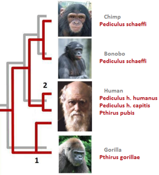
Simian foamy virus (SFV) is a species of the genus Spumavirus that belongs to the family of Retroviridae. It has been identified in a wide variety of primates, including prosimians, New World and Old World monkeys, as well as apes, and each species has been shown to harbor a unique (species-specific) strain of SFV, including African green monkeys, baboons, macaques, and chimpanzees. As it is related to the more well-known retrovirus human immunodeficiency virus (HIV), its discovery in primates has led to some speculation that HIV may have been spread to the human species in Africa through contact with blood from apes, monkeys, and other primates, most likely through bushmeat-hunting practices.

<span class="mw-page-title-main">Endoplasmic-reticulum-associated protein degradation</span>

Endoplasmic-reticulum-associated protein degradation (ERAD) designates a cellular pathway which targets misfolded proteins of the endoplasmic reticulum for ubiquitination and subsequent degradation by a protein-degrading complex, called the proteasome.

Env is a viral gene that encodes the protein forming the viral envelope. The expression of the env gene enables retroviruses to target and attach to specific cell types, and to infiltrate the target cell membrane.

Visna-maedi virus from the genus Lentivirus and subfamily Orthoretrovirinae, is a retrovirus that causes encephalitis and chronic pneumonitis in sheep. It is known as visna when found in the brain, and maedi when infecting the lungs. Lifelong, persistent infections in sheep occur in the lungs, lymph nodes, spleen, joints, central nervous system, and mammary glands; The condition is sometimes known as ovine progressive pneumonia (OPP), particularly in the United States, or Montana sheep disease. White blood cells of the monocyte/macrophage lineage are the main target of the virus.

<span class="mw-page-title-main">CD81</span>

CD81 molecule, also known as CD81, is a protein which in humans is encoded by the CD81 gene. It is also known as 26 kDa cell surface protein, TAPA-1, and Tetraspanin-28 (Tspan-28).

<span class="mw-page-title-main">AP1G2</span> Protein-coding gene in the species Homo sapiens

AP-1 complex subunit gamma-like 2 is a protein that in humans is encoded by the AP1G2 gene.

<span class="mw-page-title-main">AP1S2</span> Protein-coding gene in humans

AP-1 complex subunit sigma-2 is a protein that in humans is encoded by the AP1S2 gene.

<span class="mw-page-title-main">IFITM1</span> Protein-coding gene in the species Homo sapiens

Interferon-induced transmembrane protein 1 is a protein that in humans is encoded by the IFITM1 gene. IFITM1 has also recently been designated CD225. This protein has several additional names: fragilis, IFI17 [interferon-induced protein 17], 9-27 [Interferon-inducible protein 9-27] and Leu13.

<span class="mw-page-title-main">Tetherin</span> Mammalian protein found in Homo sapiens

Tetherin, also known as bone marrow stromal antigen 2, is a lipid raft associated protein that in humans is encoded by the BST2 gene. In addition, tetherin has been designated as CD317. This protein is constitutively expressed in mature B cells, plasma cells and plasmacytoid dendritic cells, and in many other cells, it is only expressed as a response to stimuli from IFN pathway.

<span class="mw-page-title-main">Rev (HIV)</span> HIV-1 regulating protein

Rev is a transactivating protein that is essential to the regulation of HIV-1 protein expression. A nuclear localization signal is encoded in the rev gene, which allows the Rev protein to be localized to the nucleus, where it is involved in the export of unspliced and incompletely spliced mRNAs. In the absence of Rev, mRNAs of the HIV-1 late (structural) genes are retained in the nucleus, preventing their translation.

2F5 is a broadly neutralizing human monoclonal antibody (mAb) that has been shown to bind to and neutralize HIV-1 in vitro, making it a potential candidate for use in vaccine synthesis. 2F5 recognizes an epitope in the membrane-proximal external region (MPER) of HIV-1 gp41. 2F5 then binds to this epitope and its constant region interacts with the viral lipid membrane, which neutralizes the virus.

Vpx is a virion-associated protein encoded by human immunodeficiency virus type 2 HIV-2 and most simian immunodeficiency virus (SIV) strains, but that is absent from HIV-1. It is similar in structure to the protein Vpr that is carried by SIV and HIV-2 as well as HIV-1. Vpx is one of five accessory proteins carried by lentiviruses that enhances viral replication by inhibiting host antiviral factors.

Éric A. Cohen is a Canadian molecular virologist whose research is focused on human immunodeficiency virus (HIV)-host interactions that govern viral replication and persistence.

<span class="mw-page-title-main">Host switch</span> Evolutionary change of the host specificity of a parasite or pathogen

In parasitology and epidemiology, a host switch is an evolutionary change of the host specificity of a parasite or pathogen. For example, the human immunodeficiency virus used to infect and circulate in non-human primates in West-central Africa, but switched to humans in the early 20th century.

<span class="mw-page-title-main">SERINC5</span> Protein

Serine incorporator 5 is a protein that in humans is encoded by the SERINC5 gene.

References

  1. Bour S, Schubert U, Strebel K (March 1995). "The human immunodeficiency virus type 1 Vpu protein specifically binds to the cytoplasmic domain of CD4: implications for the mechanism of degradation". Journal of Virology. 69 (3): 1510–20. doi:10.1128/JVI.69.3.1510-1520.1995. PMC   188742 . PMID   7853484.
  2. Estrabaud E, Le Rouzic E, Lopez-Vergès S, Morel M, Belaïdouni N, Benarous R, Transy C, Berlioz-Torrent C, Margottin-Goguet F (July 2007). "Regulated degradation of the HIV-1 Vpu protein through a betaTrCP-independent pathway limits the release of viral particles". PLOS Pathog. 3 (7): e104. doi:10.1371/journal.ppat.0030104. PMC   1933454 . PMID   17676996.
  3. Hussain A, Wesley C, Khalid M, Chaudhry A, Jameel S (January 2008). "Human immunodeficiency virus type 1 Vpu protein interacts with CD74 and modulates major histocompatibility complex class II presentation" (PDF). Journal of Virology. 82 (2): 893–902. doi:10.1128/JVI.01373-07. PMC   2224584 . PMID   17959659.
  4. Ewart GD, Sutherland T, Gage PW, Cox GB (October 1996). "The Vpu protein of human immunodeficiency virus type 1 forms cation-selective ion channels". Journal of Virology. 70 (10): 7108–15. doi:10.1128/JVI.70.10.7108-7115.1996. PMC   190763 . PMID   8794357.
  5. Gonzalez ME, Carrasco L (September 1998). "The human immunodeficiency virus type 1 Vpu protein enhances membrane permeability". Biochemistry. 37 (39): 13710–9. doi:10.1021/bi981527f. hdl: 20.500.12105/7976 . PMID   9753459.
  6. Gonzalez ME, Carrasco L (Sep 2003). "Viroporins". FEBS Letters. 552 (1): 28–34. doi: 10.1016/S0014-5793(03)00780-4 . hdl: 20.500.12105/7778 . PMID   12972148. S2CID   209557930.
  7. Göttlinger HG, Dorfman T, Cohen EA, Haseltine WA (August 1993). "Vpu protein of human immunodeficiency virus type 1 enhances the release of capsids produced by gag gene constructs of widely divergent retroviruses". Proc. Natl. Acad. Sci. U.S.A. 90 (15): 7381–5. Bibcode:1993PNAS...90.7381G. doi: 10.1073/pnas.90.15.7381 . PMC   47141 . PMID   8346259.
  8. 1 2 Strebel K (December 1996). "Structure and Function of HIV-1 Vpu" (PDF). Los Alamos National Laboratory.
  9. Andrew A, Strebel K (October 2010). "HIV-1 Vpu targets cell surface markers CD4 and BST-2 through distinct mechanisms". Mol. Aspects Med. 31 (5): 407–17. doi:10.1016/j.mam.2010.08.002. PMC   2967615 . PMID   20858517.
  10. Andrew AJ, Miyagi E, Strebel K (March 2011). "Differential effects of human immunodeficiency virus type 1 Vpu on the stability of BST-2/tetherin". J. Virol. 85 (6): 2611–9. doi:10.1128/JVI.02080-10. PMC   3067951 . PMID   21191020.
  11. Douglas JL, Viswanathan K, McCarroll MN, Gustin JK, Früh K, Moses AV (August 2009). "Vpu directs the degradation of the human immunodeficiency virus restriction factor BST-2/Tetherin via a {beta}TrCP-dependent mechanism". J. Virol. 83 (16): 7931–47. doi:10.1128/JVI.00242-09. PMC   2715753 . PMID   19515779.
  12. Iwabu Y, Fujita H, Kinomoto M, Kaneko K, Ishizaka Y, Tanaka Y, Sata T, Tokunaga K (December 2009). "HIV-1 accessory protein Vpu internalizes cell-surface BST-2/tetherin through transmembrane interactions leading to lysosomes". J. Biol. Chem. 284 (50): 35060–72. doi: 10.1074/jbc.M109.058305 . PMC   2787367 . PMID   19837671.
  13. Nomaguchi M, Fujita M, Adachi A (July 2008). "Role of HIV-1 Vpu protein for virus spread and pathogenesis". Microbes Infect. 10 (9): 960–7. doi:10.1016/j.micinf.2008.07.006. PMID   18672082.
  14. Cohen EA, Terwilliger EF, Sodroski JG, Haseltine WA (August 1988). "Identification of a protein encoded by the vpu gene of HIV-1". Nature. 334 (6182): 532–4. Bibcode:1988Natur.334..532C. doi:10.1038/334532a0. PMID   3043230. S2CID   4372649.
This article incorporates text from the public domain Pfam and InterPro: IPR008187