Allantoicase

Last updated
allantoicase
1sg3.jpg
Allantoicase homohexamer, Saccharomyces cerevisiae
Identifiers
EC no. 3.5.3.4
CAS no. 9025-21-2
Databases
IntEnz IntEnz view
BRENDA BRENDA entry
ExPASy NiceZyme view
KEGG KEGG entry
MetaCyc metabolic pathway
PRIAM profile
PDB structures RCSB PDB PDBe PDBsum
Gene Ontology AmiGO / QuickGO
Search
PMC articles
PubMed articles
NCBI proteins
Allantoicase
PDB 1sg3 EBI.jpg
structure of allantoicase
Identifiers
SymbolAllantoicase
Pfam PF03561
Pfam clan CL0202
InterPro IPR015908
SCOP2 1sg3 / SCOPe / SUPFAM
Available protein structures:
Pfam   structures / ECOD  
PDB RCSB PDB; PDBe; PDBj
PDBsum structure summary

Allantoicase is an enzyme (EC 3.5.3.4) that in humans is encoded by the ALLC gene. Allantoicase catalyzes the chemical reaction

Contents

allantoate + H2O (S)-ureidoglycolate + urea

Thus, the two substrates of this enzyme are allantoate and H2O, whereas its two products are (S)-ureidoglycolate and urea.

This enzyme belongs to the family of hydrolases, those acting on carbon-nitrogen bonds other than peptide bonds, specifically in linear amidines. The systematic name of this enzyme class is allantoate amidinohydrolase. This enzyme participates in purine metabolism by facilitating the utilization of purines as secondary nitrogen sources under nitrogen-limiting conditions. While purine degradation converges to uric acid in all vertebrates, its further degradation varies from species to species. Uric acid is excreted by birds, reptiles, and some mammals that do not have a functional uricase gene, whereas other mammals produce allantoin. Amphibians and microorganisms produce ammonia and carbon dioxide using the uricolytic pathway. Allantoicase performs the second step in this pathway catalyzing the conversion of allantoate into ureidoglycolate and urea.

Structural studies

As of late 2007, two structures have been solved for this class of enzymes, with PDB accession codes 1O59 and 1SG3.

The structure of allantoicase is best described as being composed of two repeats (the allantoicase repeats: AR1 and AR2), which are connected by a flexible linker. The crystal structure, resolved at 2.4A resolution, reveals that AR1 has a very similar fold to AR2, both repeats being jelly-roll motifs, composed of four-stranded and five-stranded antiparallel beta-sheets. [1] Each jelly-roll motif has two conserved surface patches that probably constitute the active site. [2]

Related Research Articles

<span class="mw-page-title-main">Uric acid</span> Organic compound

Uric acid is a heterocyclic compound of carbon, nitrogen, oxygen, and hydrogen with the formula C5H4N4O3. It forms ions and salts known as urates and acid urates, such as ammonium acid urate. Uric acid is a product of the metabolic breakdown of purine nucleotides, and it is a normal component of urine. High blood concentrations of uric acid can lead to gout and are associated with other medical conditions, including diabetes and the formation of ammonium acid urate kidney stones.

<span class="mw-page-title-main">Xanthine oxidase</span> Class of enzymes

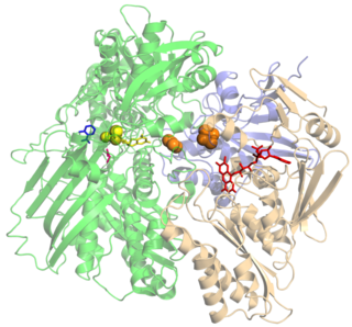
Xanthine oxidase is a form of xanthine oxidoreductase, a type of enzyme that generates reactive oxygen species. These enzymes catalyze the oxidation of hypoxanthine to xanthine and can further catalyze the oxidation of xanthine to uric acid. These enzymes play an important role in the catabolism of purines in some species, including humans.

<span class="mw-page-title-main">Urate oxidase</span> Pseudogene in the species Homo sapiens

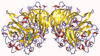
The enzyme urate oxidase (UO), uricase or factor-independent urate hydroxylase, absent in humans, catalyzes the oxidation of uric acid to 5-hydroxyisourate:

Purine metabolism refers to the metabolic pathways to synthesize and break down purines that are present in many organisms.

<span class="mw-page-title-main">GMP synthase</span>

Guanosine monophosphate synthetase, also known as GMPS is an enzyme that converts xanthosine monophosphate to guanosine monophosphate.

<span class="mw-page-title-main">Xanthine dehydrogenase</span> Protein-coding gene in the species Homo sapiens

Xanthine dehydrogenase, also known as XDH, is a protein that, in humans, is encoded by the XDH gene.

<span class="mw-page-title-main">Homoserine dehydrogenase</span> Enzyme

In enzymology, a homoserine dehydrogenase (EC 1.1.1.3) is an enzyme that catalyzes the chemical reaction

In enzymology, an ureidoglycolate dehydrogenase (EC 1.1.1.154) is an enzyme that catalyzes the chemical reaction

Alanine dehydrogenase (EC 1.4.1.1) is an enzyme that catalyzes the chemical reaction

In enzymology, a ferredoxin—nitrate reductase (EC 1.7.7.2) is an enzyme that catalyzes the chemical reaction

In enzymology, an allantoate deiminase (EC 3.5.3.9) is an enzyme that catalyzes the chemical reaction

In enzymology, an aminoacylase (EC 3.5.1.14) is an enzyme that catalyzes the chemical reaction

In enzymology, an ureidoglycolate hydrolase (EC 3.5.3.19) is an enzyme that catalyzes the chemical reaction

<span class="mw-page-title-main">Carnitine O-acetyltransferase</span> Enzyme

Carnitine O-acetyltransferase also called carnitine acetyltransferase is an enzyme that encoded by the CRAT gene that catalyzes the chemical reaction

In enzymology, a 3-deoxy-8-phosphooctulonate synthase (EC 2.5.1.55) is an enzyme that catalyzes the chemical reaction

<span class="mw-page-title-main">Methanol dehydrogenase (cytochrome c)</span>

Methanol dehydrogenase (cytochrome c) (EC 1.1.2.7, methanol dehydrogenase, MDH) is an enzyme with systematic name methanol:cytochrome c oxidoreductase. This enzyme catalyses the following chemical reaction

Catalase-peroxidase (EC 1.11.1.21, katG (gene)) is an enzyme with systematic name donor:hydrogen-peroxide oxidoreductase. This enzyme catalyses the following chemical reaction

  1. donor + H2O2 ⇌ oxidized donor + 2 H2O
  2. 2 H2O2 ⇌ O2 + 2 H2O

Beta-carotene 3-hydroxylase (EC 1.14.13.129, beta-carotene 3,3'-monooxygenase, CrtZ) is an enzyme with systematic name beta-carotene,NADH:oxygen 3-oxidoreductase . This enzyme catalyses the following chemical reaction

Phospholipase C (EC 3.1.4.3, lipophosphodiesterase I, Clostridium welchii α-toxin, Clostridium oedematiens β- and γ-toxins, lipophosphodiesterase C, phosphatidase C, heat-labile hemolysin, α-toxin) is an enzyme with systematic name phosphatidylcholine cholinephosphohydrolase. This enzyme catalyses the following chemical reaction

The enzyme exo-(1→4)-α-D-glucan lyase (EC 4.2.2.13, α-(1→4)-glucan 1,5-anhydro-D-fructose eliminase, α-1,4-glucan exo-lyase, α-1,4-glucan lyase, GLase) is an enzyme with systematic name (1→4)-α-D-glucan exo-4-lyase (1,5-anhydro-D-fructose-forming). This enzyme catalyses the following chemical reaction

References

  1. Xu Q, Schwarzenbacher R, Page R, Sims E, Abdubek P, Ambing E, Biorac T, Brinen LS, Cambell J, Canaves JM, Chiu HJ, Dai X, Deacon AM, DiDonato M, Elsliger MA, Floyd R, Godzik A, Grittini C, Grzechnik SK, Hampton E, Jaroszewski L, Karlak C, Klock HE, Koesema E, Kovarik JS, Kreusch A, Kuhn P, Lesley SA, Levin I, McMullan D, McPhillips TM, Miller MD, Morse A, Moy K, Ouyang J, Quijano K, Reyes R, Rezezadeh F, Robb A, Spraggon G, Stevens RC, van den Bedem H, Velasquez J, Vincent J, von Delft F, Wang X, West B, Wolf G, Hodgson KO, Wooley J, Wilson IA (August 2004). "Crystal structure of an allantoicase (YIR029W) from Saccharomyces cerevisiae at 2.4 A resolution". Proteins. 56 (3): 619–24. doi: 10.1002/prot.20164 . PMID   15229895. S2CID   5688375.
  2. Leulliot N, Quevillon-Cheruel S, Sorel I, Graille M, Meyer P, Liger D, Blondeau K, Janin J, van Tilbeurgh H (May 2004). "Crystal structure of yeast allantoicase reveals a repeated jelly roll motif". The Journal of Biological Chemistry. 279 (22): 23447–52. doi: 10.1074/jbc.M401336200 . PMID   15020593.

Further reading

This article incorporates text from the public domain Pfam and InterPro: IPR015908