Alpha sheet

Last updated
Diagram of the hydrogen bonding patterns in the alpha sheet structure. Oxygen atoms are shown in red and nitrogen in blue; dotted lines represent hydrogen bonds. R groups represent the amino acid side chains. Alpha sheet bonding schematic-color.svg
Diagram of the hydrogen bonding patterns in the alpha sheet structure. Oxygen atoms are shown in red and nitrogen in blue; dotted lines represent hydrogen bonds. R groups represent the amino acid side chains.
A stick representation of a peptide chain in an alpha-sheet configuration. Alpha strand 50 50 vertical.png
A stick representation of a peptide chain in an alpha-sheet configuration.

Alpha sheet (also known as alpha pleated sheet or polar pleated sheet) is an atypical secondary structure in proteins, first proposed by Linus Pauling and Robert Corey in 1951. [1] [2] [3] The hydrogen bonding pattern in an alpha sheet is similar to that of a beta sheet, but the orientation of the carbonyl and amino groups in the peptide bond units is distinctive; in a single strand, all the carbonyl groups are oriented in the same direction on one side of the pleat, and all the amino groups are oriented in the same direction on the opposite side of the sheet. Thus the alpha sheet accumulates an inherent separation of electrostatic charge, with one edge of the sheet exposing negatively charged carbonyl groups and the opposite edge exposing positively charged amino groups. Unlike the alpha helix and beta sheet, the alpha sheet configuration does not require all component amino acid residues to lie within a single region of dihedral angles; instead, the alpha sheet contains residues of alternating dihedrals in the traditional right-handed (αR) and left-handed (αL) helical regions of Ramachandran space. Although the alpha sheet is only rarely observed in natural protein structures, it has been speculated to play a role in amyloid disease [4] and it was found to be a stable form for amyloidogenic proteins in molecular dynamics simulations. [5] [6] Alpha sheets have also been observed in X-ray crystallography structures of designed peptides. [4]

Contents

The regular formation of alpha-sheet by unfolded proteins inevitably involves many L amino acid residues readily adopting the alphaL conformation, which appears at first sight to go against textbook chemistry, which is that, of the 20 amino acids, it is glycine that strongly favours this conformation. The conundrum is resolved by realizing that the alphaL region comprises two overlapping areas, here called γL and αL, which should be considered separately. It turns out that, while the γL conformation is adopted, almost exclusively, by glycine, the αL conformation of alpha-sheet is more commonly, or about as commonly, adopted by any of 15 L-amino acids compared to glycine, the exceptions being proline, threonine, valine and isoleucine, which are rare at this conformation. [7] Hence, of the 20 amino acids, 16 readily adopt the αL conformation.

Experimental evidence

When Pauling and Corey first proposed the alpha sheet, they suggested that it agreed well with fiber diffraction results from beta-keratin fibers. [2] However, since the alpha sheet did not appear to be energetically favorable, they argued that beta sheets would occur more commonly among normal proteins, [3] and subsequent demonstration that beta-keratin is made of beta sheets consigned the alpha sheet proposal to obscurity. However the alpha strand conformation is observed in isolated instances in native state proteins as solved by X-ray crystallography or protein NMR, although an extended alpha sheet is not identified in any known natural protein. Native proteins containing alpha-strand regions or alpha-sheet-patterned hydrogen bonding include synaptotagmin, lysozyme, and potassium channels, where the alpha-strands line the ion-conducting pore. [4]

Evidence for the existence of alpha-sheet in a mutant form of transthyretin has been presented. [8] Alpha-sheet conformations have been observed in crystal structures of short non-natural peptides, especially those containing a mixture of L and D amino acids. The first crystal structure containing an alpha sheet was observed in the capped tripeptide BocAla La-Ile DIle LO Me. [9] Other peptides that assume alpha-sheet structures include capped diphenyl-glycine-based dipeptides [10] and tripeptides. [11]

Role in amyloidogenesis

The alpha sheet has been proposed as a possible intermediate state in the conformational change in the formation of amyloid fibrils by peptides and proteins such as amyloid beta, poly-glutamine repeats, lysozyme, prion proteins, and transthyretin repeats, all of which are associated with protein misfolding disease. For example, amyloid beta is a major component of amyloid plaques in the brains of Alzheimer's disease patients, [6] and polyglutamine repeats in the huntingtin protein are associated with Huntington's disease. [12] These proteins undergo a conformational change from largely random coil or alpha helix structures to the highly ordered beta sheet structures found in amyloid fibrils. Most beta sheets in known proteins are "twisted" about 15° for optimal hydrogen bonding and steric packing; however, some evidence from electron crystallography suggests that at least some amyloid fibrils contain "flat" sheets with only 1–2.5° of twist. [13] An alpha-sheet amyloid intermediate is suggested to explain some anomalous features of the amyloid fibrillization process, such as the evident amino acid sequence dependence of amyloidogenesis despite the belief that the amyloid fold is mainly stabilized by the protein backbone. [14] [15]

Xu, [16] using atomic force microscopy, has shown that formation of amyloid fibers is a two-step process in which proteins first aggregate into colloidal spheres of ≈20 nm diameter. The spheres then join together spontaneously to form linear chains, which evolve into mature amyloid fibers. The formation of these linear chains appears to be driven by the development of an electrostatic dipole in each of the colloidal spheres strong enough to overcome coulomb repulsion. This suggests a possible mechanism by which alpha sheet may promote amyloid aggregation; the peptide bond has a relatively large intrinsic electrostatic dipole, but normally the dipoles of nearby bonds cancel each other out. In the alpha sheet, unlike other conformations, the peptide bonds are oriented in parallel so that the dipoles of the individual bonds can add up to create a strong overall electrostatic dipole.

Notably, the protein lysozyme is among the few native-state proteins shown to contain an alpha-strand region; lysozyme from both chickens and humans contains an alpha strand located close to the site of a mutation known to cause hereditary amyloidosis in humans, usually an autosomal dominant genetic disease. [4] Molecular dynamics simulations of the mutant protein reveal that the region around the mutation assumes an alpha strand conformation. [6] Lysozyme is among the naturally occurring proteins known to form amyloid fibers under experimental conditions, and both natively alpha-strand region and the mutation site fall within the larger region identified as the core of lysozyme amyloid fibrillogenesis. [17] [18]

A mechanism for direct alpha sheet and beta sheet interconversion has also been suggested, based on peptide plane flipping in which the αRαL dipeptide inverts to produce a ββ dihedral angle conformation. This process has also been observed in simulations of transthyretin [19] and implicated as occurring naturally in certain protein families by examination of their dihedral angle conformations in crystal structures. [20] [21] It is suggested that alpha-sheet folds into multi-strand solenoids. [22]

Evidence employing retro-enantio N-methylated peptides, or those with alternating L and D amino acids, as inhibitors of beta-amyloid aggregation is consistent with alpha-sheet being the main material of the amyloid precursor. [23] [24] [25] [26] [27]

Related Research Articles

<span class="mw-page-title-main">Alpha helix</span> Type of secondary structure of proteins

An alpha helix is a sequence of amino acids in a protein that are twisted into a coil.

<span class="mw-page-title-main">Beta sheet</span> Protein structural motif

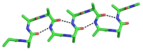
The beta sheet, (β-sheet) is a common motif of the regular protein secondary structure. Beta sheets consist of beta strands (β-strands) connected laterally by at least two or three backbone hydrogen bonds, forming a generally twisted, pleated sheet. A β-strand is a stretch of polypeptide chain typically 3 to 10 amino acids long with backbone in an extended conformation. The supramolecular association of β-sheets has been implicated in the formation of the fibrils and protein aggregates observed in amyloidosis, Alzheimer's disease and other proteinopathies.

<span class="mw-page-title-main">Protein secondary structure</span> General three-dimensional form of local segments of proteins

Protein secondary structure is the local spatial conformation of the polypeptide backbone excluding the side chains. The two most common secondary structural elements are alpha helices and beta sheets, though beta turns and omega loops occur as well. Secondary structure elements typically spontaneously form as an intermediate before the protein folds into its three dimensional tertiary structure.

<span class="mw-page-title-main">Protein folding</span> Change of a linear protein chain to a 3D structure

Protein folding is the physical process where a protein chain is translated into its native three-dimensional structure, typically a "folded" conformation, by which the protein becomes biologically functional. Via an expeditious and reproducible process, a polypeptide folds into its characteristic three-dimensional structure from a random coil. Each protein exists first as an unfolded polypeptide or random coil after being translated from a sequence of mRNA into a linear chain of amino acids. At this stage, the polypeptide lacks any stable three-dimensional structure. As the polypeptide chain is being synthesized by a ribosome, the linear chain begins to fold into its three-dimensional structure.

<span class="mw-page-title-main">Protein structure prediction</span> Type of biological prediction

Protein structure prediction is the inference of the three-dimensional structure of a protein from its amino acid sequence—that is, the prediction of its secondary and tertiary structure from primary structure. Structure prediction is different from the inverse problem of protein design. Protein structure prediction is one of the most important goals pursued by computational biology; and it is important in medicine and biotechnology.

<span class="mw-page-title-main">Amyloid</span> Insoluble protein aggregate with a fibrillar morphology

Amyloids are aggregates of proteins characterised by a fibrillar morphology of typically 7–13 nm in diameter, a β-sheet secondary structure and ability to be stained by particular dyes, such as Congo red. In the human body, amyloids have been linked to the development of various diseases. Pathogenic amyloids form when previously healthy proteins lose their normal structure and physiological functions (misfolding) and form fibrous deposits within and around cells. These protein misfolding and deposition processes disrupt the healthy function of tissues and organs.

<span class="mw-page-title-main">Transthyretin</span> Serum protein related to amyloid diseases

Transthyretin (TTR or TBPA) is a transport protein in the plasma and cerebrospinal fluid that transports the thyroid hormone thyroxine (T4) and retinol to the liver. This is how transthyretin gained its name: transports thyroxine and retinol. The liver secretes TTR into the blood, and the choroid plexus secretes TTR into the cerebrospinal fluid.

<span class="mw-page-title-main">Protein structure</span> Three-dimensional arrangement of atoms in an amino acid-chain molecule

Protein structure is the three-dimensional arrangement of atoms in an amino acid-chain molecule. Proteins are polymers – specifically polypeptides – formed from sequences of amino acids, which are the monomers of the polymer. A single amino acid monomer may also be called a residue, which indicates a repeating unit of a polymer. Proteins form by amino acids undergoing condensation reactions, in which the amino acids lose one water molecule per reaction in order to attach to one another with a peptide bond. By convention, a chain under 30 amino acids is often identified as a peptide, rather than a protein. To be able to perform their biological function, proteins fold into one or more specific spatial conformations driven by a number of non-covalent interactions, such as hydrogen bonding, ionic interactions, Van der Waals forces, and hydrophobic packing. To understand the functions of proteins at a molecular level, it is often necessary to determine their three-dimensional structure. This is the topic of the scientific field of structural biology, which employs techniques such as X-ray crystallography, NMR spectroscopy, cryo-electron microscopy (cryo-EM) and dual polarisation interferometry, to determine the structure of proteins.

Site-directed spin labeling (SDSL) is a technique for investigating the structure and local dynamics of proteins using electron spin resonance. The theory of SDSL is based on the specific reaction of spin labels with amino acids. A spin label's built-in protein structure can be detected by EPR spectroscopy. SDSL is also a useful tool in examinations of the protein folding process.

<span class="mw-page-title-main">Amylin</span> Peptide hormone that plays a role in glycemic regulation

Amylin, or islet amyloid polypeptide (IAPP), is a 37-residue peptide hormone. It is co-secreted with insulin from the pancreatic β-cells in the ratio of approximately 100:1 (insulin:amylin). Amylin plays a role in glycemic regulation by slowing gastric emptying and promoting satiety, thereby preventing post-prandial spikes in blood glucose levels.

<span class="mw-page-title-main">Fungal prion</span> Prion that infects fungal hosts

A fungal prion is a prion that infects hosts which are fungi. Fungal prions are naturally occurring proteins that can switch between multiple, structurally distinct conformations, at least one of which is self-propagating and transmissible to other prions. This transmission of protein state represents an epigenetic phenomenon where information is encoded in the protein structure itself, instead of in nucleic acids. Several prion-forming proteins have been identified in fungi, primarily in the yeast Saccharomyces cerevisiae. These fungal prions are generally considered benign, and in some cases even confer a selectable advantage to the organism.

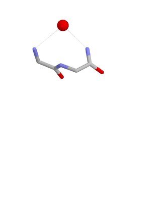
A beta bulge can be described as a localized disruption of the regular hydrogen bonding of beta sheet by inserting extra residues into one or both hydrogen bonded β-strands.

3<sub>10</sub> helix Type of secondary structure

A 310 helix is a type of secondary structure found in proteins and polypeptides. Of the numerous protein secondary structures present, the 310-helix is the fourth most common type observed; following α-helices, β-sheets and reverse turns. 310-helices constitute nearly 10–15% of all helices in protein secondary structures, and are typically observed as extensions of α-helices found at either their N- or C- termini. Because of the α-helices tendency to consistently fold and unfold, it has been proposed that the 310-helix serves as an intermediary conformation of sorts, and provides insight into the initiation of α-helix folding.

Peptide plane flipping is a type of conformational change that can occur in proteins by which the dihedral angles of adjacent amino acids undergo large-scale rotations with little displacement of the side chains. The plane flip is defined as a rotation of the dihedral angles φ,ψ at amino acids i and i+1 such that the resulting angles remain in structurally stable regions of Ramachandran space. The key requirement is that the sum of the ψi angle of residue i and the φi+1 angle of residue i+1 remain roughly constant; in effect, the flip is a crankshaft move about the axis defined by the Cα-C¹ and N-Cα bond vectors of the peptide group, which are roughly parallel. As an example, the type I and type II beta turns differ by a simple flip of the central peptide group of the turn.

<span class="mw-page-title-main">Collagen, type XXV, alpha 1</span> Mammalian protein found in Homo sapiens

Collagen alpha-1(XXV) chain is a protein that in humans is encoded by the COL25A1 gene.

<span class="mw-page-title-main">GCM transcription factors</span> Protein family

In molecular biology, the GCM transcription factors are a family of proteins which contain a GCM motif. The GCM motif is a domain that has been identified in proteins belonging to a family of transcriptional regulators involved in fundamental developmental processes which comprise Drosophila melanogaster GCM and its mammalian homologues. In GCM transcription factors the N-terminal moiety contains a DNA-binding domain of 150 amino acids. Sequence conservation is highest in this GCM domain. In contrast, the C-terminal moiety contains one or two transactivating regions and is only poorly conserved.

<span class="mw-page-title-main">Nest (protein structural motif)</span>

The Nest is a type of protein structural motif. It is a small recurring anion-binding feature of both proteins and peptides. Each consists of the main chain atoms of three consecutive amino acid residues. The main chain NH groups bind the anions while the side chain atoms are often not involved. Proline residues lack NH groups so are rare in nests. About one in 12 of amino acid residues in proteins, on average, belongs to a nest.

<span class="mw-page-title-main">P3 peptide</span>

p3 peptide also known as amyloid β- peptide (Aβ)17–40/42 is the peptide resulting from the α- and γ-secretase cleavage from the amyloid precursor protein (APP). It is known to be the major constituent of diffuse plaques observed in Alzheimer's disease (AD) brains and pre-amyloid plaques in people affected by Down syndrome. However, p3 peptide's role in these diseases is not truly known yet.

<span class="mw-page-title-main">Beta bend ribbon</span>

The beta bend ribbon, or beta-bend ribbon, is a structural feature in polypeptides and proteins. The shortest possible has six amino acid residues arranged as two overlapping hydrogen bonded beta turns in which the carbonyl group of residue i is hydrogen-bonded to the NH of residue i+3 while the carbonyl group of residue i+2 is hydrogen-bonded to the NH of residue i+5. In longer ribbons, this bonding is continued in peptides of 8, 10, etc., amino acid residues. A beta bend ribbon can be regarded as an aberrant 310 helix (3/10-helix) that has lost some of its hydrogen bonds. Two websites are available to facilitate finding and examining these features in proteins: Motivated Proteins; and PDBeMotif.

Computational methods that use protein sequence and/ or protein structure to predict protein aggregation. The table below, shows the main features of software for prediction of protein aggregation

References

  1. Pauling, L. & Corey, R. B. (1951). The pleated sheet, a new layer configuration of polypeptide chains. Proc. Natl. Acad. Sci. USA 37, 251–6. PMID   14834147
  2. 1 2 Pauling, L. & Corey, R. B. (1951). The structure of feather rachis keratin. Proc. Natl. Acad. Sci. USA 37, 256–261. doi : 10.1073/pnas.37.5.256 PMID   14834148
  3. 1 2 Pauling, L. & Corey, R. B. (1951). Configurations of Polypeptide Chains With Favored Orientations Around Single Bonds: Two New Pleated Sheets. Proc. Natl. Acad. Sci. USA 37, 729–740. PMID   16578412
  4. 1 2 3 4 Daggett V. (2006). Alpha-sheet: The toxic conformer in amyloid diseases? Acc Chem Res 39(9):594-602. doi : 10.1021/ar0500719 PMID   16981675
  5. Babin V, Roland C, Sagui C. (2011). The alpha-sheet: A missing in action secondary structure. Proteins 79:937-946. doi : 10.1002/prot.22935
  6. 1 2 3 Armen RS, DeMarco ML, Alonso DO, Daggett V. (2011). Pauling and Corey's alpha-pleated sheet structure may define the prefibrillar amyloidogenic intermediate in amyloid disease. Proc Natl Acad Sci USA 101(32):11622-7. doi : 10.1073/pnas.0401781101 PMID   15280548
  7. Hayward S. (2021). Determination of amino acids that favour the alphaL region using Ramachandran propensity plots. Implications for alpha-sheet as the possible amyloid intermediate. Journal of Structural Biology 213:α107738.
  8. Hilaire MR, Ding B, Mukherjee D, Chen J, Gai F. (2018). Possible existence of alpha-sheets in the amyloid fibrils formed by a mutant form of a peptide from transthyretin. Journal of the American Chemical Society 140:629-635. doi : 10.1021/jacs.7b09262
  9. Di Blasio B, Saviano M, Fattorusso R, Lombardi A, Pedone C, Valle V, Lorenzi GP. (1994). A crystal structure with features of an antiparallel alpha-pleated sheet. Biopolymers 34(11):1463-8. PMID   7827259
  10. De Simone G, Lombardi A, Galdiero S, Nastri F, Di Costanzo L, Gohda S, Sano A, Yamada T, Pavone V. (2000). The crystal structure of a Dcp-containing peptide. Biopolymers 53(2):182-8. PMID   10679622
  11. Pavone V, Lombardi A, Saviano M, Nastri F, Zaccaro L, Maglio O, Pedone C, Omote Y, Yamanaka Y, Yamada T. (1998). Conformational behaviour of C(alpha,alpha)-diphenylglycine: folded vs. extended structures in DphiG-containing tripeptides. 4(1):21-32. PMID   9523753
  12. Armen RS, Bernard BM, Day R, Alonso DO, Daggett V. (2005). Characterization of a possible amyloidogenic precursor in glutamine-repeat neurodegenerative diseases. Proc Natl Acad Sci USA 102(38):13433-8. PMID   16157882
  13. Jimenez, J. L., Nettleton, E. J., Bouchard, M., Robinson, C. V., Dobson, C. M. & Saibil, H. R. (2002). The protofilament structure of insulin amyloid fibrils. Proc. Natl. Acad. Sci. USA 99, 9196–9201. PMID   12093917
  14. Fraser, P. E., Duffy, L. K., O'Mally, M. B., Nguyen, J., Inouye, H. & Kirschner, D. A. (1991). Morphology and antibody recognition of synthetic beta-amyloid peptides. J. Neurosci. Res. 28, 474–485. PMID   1908024.
  15. Malinchik, S. B., Inouye, H., Szumowski, K. E. & Kirschner, D. A. (1998). Structural analysis of Alzheimer's beta(1-40) amyloid: protofilament assembly of tubular fibrils. Biophys. J. 74, 537–545. PMID   9449354
  16. Xu S. Aggregation drives "misfolding" in protein amyloid fiber formation. Amyloid 2007 Jun;14(2):119-31. PMID   17577685
  17. Frare, D.; Polverino de Laureto, P.; Zurdo, J.; Dobson, C. M., Fontana, A. (2004). A highly amyloidogenic region of hen lysozyme. J Mol Biol 340: 1153-1165. PMID   15236974
  18. Frare E, Mossuto MF, Polverino de Laureto P, Dumoulin M, Dobson CM, Fontana A. (2006). Identification of the core structure of lysozyme amyloid fibrils by proteolysis. J Mol Biol 361(3):551-61. PMID   16859705
  19. Yang MF, Lei M, Yordanov B, Huo SH. (2006). Peptide plane can flip in two opposite directions: implication in amyloid formation of transthyretin. J Phys Chem B 110(12):5829-33. PMID   16553385
  20. Milner-White EJ, Watson JD, Qi G, Hayward S. (2006). Amyloid formation may involve alpha- to beta sheet interconversion via peptide plane flipping. Structure 14(9):1369-76. PMID   16962968
  21. Hayward S, Milner-White EJ. (2008). The geometry of alpha-sheet: Implications for its possible function as amyloid precursor in proteins Proteins 71:425-431. PMID   17957773
  22. Hayward S, Milner-White EJ. (2011). Simulation of the β- to α-sheet transition results in a twisted sheet for antiparallel and an α-nanotube for parallel strands: implications for amyloid format. Proteins 79(11) 3193-3207. PMID   21989939
  23. Grillo-Bosch D, Carulla N, Cruz M, Pujol-Pina R, Madurga S, Rabanal F, Giralt E. (2009). Retro-enantio N-methylated peptides as inhibitors of beta-amyloid aggregation. ChemMedChem 4, 1488-1494. PMID   19591190
  24. Kellock J, Hopping G, Cauchey B, Daggett V. (2016). Peptides composed of alternating L- and D-amino acids inhibit amyloidogenesis in three distinct amyloid systems independent of sequence. Journal of Molecular Biology 428, 2317-2328
  25. Paranjapye N, Daggett V. (2018). De novo designed alpha-sheet peptides inhibit functional amyloid formation of Streptococcus mutant biofilms. Journal of Molecular Biology 430, 3764-3773
  26. Maris NL, Shea D. (2018). Chemical and Physical Variability in Structural Isomers of an L/D α‑Sheet Peptide Designed To Inhibit Amyloidogenesis. Biochemistry 57, 507-510. doi : 10.1021/acs.biochem.7b00345
  27. Shea D, Hsu C-C. (2019). α-Sheet secondary structure in amyloid β-peptide drives aggregation and toxicity in Alzheimer’s disease. Proc Nat Acad Sci USA 116(18) 8895-8900. doi : 10.1073/pnas.1820585116