Collariella bostrychodes

Last updated

Collariella bostrychodes
Scientific classification OOjs UI icon edit-ltr.svg
Domain: Eukaryota
Kingdom: Fungi
Division: Ascomycota
Class: Sordariomycetes
Order: Sordariales
Family: Chaetomiaceae
Genus: Collariella
Species:
C. bostrychodes
Binomial name
Collariella bostrychodes
(Zopf) Wei Wang & Samson (2016)
Synonyms
  • Chaetomium bostrychodes Zopf (1877)

Collariella bostrychodes is a fungal decomposer of lignin and carbohydrate in the family Chaetomiaceae commonly found in soil and dung. [1] The fungus is distinguished by a darkened collar-like ostiole around the ostiolar pore, giving the fungus its name. [1] [2] The fungus is highly variable in shape and form, giving raise to the belief that there are two subclades in the species. [2] The ascospores range from lemon-shaped to nearly spherical with slightly pointed ends. [3] [4] [5] It can grow to be pale green and later turn pale bluish grey or olivaceous with age. The fungus produces the toxic secondary metabolite, chaetochromin. [6]

Contents

History and taxonomy

In 1877, the fungus was originally described by Friedrich Wilhelm Zopf in the Verhandlungen des Botanischen Vereins der Provinz Brandenburg for the Botanischer Verein der Provinz Brandenburg. [7] He described the fungus as part of the genus Chaetomium and initially being named as Chaetomium bostrychodes.C. bostrychodes was originally described to have ellipsoid ascomata by Pier Andrea Saccardo [8] in 1882, but later in 1963, Ames re-examined and described the species to have subglobose to ovoid ascomata instead. [9] The fungus was noted as being unable to produce functional antheridia, which suggested it wasn't part of the heterothallic species of Chaetomium. [10] The fungus was described as unique in the genus Chaetomium for possessing banded spores that are characteristic of no species of Chaetomium described in 1937. [4]

The fungus was believed to have two subclades designated C. bostrychodes and C. bostrychodes (A), which were considered to be macroscopically indistinguishable but closely related enough to each other that both subclades were kept as the same species. [4] The two subclades were previously suggested both to be either aberrant physiological or nearly related forms. Using phylogenetic analyses, combined with morphological comparisons to related genera and species, Wang and Samson determined C. bostrychodes to have morphological diversity specific to indoor Chaetomiaceae that required creating a new genus. [1] The genus, Collariella, was created to include for this diversity, its name refers to the dark collar-like apex that occurs around the ostiolar pore of the ascomata. [1] [2] C. bostrychodes was then transferred to the new genus and renamed Collariella bostrychodes, with the two subclades then grouped together and placed under this name.

Morphology and phylogeny

Colonies of C. bostrychodes can grow 2.5 to 3.5 mm daily, usually without developing aerial mycelium. [1] It can sometimes grow with pale green exudates and a green or uncoloured reverse. When young, C. bostrychodes appears as colourless and dextrinoid, but upon maturity, turns pale bluish grey or olivaceous with an apical germ pore . C. bostrychodes often are darkened around the 25 to 35 μm wide ostiolar pore, giving the genus its name of Collariella. [2] Chaetomium convolutum can have a disposition to develop into some forms of C. bostrychodes, developing narrowly ellipsoidal or narrowly ovoidal perithecia restricted just below the darkened periostiolar collar. [11] Similarly, C. bostrychodes appears similar morphologically to Collariella hilkhuijsenii, but C. bostrychodes can be distinguished via their larger ascospores and thicker terminal ascomatal hairs compared to those of C. hilkhuijsenii. [12] The ascomatal walls are brown or ochraceous and composed of angular 5 to 12 μm cells. These tend to fracture underneath the terminal hairs, which then break away as a unit. Brown ascomatal hairs grow mainly from the apical disc, usually appearing as helically coiled in the apical region with little branching. The hairs are either verrucose or warty, which are about 4.5 to 6.5 μm thick with occasionally coiled branches. Lateral seta-like hairs are often also present and have tapered or clavate terminal ends with septate. [13] These hairs break away at maturity with no aerial mycelium. C. bostrychodes develops perithecia late and possesses tightly coiled black terminal setae. The terminal hairs are branched, forming one spiral on another. [4] [5] The ascospores are considerably larger on average than those of C. robustum. [11]

The asci are often surrounded by paraphyses-like, broad, evanescent filaments with a long, evanescent stalk that is 8 spored and 11 to 14 μm wide. [1] The ascomata matures in 10 to 17 days, taking on a silvery appearance when young but then turns dark grey to black when mature. The apically flattened ascomata can be obovate, turbinate, ampulliform or cylindrical with its width ranging from 140 to 250 μm wide and ranging from 200 to 400 μm high. [1] The fungus is recorded having touter, subglobose or ovoidal, ascomata but it is also recorded having slender, narrowly ellipsoid ascomata with 1 to 2 rows of textura prismatica. [11] The breakdown of asci begins within the perithecia, and ascospores are later ejected in a slime consisting of the freed ascospores. [14] [15] The filaments grow upward and inward to exert pressure against each other to create a central cavity which asci push their way up. The asci deliquesce and then oozes out of the perithecial neck and are not violently discharged. [14] C. bostrychodes fruits in the spring and early summer. [3]

The two subclades are divided by phylogenetic and morphological evidence, but high statistical support was found to suggest they share a recent common ancestor and are closely related. [1] The two subclades differ in setae, variability in length and color of terminal hairs, variety in ascospores' shape, with extreme variability between the two in the species. C. bostrychodes can vary in shape, such as possessing mostly ellipsoidal or broadly ellipsoid perithecia. [11] However, both subclades have a specific banding of the spores that appears which is characteristic of C. bostrychodes only. [4] There are differences on what the two subclades will reproduce on, with C. bostrychodes (A) being able to reproduce on asparagin and alcohol-glucose cultures, whereas C. bostrychodes can reproduce on blood fibrin, starch, phenol red, eosin methylene blue, and dextrine-FeClh instead. [1] [4] In C. bostrychodes (A), anastomoses of the terminal hairs are common, but not found in C. bostrychodes.

Habitat and ecology

Collariella bostrychodes is considered a lignin, lignocellose, and carbohydrate decomposer but shows a significant preference for lignin, with a high frequency of occurrence in the fermentation layers of the forest. [16] It can be isolated from soil, dung, decaying stems, roots, and seeds, but very rarely in woody materials. [17] It has been isolated and found to be saprobic both in and on dung or excretions of dung of Oryctolagus cuniculu. [18] C. bostrychodes is not limited to forest ecosystems. While not a marine fungi, it has been isolated in the ocean and sediments in the marine muds, which suggests that the fungus can be wind-carried. [19] C. bostrychodes has been rarely isolated from food products, but it has been isolated in contaminated oat grains, okra, pea seeds and pepper. When C. bostrychodes is found in food products, it is typically a result of simple contamination. [20]

Health significance

A toxic pigment, chaetochromin, has now been proven to be widely associated with Chaetomium fungi., including C. bostrychodes. [6] Oral and intraperitoneal administration of chaetochromin to mice may result in pronounced toxicity, which can lead to potential detrimental effects like selective inhibition of haematopoiesis. [21] C. bostrychodes has been isolated from industrially polluted soils of the Kola Peninsula in ecosystems, specifically in ground cover and in areas of pine forests that have associations of moss shrubs and lichens shrubs, it believed to potentially cause diseases like Onychomycosis, skin infections, and peritonitis. [6] However, such cases are rare and therefore not well studied.

Related Research Articles

An ascocarp, or ascoma, is the fruiting body (sporocarp) of an ascomycete phylum fungus. It consists of very tightly interwoven hyphae and millions of embedded asci, each of which typically contains four to eight ascospores. Ascocarps are most commonly bowl-shaped (apothecia) but may take on a spherical or flask-like form that has a pore opening to release spores (perithecia) or no opening (cleistothecia).

<i>Chaetomium</i> Genus of fungi

Chaetomium is a genus of fungi in the Chaetomiaceae family. It is a dematiaceous (dark-walled) mold normally found in soil, air, cellulose and plant debris. According to the Dictionary of the Fungi, there are about 95 species in the widespread genus.

Farrowia is a genus of fungi within the Chaetomiaceae family.

<i>Chlamydosauromyces</i> Genus of fungi

Chlamydosauromyces punctatus is the sole species in the monotypic genus of fungi, Chlamydosauromyces in the family, Onygenaceae. It was found in the skin shed from frilled lizard. This fungus is mesophilic and digests hair. It reproduces both sexually and asexually. The fungus has so far not been reported to be pathogenic.

Polytolypa is a monotypic genus of fungus containing the single species Polytolypa hystricis. First classified in the Onygenaceae family, as of 2008 it is considered to be in the Ajellomycetaceae, although there is still uncertainty as to its phylogenetic relationships with other similar genera. This species is only known from a single specimen derived in the laboratory from a specimen of dung of the North American porcupine, Erethizon dorsatum, collected in Ontario, Canada. Polytolypa hystricis contains bioactive compounds that have antifungal activity.

Chaetomium cupreum is a fungus in the family Chaetomiaceae. It is able to decay in manufactured cellulosic materials, and is known to antagonize a wide range of soil microorganisms. This species is component of the biocontrol agent, Ketomium, a commercial biofungicide. It has also been investigated for use in the production of natural dyes. Chaetomium cupreum is mesophilic and known to occur in harsh environments and can rapidly colonize organic substrates in soil. Laboratory cultures of C. cupreum can be propagated on a range of common growth media including potato dextrose at ambient or higher than ambient temperature producing cottony white colonies with a reddish reverse.

Massarina carolinensis is a species of fungus in the Lophiostomataceae family. The species is found exclusively on the lower parts of the culms of the saltmarsh Juncus roemerianus on the Atlantic Coast of North Carolina.

<i>Paraphaeosphaeria pilleata</i> Species of fungus

Paraphaeosphaeria pilleata is a species of fungus in the Lophiostomataceae family. The species fruits exclusively in the lower parts of the culms of the black needlerush. It is found on the Atlantic Coast of North Carolina.

<i>Aphanoascus fulvescens</i> Species of fungus

Aphanoascus fulvescens is a mould fungus that behaves as a keratinophilic saprotroph and belongs to the Ascomycota. It is readily isolated from soil and dung containing keratin-rich tissues that have been separated from their animal hosts. This organism, distributed worldwide, is most commonly found in areas of temperate climate, in keeping with its optimal growth temperature of 28 °C (82 °F). While A. fulvescens is recognized as a geophilic fungal species, it is also a facultative opportunistic pathogen. Although it is not a dermatophyte, A. fulvescens has occasionally been shown to cause onychomycosis infections in humans. Its recognition in the laboratory is clinically important for correct diagnosis and treatment of human dermal infections.

<i>Chaetomium globosum</i> Species of fungus

Chaetomium globosum is a well-known mesophilic member of the mold family Chaetomiaceae. It is a saprophytic fungus that primarily resides on plants, soil, straw, and dung. Endophytic C. globosum assists in cellulose decomposition of plant cells. They are found in habitats ranging from forest plants to mountain soils across various biomes. C. globosum colonies can also be found indoors and on wooden products.

Thielavia subthermophila is a ubiquitous, filamentous fungus that is a member of the phylum Ascomycota and order Sordariales. Known to be found on plants of arid environments, it is an endophyte with thermophilic properties, and possesses dense, pigmented mycelium. Thielavia subthermophila has rarely been identified as a human pathogen, with a small number of clinical cases including ocular and brain infections. For treatment, antifungal drugs such as amphotericin B have been used topically or intravenously, depending upon the condition.

Chaetomium atrobrunneum is a darkly pigmented mould affiliated with the fungal division, Ascomycota. This species is predominantly saprotrophic, although it has been known to infect animals including humans, showing a proclivity for the tissues of the central nervous system. Chaetomium atrobrunneum was described in 1949 from a mouldy military mattress cover obtained from the island of Guadalcanal.

Botryotrichum murorum is a common soil and indoor fungus resembling members of the genus Chaetomium. The fungus has no known asexual state, and unlike many related fungi, is intolerant of high heat exhibiting limited growth when incubated at temperatures over 35 °C. In rare cases, the fungus is an opportunistic pathogen of marine animals and humans causing cutaneous and subcutaneous infection.

<i>Chaetomium elatum</i> Species of fungus

Chaetomium elatum is a very common and widely distributed saprotrophic fungus of the Chaetomiaceae family of molds which has been found to grow on many different substances all over the world. It was first established by Gustav Kunze after he observed it growing on dead leaves. Its defining features that distinguish it from other Chaetomium species are its extremely coarse terminal hairs and the lemon-shaped morphology of its ascospores. It produces many metabolites with potential biotechnology uses including one with promise against the rice blast disease fungus, Magnaporthe grisea. It shows very little pathogenic ability causing confirmed disease in only a few plant species.

<i>Chaetomium subspirale</i> Species of fungus

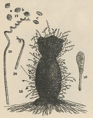
Chaetomium subspirale is a fungus from the phylum Ascomycota. It was described by A. H. Chivers in 1912 in America. The species has sexual fruiting bodies that are ornamented with characteristic, coiled hairs giving it a wooly appearance. C. subspirale colonies are brown, which the characteristic hairs are also responsible for. It is commonly found in various soil and dung samples. C. subspirale produces the mycotoxin, oxaspirodion, which inhibits inducible TNF-a expression and inhibits the activation of the transcription factor NF-kappaB.

Botryotrichum piluliferum is a fungal species first identified in 1885 by Saccardo and Marchal. It was discovered to be the asexual state of a member of the ascomycete genus, Chaetomium. The name B. piluliferum now applies to the fungus in all its states. B. piluliferum has been found worldwide in a wide range of habitats such as animal dung and vegetation. The colonies of this fungus start off white and grow rapidly to a brown colour. The conidia are smooth and white. B. piluliferum grows optimally at a temperature of 25-30 °C and a pH of 5.5.

Arcopilus aureus is a plant and soil fungus in the genus Arcopilus. It was first identified by A. H. Chivers in 1912, who named it Chaetomium aureum. It was later transferred to the genus Arcopilus by Wang and colleagues. The fungus has recently been recognized to have industrial use for the production of the metabolites resveratrol. and sclerotiorin Additionally, A. aureus has high lead tolerance and clearance, suggesting a potential role in environmental biotechnology.

<i>Myxotrichum chartarum</i> Species of fungus

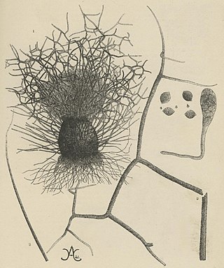
Myxotrichum chartarum is a psychrophilic and cellulolytic fungus first discovered in Germany by Gustav Kunze in 1823. Its classification has changed many times over its history to better reflect the information available at the time. Currently, M. chartarum is known to be an ascomycete surrounded by a gymnothecium composed of ornate spines and releases asexual ascospores. The presence of cellulolytic processes are common in fungi within the family Myxotrichaceae. M. chartarum is one of many Myxotrichum species known to degrade paper and paper products. Evidence of M. chartarum "red spot" mold formation, especially on old books, can be found globally. As a result, this fungal species and other cellulolytic molds are endangering old works of art and books. Currently, there is no evidence that suggests that species within the family Myxotrichaceae are pathogenic.

Pseudothielavia terricola is a fungal species of the phylum Ascomycota, family Chaetomiaceae, and genus Pseudothielavia. Pseudothielavia terricola is widely distributed, especially in the tropical region of the world – with documented appearances in Africa, Southern Europe, and Asia. The species is mainly found in soil, but can also be found on other materials such as animal dung. The species was first assigned to the genus Coniothyrium in 1927, but was soon re-assigned to the genus Thielavia which endured for almost 90 years. Through intensive phylogenetic research and reassessment, the species was designated to a new genus, Pseudothielavia; the etymology of Pseudothielavia means similar to the genus Thielavia – the high resemblance was what contributed to the species assignment to the genus Thielavia nine decades ago. The fungus is mesophilic, grows abundantly in a pH level between 3.9–6, and is able to utilize multiple carbohydrates to support its growth. Mature Pseudothielavia terricola colonies in culture is dark brown in colour and spread out. Pseudothielavia terricola synthesizes a variety of compounds, two of which are thielavin A & B. These compounds were determined to be strong inhibitors of prostaglandin synthesis.

Cercophora areolata is a member of the Ascomycota division, and is grouped into the Lasiosphaeriaceae family based on morphology. C. areolata is a coprophilous fungus that has been most recently isolated from porcupine dung. Defining features of C. areolata include: 1) ovoid-conical, glabrous ascomata, 2) black, carbonaceous, areolate peridium and 3) clavate-shaped, single-walled asci. From studies on C. areolata, this fungus produces multiple antifungal compounds, which inhibit other competitor fungi.

References

  1. 1 2 3 4 5 6 7 8 9 Wang, X.W.; Houbraken, J.; Groenewald, J.Z.; Meijer, M.; Andersen, B.; Nielsen, K.F.; Crous, P.W.; Samson, R.A. (2016). "Diversity and taxonomy of Chaetomium and chaetomium-like fungi from indoor environments". Studies in Mycology. 84: 145–224. doi:10.1016/j.simyco.2016.11.005. ISSN   0166-0616. PMC   5226397 . PMID   28082757.
  2. 1 2 3 4 Wang, X.W.; Yang, F.Y.; Meijer, M.; Kraak, B.; Sun, B.D.; Jiang, Y.L.; Wu, Y.M.; Bai, F.Y.; Seifert, K.A. (2019). "Redefining Humicola sensu stricto and related genera in the Chaetomiaceae". Studies in Mycology. 93: 65–153. doi:10.1016/j.simyco.2018.07.001. ISSN   0166-0616. PMC   6133331 . PMID   30210181.
  3. 1 2 Cooke, R. C.; Rayner, A.D.M. (1984). Ecology of saprotrophic fungi. London: Longman. ISBN   978-0582442603. OCLC   9370828.
  4. 1 2 3 4 5 6 Tschudy, R. H. (1937). "Experimental Morphology of Some Species of Chaetomium. I. Use of Cultural Reactions in Determining Species Characteristics". American Journal of Botany. 24 (7): 472–480. doi:10.2307/2436435. ISSN   0002-9122. JSTOR   2436435.
  5. 1 2 Rippon, John Willard (1988). Medical mycology: the pathogenic fungi and the pathogenic actinomycetes (3rd ed.). Philadelphia, PA: Saunders. ISBN   978-0721624440.
  6. 1 2 3 Korneykova, Maria V.; Lebedeva, Elena V. (2018). "Opportunistic Fungi In The Polluted Soils Of Kola Peninsula" (PDF). Geography, Environment, Sustainability. 11 (2): 125–137. doi: 10.24057/2071-9388-2018-11-2-125-137 .
  7. Brandenburg, Botanischer Verein der Provinz (1877). "Verhandlungen des Botanischen Vereins für die Provinz Brandenburg". Jahrg. 19–20: 1877–1878.
  8. Saccardo, Pier Andrea (1882). Sylloge Pyrenomycetum, Vol. I. Patavii: Typis seminarii.
  9. Ames, L.M. (1961). A monograph of the Chaetomiaceae. U.S. Army Research and Development.
  10. Page, W.M. (1939). "Contributions to the life History of certain coprophilous fungi". Transactions of the British Mycological Society. 23: 253–268. doi:10.1016/s0007-1536(39)80033-x. ISSN   0007-1536.
  11. 1 2 3 4 Doveri, Francesco (2016-11-01). "Chaetomium convolutum and C. hexagonosporum from Italy with a morphological revision of the group-bostrychodes". Ascomycete.org. 8: 185–198.
  12. Crous, P. W.; Wingfield, M. J.; Burgess, T. I.; Carnegie, A. J.; Hardy, G. E. St J.; Smith, D.; Summerell, B. A.; Cano-Lira, J. F.; Guarro, J. (2017). "Fungal Planet description sheets: 625-715". Persoonia. 39: 270–467. doi:10.3767/persoonia.2017.39.11. ISSN   0031-5850. PMC   5832955 . PMID   29503478.
  13. Eriksson, Ove E (2009). The non-lichenized ascomycetes of Sweden. Umeå: Department of Ecology and Environmental Science, Umeå University. ISBN   9789172648982. OCLC   932131024.
  14. 1 2 Moore-Landecker, Elizabeth (1996). Fundamentals of the fungi (4th ed.). Upper Saddle River, N.J.: Prentice Hall. ISBN   978-0133768640. OCLC   33863914.
  15. Webster, John (2007). Introduction to fungi. Weber, Roland, 1968- (3rd ed.). Cambridge, UK: Cambridge University Press. pp. 226–249. ISBN   9780511648953. OCLC   593239922.
  16. Song, Fuqiang; Tian, Xingjun; Fan, Xiaoxu; He, Xingbing (2010). "Decomposing ability of filamentous fungi on litter is involved in a subtropical mixed forest". Mycologia. 102 (1): 20–26. doi:10.3852/09-047. ISSN   0027-5514. PMID   20120224. S2CID   13241829.
  17. Misra, J. K.; Deshmukh, S. K. (2009). Fungi from different environments. Enfield, NH: Science Publishers. ISBN   9781578085781. OCLC   259754315.
  18. Dighton, John (2016). Fungi in ecosystem processes (2nd ed.). Boca Raton, FL: CRC Press. pp. 349–352. ISBN   9781482249064. OCLC   945735569.
  19. Sparrow, F.K.; Johnson, T.W. (1961). Fungi in Oceans and Estuaries. New York, N.Y.: Wheldon. ISBN   978-0854860609.
  20. Paterson, R. Russell M.; Lima, Nelson (2015). Molecular biology of food and water borne mycotoxigenic and mycotic fungi. Boca Raton: CRC Press. ISBN   9781466559882. OCLC   890164066.
  21. Sekita, S.; Yoshihira, K.; Natori, S.; Udagawa, S.; Muroi, T.; Sugiyama, Y.; Kurata, H.; Umeda, M. (1981). "Mycotoxin production by Chaetomium spp. and related fungi". Canadian Journal of Microbiology. 27 (8): 766–772. doi:10.1139/m81-119. ISSN   0008-4166. PMID   7296410.