Dracunculus (nematode)

Last updated

Contents

Dracunculus
Dracunculus medinensis larvae.jpg
Dracunculus medinensis larvae
Scientific classification OOjs UI icon edit-ltr.svg
Domain: Eukaryota
Kingdom: Animalia
Phylum: Nematoda
Class: Secernentea
Order: Camallanida
Family: Dracunculidae
Genus: Dracunculus
Reichard, 1759
Species

Dracunculus alii
Dracunculus dahomensis
Dracunculus fuelliborni
Dracunculus globocephalus
Dracunculus insignis
Dracunculus lutrae
Dracunculus medinensis
Dracunculus ophidensis

Dracunculus is a genus of spiruroid nematode parasites in the family Dracunculidae.

The worms can reach a metre in length. If one simply pulls off the protruding head of the worm, the worm will break and leak high levels of foreign antigen which can lead to anaphylactic shock and fast death of the host. Hence it is important to remove the worm slowly (over a period of weeks). This is typically undertaken by winding the worm onto a stick (say, a matchstick), by a few centimetres each day.

Life cycle

All members of Dracunculus are obligate parasites of mammals or reptiles. Adult females reside just under the skin, and eventually form a blister in the host's skin through which they access the environment. When the blister comes into contact with water, the female releases several hundred thousand first-stage ("L1") larvae. L1 larvae must be ingested by a cyclopoid copepod, which serves as an intermediate host. Inside the copepod, the larvae develop to the third-stage ("L3"). Definitive hosts acquire Dracunculus by incidentally ingesting infected copepods while drinking water, or by consuming a paratenic host (e.g. a frog or fish) that has itself consumed a copepod. Inside the definitive host, the L3 larvae leave the digestive tract and migrate to deeper tissues, where within 60–70 days they undergo their final two molts to form sexually mature adults. Male and female adult worms then mate, and pregnant females migrate back to the host's skin – typically to an extremity – and form a blister to repeat the cycle. [1] Following the release of her larvae, the female worm dies, and is either extracted by the affected animal, or falls back into the tissue and is calcified. [1]

Description

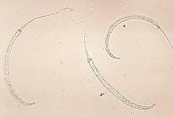
Once released, the L1 larvae measure 0.3–0.9 millimeters in length and feature a very long tapered tail. As they develop into L3 larvae, they lose the tapered tail, broaden, and develop a tri-lobed tail; the lengths of most L3 larvae are unknown. [1] Adults of both sexes are narrow yellow-white colored worms, with a rounded front-end, and a conical tail-end with a pointed tip. Females of different species within the genus tend to look similar, and can rarely be distinguished on morphology alone. [1] The body of a fertilized adult female is almost completely filled by its uterus, distended with L1 larvae. [1] Adult female Dracunculus worms are noted for their extraordinary length, with some growing up to 100 centimeters long. Males are much smaller (16 – 40 millimeters) and are relatively rare – in some species the male has never been described. [1]

Distribution

Dracunculus worms are distributed globally, though each species has a narrower range. The majority of Dracunculus species described infect reptiles – predominantly snakes. [1] These are spread across the globe, with D. ophidensis in the United States, D. brasiliensis in Brazil, D. coluberensis and D. alii in India, D. houdemeri in Vietnam, D. doi in Madagascar, D. dahomensis in Benin, D. oesophageus in Italy, and D. mulbus in Australia and Papua New Guinea. The only species known to infect a non-snake reptile is D. globocephalus which has been described in snapping turtles in the United States and Costa Rica. [1]

Most mammal-infecting species are in the Americas, with D. insignis infecting several wild and domestic mammals in the United States and Canada, D. lutrae infecting river otters in the United States and Canada, and D. fuelleborni infecting big-eared opposums in Brazil. [1] The major exception is D. medinensis, also known as Guinea Worm Disease, as it is by far the most studied Dracunculus as it infects humans. [2] [3] D. medinensis was historically widespread in sub-saharan Africa and South Asia, [1] but is now limited to dozens of cases annually in humans and domestic dogs, and may soon be driven to extinction due to eradication efforts. [4] [5]

Species

Reptile-infecting species

There are 14 accepted Dracunculus species, 10 of which infect reptiles. Eurasia hosts several reptile-infecting Dracunculus species. D. oesophageus was originally described from the esophagus of the viperine water snake , and has been described several times since. The remaining three Eurasian reptile-infecting species have been described a single time each: D. coluberensis from an Indian trinket snake, and D. alii and D. houdemeri from Checkered keelback snakes in India and Vietnam respectively. [1]

The only snake-infecting Dracunculus species known in North America is D. ophidensis. It was originally described in garter snakes in Michigan and Minnesota by Sterling Brackett in 1938, and has since been reported in blackbelly garter snakes from Mexico, as well as northern water snakes and a plain-bellied water snake in Michigan. [1] D. brasiliensis is the only described snake-infecting Dracunculus in South America. It was described in 2009 based on a single female worm from an anaconda in Brazil, and has since also been found in a Brazilian brown-banded water snake. [1] Several worms that appear to be from the genus Dracunculus have been described in Central America, South America, and the Caribbean; however, they have not been described in sufficient detail to assign them to a species. [1] The Americas are also home to the only known turtle-infecting Dracunculus (also the only species that infects a non-snake reptile), D. globocephalus. First described in 1927 in Oklahoma and Illinois, it is now found in snapping turtles across the United States, as well as the South American snapping turtle in Costa Rica.

Africa hosts two known snake-infecting species. Both male and female D. doi were described from Madagascar ground boas in 1960 and 1973 respectively. D. dahomensis has been described only from a captive African rock python. [1]

In Australia, the only known snake-infecting Dracunculus is D. mulbus, described from numerous water pythons in Northern Australia in 2007. It has since been described in Papua New Guinea's Papuan olive python as well. [1]

Mammal-infecting species

The life cycle of Dracunculus medinensis Drac life cycle.png
The life cycle of Dracunculus medinensis

Just four Dracunculus species are known to infect mammals, of which the best known is the human parasite D. medinensis. Historically spread across Africa and South Asia, a major eradication effort has restricted D. medinensis to just Chad, Ethiopia, Mali, and South Sudan. [1] Case numbers have similarly fallen, from an estimated 3.5 million per year at the 1986 start of the eradication program, to just 15 in 2021. [6] [7] D. medinensis is now most common in dogs, particularly in Chad, where it may spread via fish or frogs as paratenic hosts. [8]

D. insignis infects dogs and wild carnivores, causing cutaneous lesions, ulcers, and sometimes heart and vertebral column lesions. Like D. medinensis, it is also known as Guinea worm, as well as Dragon or Fiery Dragon. The range of D. insignis is limited to North America.

D. fuelliborni parasitizes opossum, D. lutrae parasitizes otters, and D. ophidensis parasitizes reptiles.

Related Research Articles

<i>Strongyloides stercoralis</i> Species of worm

Strongyloides stercoralis is a human pathogenic parasitic roundworm causing the disease strongyloidiasis. Its common name in the US is threadworm. In the UK and Australia, however, the term threadworm can also refer to nematodes of the genus Enterobius, otherwise known as pinworms.

<span class="mw-page-title-main">Copepod</span> Subclass of crustaceans

Copepods are a group of small crustaceans found in nearly every freshwater and saltwater habitat. Some species are planktonic, some are benthic, a number of species have parasitic phases, and some continental species may live in limnoterrestrial habitats and other wet terrestrial places, such as swamps, under leaf fall in wet forests, bogs, springs, ephemeral ponds, puddles, damp moss, or water-filled recesses of plants (phytotelmata) such as bromeliads and pitcher plants. Many live underground in marine and freshwater caves, sinkholes, or stream beds. Copepods are sometimes used as biodiversity indicators.

<span class="mw-page-title-main">Dracunculiasis</span> Infection by the Guinea worm

Dracunculiasis, also called Guinea-worm disease, is a parasitic infection by the Guinea worm, Dracunculus medinensis. A person becomes infected by drinking water contaminated with Guinea-worm larvae that reside inside copepods. Stomach acid digests the copepod and releases the Guinea worm, which penetrates the digestive tract and escapes into the body. Around a year later, the adult female migrates to an exit site – usually the lower leg – and induces an intensely painful blister on the skin. The blister eventually bursts to form a painful wound, out of which the worm slowly crawls over several weeks. The wound remains painful throughout the worm's emergence, disabling the affected person for the three to ten weeks it takes the worm to emerge.

<i>Haemonchus contortus</i> Species of roundworm

Haemonchus contortus, also known as the barber's pole worm, is a very common parasite and one of the most pathogenic nematodes of ruminants. Adult worms attach to abomasal mucosa and feed on the blood. This parasite is responsible for anemia, oedema, and death of infected sheep and goats, mainly during summer in warm, humid climates.

<i>Dirofilaria immitis</i> Species of worm that causes parasitic disease in animals

Dirofilaria immitis, also known as heartworm or dog heartworm, is a parasitic roundworm that is a type of filarial worm, a small thread-like worm, and which causes dirofilariasis. It is spread from host to host through the bites of mosquitoes. Four genera of mosquitoes transmit dirofilariasis, Aedes, Culex, Anopheles, and Mansonia. The definitive host is the dog, but it can also infect cats, wolves, coyotes, jackals, foxes, ferrets, bears, seals, sea lions and, under rare circumstances, humans.

<i>Dictyocaulus</i> Genus of nematode parasites of the bronchial tree of horses, sheep, goats, deer, and cattle

Dictyocaulus is a genus of nematode parasites of the bronchial tree of horses, sheep, goats, deer, and cattle. Dictyocaulus arnfieldi is the lungworm of horses, and Dictyocaulus viviparus is the lungworm affecting ruminants.

Spirometra erinaceieuropaei is a parasitic tapeworm that infects domestic animals and humans. The medical term for this infection in humans and other animals is sparganosis. Morphologically, these worms are similar to other worms in the genus Spirometra. They have a long body consisting of three sections: the scolex, the neck, and the strobilia. They have a complex life cycle that consists of three hosts, and can live in varying environments and bodily tissues. Humans can contract this parasite in three main ways. Historically, humans are considered a paratenic host; however, the first case of an adult S. erinaceieuropaei infection in humans was reported in 2017. Spirometra tapeworms exist worldwide and infection is common in animals, but S. erinaceieuropaei infections are rare in humans. Treatment for infection typically includes surgical removal and anti-worm medication.

Sparganosis is a parasitic infection caused by the plerocercoid larvae of the genus Spirometra including S. mansoni, S. ranarum, S. mansonoides and S. erinacei. It was first described by Patrick Manson in 1882, and the first human case was reported by Charles Wardell Stiles from Florida in 1908. The infection is transmitted by ingestion of contaminated water, ingestion of a second intermediate host such as a frog or snake, or contact between a second intermediate host and an open wound or mucous membrane. Humans are the accidental hosts in the life cycle, while dogs, cats, and other mammals are definitive hosts. Copepods are the first intermediate hosts, and various amphibians and reptiles are second intermediate hosts.

<i>Dracunculus medinensis</i> Species of parasitic worm

Dracunculus medinensis is a nematode that causes dracunculiasis, also known as guinea worm disease. The disease is caused by the female which, at around 80 centimetres in length, is among the longest nematodes infecting humans. In contrast, the longest recorded male Guinea worm is only 4 cm.

<span class="mw-page-title-main">Strongylida</span> Suborder of roundworms

The Strongylida suborder includes many of the important nematodes found in the gastrointestinal tracts of ruminants, horses, and swine, as well as the lungworms of ruminants and the hookworms of dogs and cats.

In population ecology, density-dependent processes occur when population growth rates are regulated by the density of a population. This article will focus on density dependence in the context of macroparasite life cycles.

<span class="mw-page-title-main">Microfilaria</span> Early stage in the life cycle of certain parasitic nematodes in the family Onchocercidae

The microfilaria is an early stage in the life cycle of certain parasitic nematodes in the family Onchocercidae. In these species, the adults live in a tissue or the circulatory system of vertebrates. They release microfilariae into the bloodstream of the vertebrate host. The microfilariae are taken up by blood-feeding arthropod vectors. In the intermediate host the microfilariae develop into infective larvae that can be transmitted to a new vertebrate host.

<i>Thelazia callipaeda</i> Species of worm

Thelazia callipaeda is a parasitic nematode, and the most common cause of thelaziasis in humans, dogs and cats. It was first discovered in the eyes of a dog in China in 1910. By 2000, over 250 human cases had been reported in the medical literature.

<span class="mw-page-title-main">Thelaziasis</span> Medical condition

Thelaziasis is the term for infestation with parasitic nematodes of the genus Thelazia. The adults of all Thelazia species discovered so far inhabit the eyes and associated tissues of various mammal and bird hosts, including humans. Thelazia nematodes are often referred to as "eyeworms".

<i>Trichostrongylus</i> Genus of roundworms

Trichostrongylus species are nematodes, which are ubiquitous among herbivores worldwide, including cattle, sheep, donkeys, goats, deer, and rabbits. At least 10 Trichostrongylus species have been associated with human infections. Infections occur via ingestion of infective larvae from contaminated vegetables or water. Epidemiological studies indicate a worldwide distribution of Trichostrongylus infections in humans, with the highest prevalence rates observed in individuals from regions with poor sanitary conditions, in rural areas, or who are farmers / herders. Human infections are most prevalent in the Middle East and Asia, with a worldwide estimated prevalence of 5.5 million people.

<i>Angiostrongylus vasorum</i> Species of roundworm

Angiostrongylus vasorum, also known as French heartworm, is a species of parasitic nematode in the family Metastrongylidae. It causes the disease canine angiostrongylosis in dogs. It is not zoonotic, that is, it cannot be transmitted to humans.

Gnathostoma hispidum is a nematode (roundworm) that infects many vertebrate animals including humans. Infection of Gnathostoma hispidum, like many species of Gnathostoma causes the disease gnathostomiasis due to the migration of immature worms in the tissues.

<span class="mw-page-title-main">Eradication of dracunculiasis</span> Permanent global eradication from humans

Eradication of dracunculiasis is an ongoing program. Dracunculiasis, or Guinea worm disease, is an infection by the Guinea worm. In 1986, there were an estimated 3.5 million cases of Guinea worm in 20 endemic nations in Asia and Africa. Ghana alone reported 180 000 cases in 1989. The number of cases has since been reduced by more than 99.999% to 14 in 2023 in five remaining endemic states: South Sudan, Chad, Mali, Ethiopia, Angola and Central African Republic.

<span class="mw-page-title-main">Hookworm</span> Intestinal, blood-feeding, parasitic roundworms that cause types of infection known as helminthiases

Hookworms are intestinal, blood-feeding, parasitic roundworms that cause types of infection known as helminthiases. Hookworm infection is found in many parts of the world, and is common in areas with poor access to adequate water, sanitation, and hygiene. In humans, infections are caused by two main species of roundworm, belonging to the genera Ancylostoma and Necator. In other animals the main parasites are species of Ancylostoma. Hookworm is closely associated with poverty because it is most often found in impoverished areas, and its symptoms promote poverty through the educational and health effects it has on children. It is the leading cause of anemia and undernutrition in developing countries, while being one of the most commonly occurring diseases among poor people. Hookworm thrives in areas where rainfall is sufficient and keeps the soil from drying out, and where temperatures are higher, making rural, coastal areas prime conditions for the parasite to breed.

<span class="mw-page-title-main">Nematode infection in dogs</span> Threadworm infections of dogs are frequent

Nematode infection in dogs - the infection of dogs with parasitic nemamotodes - are, along with tapeworm infections and infections with protozoa, frequent parasitoses in veterinary practice. Nematodes, as so-called endoparasites, colonize various internal organs - most of them the digestive tract - and the skin. To date, about 30 different species of nematode have been identified in domestic dogs; they are essentially also found in wild dog species. However, the majority of them often cause no or only minor symptoms of disease in adult animals. The infection therefore does not necessarily have to manifest itself in a worm disease (helminthosis). For most nematodes, an infection can be detected by examining the feces for eggs or larvae. Roundworm infection in dogs and the hookworm in dogs is of particular health significance in Central Europe, as they can also be transmitted to humans (zoonosis). Regular deworming can significantly reduce the frequency of infection and thus the risk of infection for humans and dogs.

References

  1. 1 2 3 4 5 6 7 8 9 10 11 12 13 14 15 16 17 Cleveland CA, Garrett KB, Cozad RA, Williams BM, Murray MH, Yabsley MJ (December 2018). "The wild world of Guinea Worms: A review of the genus Dracunculus in wildlife". Int J Parasitol Parasites Wildl. 7 (3): 289–300. doi:10.1016/j.ijppaw.2018.07.002. PMC   6072916 . PMID   30094178.
  2. Hopkins, Donald R. (2023). "Progress Toward Eradication of Dracunculiasis — Worldwide, January 2022–June 2023". MMWR. Morbidity and Mortality Weekly Report. 72 (45): 1230–1236. doi:10.15585/mmwr.mm7245a4. ISSN   0149-2195. PMC   10651320 . PMID   37943706.
  3. Yabsley, Michael J.; Garrett, Kayla B.; Thompson, Alec T.; Box, Erin K.; Giner, Madeline R.; Haynes, Ellen; Barron, Heather; Schneider, Renata M.; Coker, Sarah M.; Beasley, James C.; Borchert, Ernest J.; Tumlison, Renn; Surf, Allison; Dukes, Casey G.; Olfenbuttel, Colleen (April 2024). "Otterly diverse - A high diversity of Dracunculus species (Spirurida: Dracunculoidea) in North American river otters (Lontra canadensis)". International Journal for Parasitology: Parasites and Wildlife. 23: 100922. doi:10.1016/j.ijppaw.2024.100922. ISSN   2213-2244. PMC   10955650 . PMID   38516639.
  4. Yabsley, Michael J.; Garrett, Kayla B.; Thompson, Alec T.; Box, Erin K.; Giner, Madeline R.; Haynes, Ellen; Barron, Heather; Schneider, Renata M.; Coker, Sarah M.; Beasley, James C.; Borchert, Ernest J.; Tumlison, Renn; Surf, Allison; Dukes, Casey G.; Olfenbuttel, Colleen (April 2024). "Otterly diverse - A high diversity of Dracunculus species (Spirurida: Dracunculoidea) in North American river otters (Lontra canadensis)". International Journal for Parasitology: Parasites and Wildlife. 23: 100922. doi:10.1016/j.ijppaw.2024.100922. ISSN   2213-2244. PMC   10955650 . PMID   38516639.
  5. "Dracunculiasis (guinea-worm disease)". World Health Organization. 10 January 2022. Retrieved 5 September 2022.
  6. Despommier DD, Griffin DO, Gwadz RW, Hotez PJ, Knirsch CA (2019). "25. Dracunculus medinensis". Parasitic Diseases (PDF) (7 ed.). New York: Parasites Without Borders. pp. 285–290. Retrieved 26 January 2021.
  7. Dracunculiasis Eradication: Global Surveillance Summary, 2021 (Report). World Health Organization. 27 May 2022. Retrieved 11 October 2022.
  8. Molyneux D, Sankara DP (April 2017). "Guinea worm eradication: Progress and challenges- should we beware of the dog?". PLOS Negl Trop Dis. 11 (4): e0005495. doi: 10.1371/journal.pntd.0005495 . PMC   5398503 . PMID   28426663.